
Research article · 2025 · Full text
International Journal of Veterinary Sciences and Animal HusbandrySurgical management of testicular tumour in a senior dog
Volume 10(1) · Pages 04–07 · DOI: 10.22271/veterinary.2025.v10.i1a.1981 ↗
Research areasTesticular diseases and treatmentsDiet and metabolism studiesVeterinary Medicine and Surgery
Abstract
Author-writtenThis case report details the surgical management of testicular tumour in a 9 year old male Spitz dog. The animal was presented at DVC, Kozhikode with a history of a fluctuating pendulous swelling at caudo-ventral abdominal region since the past few months. There was generalized alopecia without signs of pruritus except for the head. Palpation of the mass did not elicit any pain reflex and the mass appeared to be hard, but mildly fluctuating. Ultrasonography showed that the mass was an undescended testicle. Fine needle aspiration cytology of the mass revealed spermatozoa and abnormal cells. The condition was diagnosed as testicular tumour of the undescended testicle. Surgical removal of the testicular mass under general anaesthesia was done and the dilated inguinal hernia ring was apposed using PGA No. 0.On histological examination, the tumour was confirmed as Sertoli cell tumour. Post-operative antibiotics and pain medications were administered for 7 days. The surgical wound was cleaned and dressed aseptically. Sutures were removed 14 days post the surgery. The animal had an uneventful recovery. Clinical signs of feminization were found to be disappearing with regrowth of body hairs.
Summary
Source-checkedPlain-language reading of the full paper, prepared with AI and checked against the text. Each point links to the authors' own words.
Why it matters
- Primary testicular tumors are the most common neoplasms found in the male genitalia of dogs, representing about 90% of such cases. View evidence ↗
- Cryptorchidism, where testes fail to descend, significantly increases the risk of developing testicular tumors. View evidence ↗
- Sertoli cell tumors can cause feminization syndrome in 25-50% of cases due to the excessive secretion of estrogen. View evidence ↗
The question
- The study aims to detail the diagnosis and surgical management of a Sertoli cell tumor in an older male Spitz dog. View evidence ↗
What they did
- The subject was a 9-year-old male Spitz dog weighing 8.5 kg presenting with a pendulous abdominal mass and generalized alopecia. View evidence ↗
- Diagnosis was achieved using abdominal ultrasonography to identify the undescended testicle and fine needle aspiration cytology to detect abnormal cells and spermatozoa. View evidence ↗
- The tumor was surgically removed via an inguinal approach, with the vascular bundle and spermatic cord double ligated using PGA No. 0 suture. View evidence ↗
- The dilated inguinal canal was corrected using a simple interrupted pattern with PGA No. 0 suture while preserving vascular structures. View evidence ↗
What they found
- Histological examination confirmed that the testicular mass was a Sertoli cell tumor. View evidence ↗
- The resected testicular tumor weighed 800 grams. View evidence ↗
- Pre-operative hematology showed a mild elevation in total leukocyte count (21 ×10^3/μL) and a low platelet count (94 ×10^3/μL). View evidence ↗
- Post-operative recovery included hair regrowth starting at one month, returning to normalcy by three months, and regaining the original pattern by six months. View evidence ↗
- The dog experienced an uneventful recovery, with the clinical signs of feminization disappearing as body hair regrew. View evidence ↗
What it means
- The authors conclude that surgical excision of the tumor successfully restored hormonal balance and alleviated hyperestrogenism, leading to complete recovery. View evidence ↗
- The authors recommend prompt veterinary care and regular follow-up monitoring to ensure the long-term health of dogs with similar conditions. View evidence ↗
Visual summary
1 visualsIllustrations explain the paper; they do not show study data.
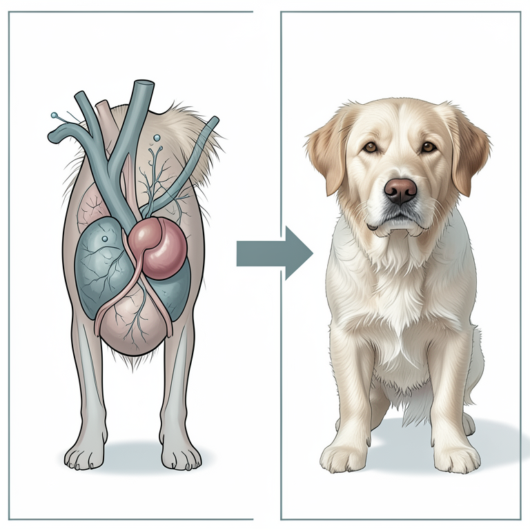
Hormonal Feedback Loop of Sertoli Cell Tumors
AI-generated conceptual illustration (senior dog with a testicular Sertoli cell tumor). It explains the idea and does not show study data.
View source evidence ↗Figures & tables
From the original paper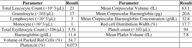 Table 1
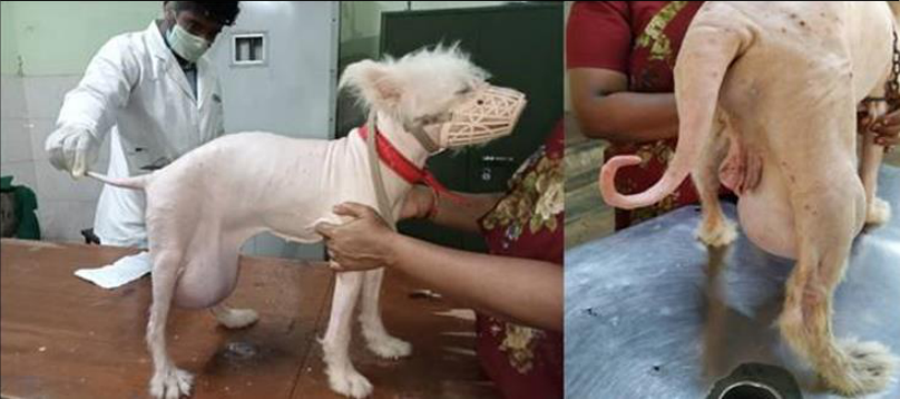 Figure 1
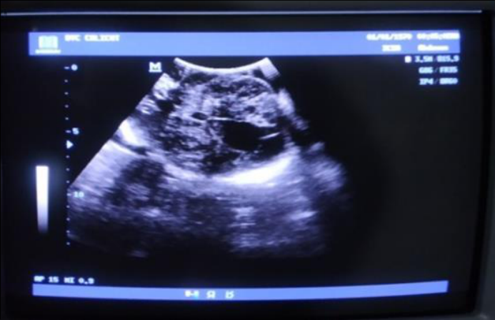 Figure 2
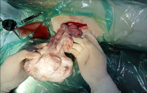 Figure 3
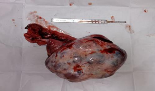 Figure 4
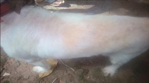 Figure 5
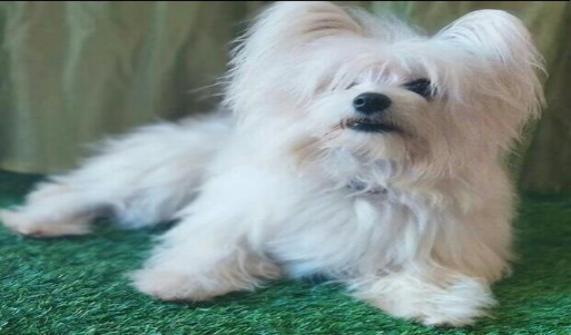 Figure 6
Introduction
Primary testicular tumors are the most prevalent neoplasms in male dogs, accounting for approximately 90% of all tumors affecting the male genitalia (Liao et al., 2009) [1] . According to the World Health Organization (WHO) classification of tumors in domestic animals, the primary types of testicular tumors in dogs include sex cord stromal tumors, germ cell tumors, and mixed germ cell sex cord stromal tumors (Nielsen and Lein, 1974) [2] . Among these, Sertoli cell tumors (SCTs) and interstitial cell tumors (ICTs) are the most common within the sex cord stromal category. Several factors contribute to the development of testicular tumors in dogs, including advanced age, specific breeds, cryptorchidism, and exposure to environmental carcinogens. Older male dogs, particularly those aged nine years or older, are at greater risk. Cryptorchidism significantly increases the likelihood of tumor development, as undescended testes are more prone to neoplasia. Certain breeds, such as Boxers, German Shepherds, Afghan Hounds, Weimaraners, and Shetland Sheepdogs, are also more predisposed to these tumors. Clinically, affected dogs often present with unilateral or bilateral testicular enlargement, along with firmness or irregularities in the testicular structure. Systemic signs include feminization syndrome, which occurs in approximately 25-50% of cases and is attributed to excessive estrogen secretion by neoplastic Sertoli cells (Padaliya et al., 2023) [3] . This dysregulated endocrine activity leads to overproduction of estrogenic hormones such as estradiol-17β. Elevated estrogen levels disrupt the hypothalamic-pituitary-gonadal (HPG) axis, suppressing testosterone production by Leydig cells through feedback inhibition. Additionally, increased inhibin secretion by Sertoli cells exacerbates the suppression of follicle-stimulating hormone (FSH), further impairing testicular function. The systemic effects of hyperestrogenism include notable skin changes, such as atrophy of sebaceous glands, alterations in hair follicle cycling, bilateral symmetrical alopecia, and hyperpigmentation (Gokulakrishnan et al., 2023) [4] .
Importantly, these clinical signs are generally reversible with the surgical excision of the tumor. The present paper details the diagnosis and surgical management of a Sertoli cell tumor in a senior male Spitz dog, highlighting the clinical presentation, diagnostic approach, and successful therapeutic intervention.
Case History and Clinical Examination
A 9 year old male Spitz dog weighing 8.5 kg was presented at DVC Kozhikode with a history of a pendulous tumour at caudo-ventral abdominal region since last few months and generalized alopecia, except for the head. Clinical examination revealed a respiratory rate of 32 breaths per minute, pulse rate of 120 beats per minute, pale roseate mucous membrane and a rectal temperature of 102.1oF. On palpation of caudal ventral abdomen, a mass could be found which was hard in consistency. Only one testicle was descended. Complete haemogram was unremarkable except for a mild elevation of total leukocyte count and low platelet count (Table ). Ultrasound examination of the abdomen was performed and the mass was identified as identified as an undescended testicle (Figure 1). The lack of testicle inside scrotum helped to confirm the mass as undescended testicle. Further the diagnosis was confirmed by a fine needle aspiration cytology of the mass which revelaed the presence of spermatozoa and abnormal cells. Based on history, clinical sign, physical examination and ultrasonography and cyutological examination findings, the condition was diagnosed as testicular tumour of the undescended testicle.
Surgical correction 3.1 Preoperative Care
The owner was advised to withhold food and water for 12 hours and 6 hours prior to surgery, respectively. The skin above the tumour mass was clipped, shaved, surgically scrubbed and aseptically prepared with tincture of iodine solution. Prior to the surgery, the animal was given Ceftriaxone tazobactum at the dose rate of 20mg/kg body weight intravenously and meloxicam at the dose rate of 0.2 mg/kg s/c. Animal was premedicated with injections Inj. Atropine at the dose rate of 0.045 mg/kg S/c and Xylazine (20mg/mL) at a dose rate of 1mg/kg body weight im. General anaesthesia was induced with Propofol at a dose rate of 4mg/kg bodyweight iv, Ketamine (50mg/mL) at a dose rate of 5mg/kg body weight iv and midazolam (1mg/mL) at a dose rate of 0.2mg/kg iv. The anaesthesia was maintained with intermittent bolus dose of ketamine and midazolam at the dose of 2.5 mg/kg and 0.1 mg/kg body weight respectively. The dog was placed on lateral recumbency, the thoracic limbs were fixed cranially and the pelvic limbs were fixed caudally in a relaxed position.
Surgical procedure
The skin incision was made using a No 22 BP blade over the inguinal region. The incision was extended to exteriorize the tumour mass (Figure 3, Figure 4), after exteriorizing the tumour, the vascular bundle and spermatic cord was separated and double ligated using suture PGA No. 0. The dilated inguinal canal was corrected using PGA No 0 in simple interrupted pattern without obliterating the vascular structures. The dead space was obliterated including muscle layer using Polyglactin (2/0) along with subcutis in a simple continuous pattern. The skin incision was apposed in horizontal mattress pattern using No 1 Nylon.
Post-Operative Recovery
Postoperatively, tincture benzoin was applied over suture line. Supportive therapry including antibacterials, analgesics, multivitamins and multi minerals were continued for 5 more days. The animal made an uneventful recovery and suture was removed on day 14 postoperatively. By one month the hairs started regrwoing and by three months (Figure 5) the hair coat had returned to normalcy. By six months post orchiectomy the animal had regained its original hair coat pattern (Figure 6).
Discussion
Sertoli cells, located within the seminiferous tubules of the testes, provide structural and nutritional support for spermatogenesis. They also maintain the blood-testis barrier, secrete inhibin to regulate follicle-stimulating hormone (FSH) levels, and produce androgen-binding protein to concentrate testosterone, essential for spermatogenesis (O'Donnell et al., 2022). Testicular tumors are among the most common neoplasms in older intact male dogs, with Sertoli cell tumors (SCTs) being one of the primary types. These tumors arise from the disordered, uncontrolled growth of Sertoli cells, which are responsible for supporting spermatogenesis. While testicular tumors involving scrotal testes are often benign, tumors in cryptorchid testes have a higher risk of malignancy (Thorup et al., 2024) [6] . Sertoli cell tumors can produce excessive estrogen, leading to feminization. This manifests as systemic effects such as bilateral alopecia, hyperpigmentation, gynecomastia, and testicular atrophy. Hyperestrogenism also disrupts the hypothalamic-pituitary-gonadal (HPG) axis, causing reduced testosterone production, atrophy of sebaceous glands, and impaired spermatogenesis (Herzog et al., 2023, Welle, 2023) [7, 8] . These systemic effects significantly impact the overall health and well-being of affected animals.
In the present case the preanaesthesia involved a combination of atropine and xylazine. The combination provides significant advantages, including sedation, analgesia, and reduced salivary and bronchial secretions, which improve the safety and comfort of the surgical procedure (Adami et al., 2023) [9] . Xylazine also offers muscle relaxation, making it easier to manage the patient during surgery (Hasan and SM Alnajjar, 2023) [10] . Induction with a combination of propofol and ketamine is advantageous for maintaining stable anesthesia with minimal cardiopulmonary depression. Propofol provides rapid induction and recovery (Walters et al., 2022) [11] , while ketamine ensures prolonged analgesia and prevents sudden arousal during surgery (Guzel et al., 2022) [12] . The intermittent bolus administration of ketamine and midazolam effectively maintained anesthesia throughout the procedure. Surgical excision via castration is the definitive treatment for testicular tumors. The surgical technique varies based on the location of the tumor. For scrotal tumors, scrotal ablation or standard orchiectomy is typically performed, while for tumors involving cryptorchid testes, inguinal or abdominal approaches are necessary. In this case, an inguinal approach was used to exteriorize and remove the tumor. Careful ligation of the vascular bundle and spermatic cord, along with correction of the inguinal canal, was critical in preventing complications. The inguinal canal was closed in a manner that preserved vascular structures, which minimized postoperative risks. Among the surgical techniques, the inguinal approach for cryptorchid testes is considered optimal due to better accessibility, minimal invasiveness, and reduced complications compared to abdominal surgery (Faulkner et al., 2024) [13] . Recovery rates following surgical correction are typically excellent, with most dogs showing significant improvement in clinical signs within weeks to months. Physiologically, the removal of the tumor eliminates the excessive production of estrogen. This cessation allows the hypothalamic-pituitary-gonadal axis to regain normal function, leading to the resumption of testosterone production and the resolution of hyperestrogenic effects. In the present case, the animal exhibited an uneventful recovery, with the surgical site healing well and hair regrowth beginning by one month postoperatively. By three months, the dog's coat had returned to normalcy, indicating complete resolution of feminization syndrome. Postoperative recovery can be assessed through the resolution of clinical signs, such as the normalization of hair growth, disappearance of hyperpigmentation, and improvement in overall health. Hormonal assays measuring estrogen and testosterone levels provide objective markers of recovery. Additionally, regular follow-ups to monitor surgical site healing and the absence of any systemic complications.
Conclusion
In conclusion, Sertoli cell tumors represent a significant health concern in older male dogs, particularly those with cryptorchidism. This case highlights the importance of early diagnosis and surgical intervention in managing testicular tumors. The successful surgical excision of the tumor not only alleviated the clinical signs associated with hyperestrogenism but also restored normal hormonal balance, leading to a complete recovery. The use of a combination of anesthetic agents ensured a safe surgical procedure, while the inguinal approach provided optimal access for tumor removal. Regular follow-up and monitoring are essential to ensure the longterm health and well-being of affected dogs, emphasizing the need for awareness and prompt veterinary care in similar cases.
Figures and tables from the paper
| Parameter | Result | Parameter | Result |
|---|---|---|---|
| Total Leucocyte Count (×10^3/μL) | 21 | Mean Corpuscular Volume (fL) | 63.1 |
| Granulocyte (×103/μL) | 17 | Mean Corpuscular Haemoglobin (pg) | 20.6 |
| Lymphocytes (×10^3/μL) | 3 | Mean Corpuscular Haemoglobin Concentration (g/dL) | 32.8 |
| Monocyte (×10^3/μL) | 1 | Red cell Distribution Width (%) | 17.7 |
| Total Erythrocyte Count (×106/μL) | 5.51 | Platelt count (×103/μL) | 94 |
| Haemoglobin (g/dL) | 11.4 | Mean Platlet Volume (fL) | 7.8 |
| Volume of Packed Red Cells (%) | 34.7 | ||
| Platletcrit (%) | 0.073 |
Table text extracted from the original PDF; superscript letters are shown inline. The original table image is below.
View original table image
References
13 in the original paper- Liao AT, Chu PY, Yeh LS, Lin CT, Liu CH. A 12-year retrospective study of canine testicular tumors. J Vet Med Sci. 2009;71:919-23.
- Nielsen SW, Lein DH. Tumours of the testis. Bull World Health Organ. 1974;50:71.
- Padaliya NR, Vadalia JV, Talekar SH, Bhatt RH, Bhadaniya AR, Kalaria VA. Surgical management of sertoli cell tumour in dogs: Report of two cases. Indian J Vet Sci Biotechnol. 2023;19:101-103.
- Gokulakrishnan M, Rasool A, Sarath T, Jeyaraja K, Ali M. Testicular tumor and associated clinical, hormonal and hematobiochemical symptoms and its successful surgical management in canines. Explor Anim Med Res, 2023, p. 13.
- O'donnell L, Smith LB, Rebourcet D. Sertoli cells as key drivers of testis function. Semin Cell Dev Biol, Elsevier, 2022, p. 2-9.
- Thorup J, Hildorf S, Hildorf AE, Baastrup JM, Mamsen LS, Andersen CY, et al. The fate of germ cells in cryptorchid testis. Front Endocrinol. 2024;14:1305428.
- Herzog F, Adams D, Blanc AS, Lee J, Langohr I, Pinto C. Canine Sertoli cell tumor: anti-Müllerian hormone, inhibin B, and estrone sulphate. Clin Theriogenol, 2023, p. 15.
- Welle MM. Canine noninflammatory alopecia: An approach to its classification and a diagnostic aid. Vet Pathol. 2023;60:748-769.
- Adami C, Bergadano A, Casoni D. Tranquilizers, sedatives, local anaesthetics and antimuscarinic agents. In: Anesthesia and Analgesia in Laboratory Animals. Elsevier, 2023.
- Hasan NH, Alnajjar SM. Comparative study between IV and IM co-administration of xylazine with ketamine on some physiological parameters in dogs. Egypt J Vet Sci. 2023;54:1237-1244.
- Walters K, Lehnus K, Liu NC, Bigby SE. Determining an optimum propofol infusion rate for induction of anaesthesia in healthy dogs: A randomized clinical trial. Vet Anaesth Analg. 2022;49:243-250.
- Guzel O, Sevim G, Kaya DA, Sezer D, Erek M, Gursel FE, et al. Ketamine or propofol anesthesia in dogs: how do they affect cytokines, antioxidants and neutrophil functions? J Hellenic Vet Med Soc. 2022;73:3783-3792.
- Faulkner EB, Kimmerlein A, Morrison J, Beal T, Kerl M, Saito EK. Serious surgical complications of canine cryptorchid castration are associated with surgical approach: A case-control study of 202 dogs. J Am Vet Med Assoc. 2024;1:1-6.