
Research article · 2025 · Full text
International Journal of Veterinary Sciences and Animal HusbandryUse of echocardiography and cardiac biomarkers for the diagnosis and therapeutic management of Mitral Valve Disease (MVD) in dogs
Volume 10(1) · Pages 01–03 · DOI: 10.22271/veterinary.2025.v10.i1a.1980 ↗
Research areasCardiovascular Conditions and TreatmentsCardiac Valve Diseases and TreatmentsMechanical Circulatory Support Devices
Abstract
Author-writtenThis case report describes the clinical presentation, diagnosis, and management of Myxomatous Mitral Valve Disease (MMVD) in an 8-year-old intact male Shih Tzu named Oreo. Oreo presented with chronic coughing, exercise intolerance, and increased respiratory effort. Diagnostics revealed a Grade IV/VI systolic murmur, cardiomegaly, pulmonary congestion, elevated NT-proBNP and Troponin I along with mitral valve abnormalities on echocardiography. A comprehensive treatment regimen including furosemide, pimobendan, an ACE inhibitor, spironolactone, and dietary modifications led to clinical improvement. This report highlights the significance of early diagnosis and multimodal therapy in managing MMVD in small-breed dogs.
Summary
Source-checkedPlain-language reading of the full paper, prepared with AI and checked against the text. Each point links to the authors' own words.
Why it matters
- Myxomatous Mitral Valve Disease (MMVD), or degenerative mitral valve disease, is the most frequent acquired heart disorder in dogs, particularly in older small-breed dogs. View evidence ↗
- The disease involves the gradual breakdown of the mitral valve leaflets and chordae tendineae, which leads to mitral regurgitation and overload of the left side of the heart. View evidence ↗
- MMVD is a major cause of illness in dogs, representing about 75% of all canine cardiovascular disorders. View evidence ↗
The question
- The study aims to describe the clinical presentation, diagnosis, and treatment of MMVD in an 8-year-old male Shih Tzu, specifically focusing on echocardiography and outcomes. View evidence ↗
What they did
- The subject was an 8-year-old male Shih Tzu treated at the Veterinary Clinical Complex of Kamdhenu University in Gujarat. View evidence ↗
- The diagnostic process utilized auscultation, thoracic X-rays, electrocardiography (ECG), and echocardiography. View evidence ↗
- Cardiac biomarkers, specifically NT-proBNP and cardiac troponin I, were measured to evaluate heart health. View evidence ↗
- The treatment regimen consisted of Benazepril (0.25 mg/kg), Spironolactone, Furosemide (2 mg/kg), Pimobendan (0.25 mg/kg), and a low-sodium diet. View evidence ↗
What they found
- Physical examination of the dog revealed a Grade IV/VI left apical systolic murmur. View evidence ↗
- The dog exhibited tachycardia and tachypnea, with a heart rate of 170 bpm, pulse rate of 165 bpm, and respiratory rate of 70 breaths per minute. View evidence ↗
- Cardiac biomarkers were significantly elevated, with NT-proBNP at 1752 pmol/L (normal < 900 pmol/L) and Troponin I at 0.7 ng/mL (normal < 0.1 ng/mL). View evidence ↗
- Echocardiography confirmed MMVD, showing an enlarged left atrium (3.15 cm), a dilated aorta (0.88 cm), and a left atrium/aorta ratio of 3.58 cm. View evidence ↗
- X-rays showed pulmonary congestion, an enlarged pulmonary vein, and left atrial enlargement, indicating fluid accumulation in the lungs. View evidence ↗
- The ECG showed sinus tachycardia at 172 bpm, P-wave abnormalities suggesting atrial enlargement, and ST segment elevation in Lead II, which may indicate myocardial injury. View evidence ↗
- The addition of a short course of Prednisolone resulted in the complete resolution of coughing and improved activity levels. View evidence ↗
What it means
- The authors conclude that early diagnosis and a multimodal treatment approach are critical for managing MMVD in dogs. View evidence ↗
- The authors recommend early intervention and personalized treatment plans to improve the quality of life for affected dogs. View evidence ↗
Visual summary
1 visualsIllustrations explain the paper; they do not show study data.
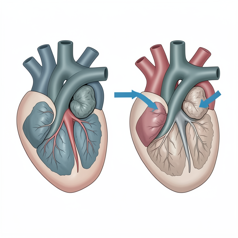
Mechanism of Mitral Valve Disease
AI-generated conceptual illustration (canine heart). It explains the idea and does not show study data.
View source evidence ↗Figures & tables
From the original paper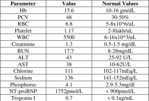 Table 1
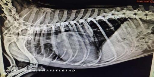 Figure 1
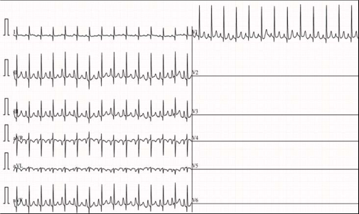 Figure 2
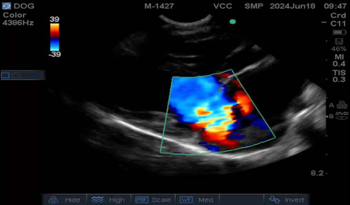 Figure 4
Introduction
Myxomatous Mitral Valve Disease (MMVD) also referred to as degenerative mitral valve disease is the most common acquired cardiac disorder in dogs predominantly affecting geriatric small-breed dogs (Atkins et al., 2009; Keene et al., 2019) [1,2] . The condition is characterized by progressive degeneration of the mitral valve leaflets and chordae tendineae resulting in mitral regurgitation (MR) and left-sided cardiac overload (Keene et al., 2019) [2] . This disease accounts for approximately 75% of canine cardiovascular disorders making it a significant contributor to morbidity in the canine population (Borgarelli & Buchanan, 2012) [3] . Clinical signs of MMVD vary with disease progression ranging from asymptomatic murmurs in early stages to congestive heart failure (CHF) in advanced cases (Häggström et al., 2005). Common clinical signs include coughing, exercise intolerance, tachypnea, and syncope which may worsen with disease severity. Diagnostic evaluation typically includes auscultation, thoracic radiography, electrocardiography (ECG) and echocardiography, the latter being the gold standard for diagnosing MR and assessing disease severity (Boon, 2011) [5] . This report describes the clinical presentation, diagnostic evaluation, and therapeutic management of MMVD in an 8-year-old male Shih Tzu, with a focus on echocardiographic findings and long-term outcomes.
Case History and Clinical Observations
An 8-year-old male Shih Tzu was presented at the Veterinary Clinical Complex (VCC), College of Veterinary Science & A.H., Kamdhenu University, Navsari, Gujarat, with a history of chronic coughing, particularly at night or following physical exertion. The dog also exhibited exercise intolerance and increased difficulty breathing at rest, with symptoms worsening over the past month. During the clinical examination, the rectal body temperature was recorded at 102.2°F, within the normal range. The mucous membranes appeared normal, and the capillary refill time (CRT) was less than 1 second, indicating adequate perfusion.
However, the heart rate was elevated at 170 beats per minute, with a pulse rate of 165 beats per minute, and the respiratory rate was increased at 70 breaths per minute. Auscultation revealed a Grade IV/VI left apical systolic murmur, and a pulse deficit was noted, suggesting potential cardiac involvement. These clinical findings raised concern for a cardiovascular condition, leading to further diagnostic evaluation.
Laboratory Findings and cardiac biomarkers evaluation
Complete blood count and serum biochemical parameters were within normal reference ranges with mildly decreased sodium (Table 1) may indicate a slight electrolyte imbalance potentially due to chronic heart disease or diuretic therapy. The elevation of biomarkers NT-proBNP (1752ng/mL) and cardiac troponin I (0.7ng/ml) were also noticed.
| Parameter | Value | Normal Values |
|---|---|---|
| Hb | 15.6 | 10-16 gm/dL |
| PCV | 48 | 30-50% |
| RBC | 6.8 | 5-8x10^6/uL |
| Platelet | 1.17 | 2-8lakh/uL |
| WBC | 5500 | 6-16x10^3/uL |
| Creatinine | 1.3 | 0.5-1.5 mg/dL |
| BUN | 17.7 | 8-28mg/dL |
| ALT | 43 | 25-92 U/L |
| AST | 38 | 10-62U/L |
| Chlorine | 111 | 102-117mEq/L |
| Sodium | 136 | 141-152mEq/L |
| Phosphorus | 4.1 | 2.9-5.3mg/dl |
| NT-proBNP | 1752pmol/L | < 900pmol/L |
| Troponin I | 0.7 | < 0.1ng/mL |
Table text extracted from the original PDF; superscript letters are shown inline. The original table image is below.
View original table image
Radiographic Findings
Thoracic radiographs revealed left atrial enlargement, an enlarged pulmonary vein and pulmonary congestion, all consistent with cardiac involvement and indicative of increased left heart pressure and impaired cardiac function with fluid accumulation in the lungs.
Clinical Management and Follow-Up Care
The therapeutic plan for managing mitral valve disease (MMVD) in this dog included a combination of pharmacologic and dietary interventions aimed at alleviating symptoms, improving cardiac function, and preventing disease progression. The medications prescribed were Benazepril (0.25 mg/kg every 12 hours orally), to reduce afterload and manage blood pressure, thereby decreasing the workload on the heart. A combination of Spironolactone and Furosemide (2 mg/kg every 12 hours orally) was used to address pulmonary edema and fluid retention, improving respiratory function and reducing symptoms of congestive heart failure. Pimobendan (0.25 mg/kg every 12 hours orally) was added to enhance cardiac contractility and reduce the heart's workload, thereby improving left ventricular efficiency. A low-sodium diet was recommended to minimize fluid retention and reduce cardiac stress, helping to control heart failure symptoms and slow disease progression. Followup care involved regular monitoring of cardiac parameters, including auscultation for murmurs and arrhythmias, echocardiography to assess valve function and ventricular dimensions, and radiographs to evaluate heart size and pulmonary congestion. Adjustments to the treatment plan were made based on clinical response and disease progression.
Outcome
After 60 days of treatment, the dog demonstrated a significant reduction in pulmonary congestion, indicating an improvement in heart failure symptoms. However, intermittent coughing persisted despite the initial therapeutic interventions. To address this, Prednisolone was added to the treatment regimen for a short duration, leading to an uneventful improvement in activity levels and complete resolution of the coughing. This adjustment helped further manage the symptoms and enhance the dog's overall quality of life.
Conclusion
This case underscores the importance of early diagnosis and a multi-modal therapeutic approach in managing dogs with Myxomatous Mitral Valve Disease (MMVD). Echocardiography plays a critical role in the assessment of cardiac parameters, providing valuable insights into the progression of the disease. Additionally, a combination of pharmacological therapy, including ACE inhibitors, diuretics, and positive inotropes, along with dietary adjustments, can significantly improve clinical outcomes. Early intervention and tailored treatment plans are essential for managing MMVD and enhancing the quality of life in affected dogs.
Acknowledgement
AcknowledgmentsThe authors thank the staff of the Veterinary Clinical Complex, Kamdhenu University, for their assistance in the management of the case
Figures and tables from the paper
References
7 in the original paper- Atkins CE, Bonagura JD, Ettinger SJ, Fox PR, Gordon SG, Häggström J, et al. Guidelines for the diagnosis and treatment of canine chronic valvular heart disease. J Vet Intern Med. 2009;23(6):1142-1150.
- Keene BW, Atkins CE, Bonagura JD, Fox PR, Häggström J, Fuentes VL, et al. ACVIM consensus guidelines for the diagnosis and treatment of myxomatous mitral valve disease in dogs. J Vet Intern Med. 2019;33(3):1127-1140.
- Borgarelli M, Buchanan JW. Historical review, epidemiology and natural history of degenerative mitral valve disease. J Vet Cardiol. 2012;14(1):93-101.
- Häggström J, Boswood A, O'Grady M, Jönsson L, Smith S, Swift S, et al. Effect of pimobendan or benazepril hydrochloride on survival times in dogs with congestive heart failure due to naturally occurring myxomatous mitral valve disease: The quest study. J Vet Intern Med. 2005;22(5):1124-1135.
- Boon JA. Veterinary Echocardiography. Hoboken, NJ: John Wiley & Sons, 2011.
- Oyama MA, Singletary GE. The use of NT-proBNP assay in the diagnosis of canine heart disease. Vet Clin North Am Small Anim Pract. 2010;40(4):545-558.
- Sleeper MM, Clifford CA, Laster LL. Cardiac troponin I in the normal dog and cat. J Vet Intern Med. 2001;15(5):501-503.