
Research article · 2024 · Full text
International Journal of Veterinary Sciences and Animal HusbandryCytological study on round cell tumors in dogs: Case report
Volume 9(6S) · Pages 299–301 · DOI: 10.22271/veterinary.2024.v9.i6se.1974 ↗
Research areasVeterinary Oncology ResearchSarcoma Diagnosis and TreatmentCancer Diagnosis and Treatment
Abstract
Author-writtenA 8 year old female dog was presented with a history of multiple dermal masses over the body and 9 year old male dog was presented with a history of masses near the neck and the shoulder region. Fine needle aspiration cytology was performed from both the case. Based on the cytology the samples were diagnosed as cuteneous TVT and mast cell tumor.
Summary
Source-checkedPlain-language reading of the full paper, prepared with AI and checked against the text. Each point links to the authors' own words.
Why it matters
- Skin and subcutaneous tissues are the most frequent locations for tumors in dogs, representing 67.5% of all neoplasms. View evidence ↗
- Round cell tumors in dogs commonly include mast cell tumors, transmissible venereal tumors (TVT), histiocytoma, lymphoma, and plasmacytoma. View evidence ↗
- Mast cell tumors are particularly prevalent, making up about one-quarter of all canine skin tumors. View evidence ↗
The question
- The study aims to report on the occurrence of cutaneous transmissible venereal tumor and mast cell tumor in dogs. View evidence ↗
What they did
- The study involved two dogs: an 8-year-old female with multiple dermal masses and a 9-year-old male with masses near the neck and shoulder. View evidence ↗
- Fine needle aspiration cytology (FNAC) was used to collect samples from both dogs. View evidence ↗
- Samples were prepared as thin smears, fixed with methanol for 5 minutes, stained with Giemsa for 30 minutes, and examined microscopically. View evidence ↗
What they found
- The 8-year-old female dog's dermal masses showed high cellularity and features characteristic of transmissible venereal tumors (TVT). View evidence ↗
- Cytology of the female dog revealed large round to oval cells with distinct borders and peripheral cytoplasmic vacuoles. View evidence ↗
- While previous reports often noted cutaneous TVT in male dogs, this study identified it in a female dog. View evidence ↗
- The 9-year-old male dog's masses near the neck and shoulder were diagnosed as a mast cell tumor, characterized by high cellularity, pleomorphic cells, and cytoplasmic metachromatic granules. View evidence ↗
What it means
- The authors conclude that diagnostic cytology successfully identified the samples as cutaneous TVT and mast cell tumor. View evidence ↗
- The authors state that diagnostic cytology is a valuable tool for identifying disease processes, including neoplasia and inflammation. View evidence ↗
Visual summary
1 visualsIllustrations explain the paper; they do not show study data.
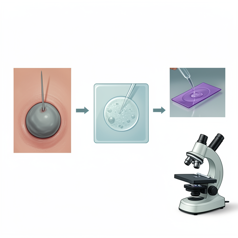
FNAC Process Flow
AI-generated conceptual illustration (canine round cell tumor mass). It explains the idea and does not show study data.
View source evidence ↗Figures & tables
From the original paper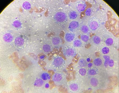 Figure 1
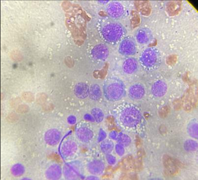 Figure 1
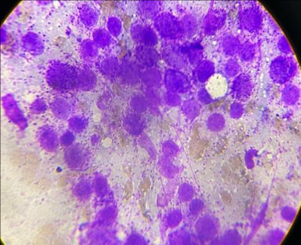 Figure 3
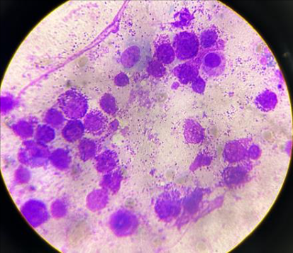 Figure 3
Introduction
Skin and subcutis are the most common sites for neoplasms in dogs accounting for 67.5% of tumors (Dorrn et al., 1968) [1] . Common round cell tumors in dogs are mast cell tumor, transmissible venereal tumor, histiocytoma, lymphoma and plasmacytoma (Fernandez et al., 2005) [2] . Mast cell tumor alone accounts for approximately one fourth of all skin tumors in dogs (Welle et al., 2008) [3] . The most common sites of mast cell tumors varied from ventral aspect of neck, lateral aspect of thigh, thoracic and abdominal regions (Madhesh et al., 2019) [4] . Histiocytomas are found in the head, neck and appendages and are usually benign. Plasmacytoma and multiple myelomas are common in older dogs and are typically malignant and TVTs (Transmissible Venereal Tumors) are sexually transmitted (Mathur et al., 2018) [5] . Apart from genitalia, transmissible venereal tumors has been reported in the skin, eyes, brain, subcutaneous tissue, lymph nodes, liver, spleen (Santos et al., 2008) [6] . The present study report on the occurrence of cuteneous transmissible venereal tumor and mast cell tumor in dog.
Materials and Methods
The current study was carried out in the Department of Veterinary Pathology, Institute of Veterinary Science and Animal Husbandry (IVS & AH), SOA University with 2 dogs presented to Veterinary Clinical Complex, IVS & AH with a history of 8 year old female dog showing multiple dermal masses over the body and 9 year old male dog showing a masses near the neck and the shoulder region. Fine needle aspiration cytology was performed from both the case. Thin smear was prepared and slide was fixed with methanol for 5 minutes and then stain by giemsa for 30 minutes and observed under the microscope.
Result and Discussion
In general, genital TVT is common in females and extragenital TVT is common in males due to its breeding behaviour (Ganguly et al., 2016) [7] . Apart from genital tract, TVT can also affect the skin, eyes, brain, subcutaneous tissue, lymph nodes, liver, spleen (Santos et al., 2008; Preet et al., 2021) [6,8] . These tumors are usually spread at the time of coitus or sniffing and licking. In the present case, 8 year old female dog was presented with a history of multiple dermal masses over the body. Fine needle aspiration cytology showed high cellularity and had similar feature of TVT tumors. The cells were large round to oval with distinct cytoplasmic borders. The cytoplasm contains distinct vacuoles arranged along the periphery of the cells (Figure 1&2). Our findings are in accordance with the earlier worker (Santos et al., 2008; Preet et al., 2021) [6,8] who had reported cuteneous TVT. Madesh et al. (2019) [4] also reported multiple subcutaneous nodules over the body in extragenital form of TVT which support our findings. Madesh et al. (2019) [4] ,
Preet et al. (2021) [8] and Simon et al. (2016) [9] reported the incidence of cuteneous TVT in male dog whereas our study showed the incidence on female dog. Mastocytoma is a round-cell tumor comprises of mast cells. Mast cells are a part of immune system and closely resemble basophils. The cytoplasmic granules consist of histamine. In the present case, a 9 year male dog was presented with a history of masses near the neck and shoulder region. Fine needle aspiration cytology revealed high cellularity along with pleomorphic cells, round shaped of nucleus with stained mass of cytoplasmic metachromatic granules (Figure 3 &4).
The findings are in accordance with the earlier work of Alleman and Bain (2000) [10] and Girish et al. (2011) [11] . Mathur et al. (2018) [5] reported incidence was more common among age group between 6 to 10 years which support with our findings. Mathur et al. (2018) [5] also reported the most common sites of mast cell tumors varied from ventral aspect of neck, lateral aspect of thigh, thoracic and abdominal regions which support our findings.
Acknowledgement
AcknowledgementThe authors would like to acknowledge the Dean, IVS & AH for providing necessary facilities to carry out the work and Veterinary Clinical Complex, Department of Surgery & Radiology for providing the sample
References
11 in the original paper- Dorn CR, Taylor DON, Schneider R, Hibbard HH, Klauber MR. Survey of animal neoplasms in Alameda and Contra Costa Counties, California. II. Cancer morbidity in dogs and cats from Alameda County. J Natl Cancer Inst. 1968;40(2):307-318.
- Fernandez NJ, West KH, Jackson ML, Kidney BA. Immunohistochemical and histochemical stains for differentiating canine cutaneous round cell tumors. Vet Pathol. 2005;42:437-445.
- Welle M, Bley CR, Howard J, Rüfenacht S. Canine mast cell tumours: A review of the pathogenesis, clinical International Journal of Veterinary Sciences and Animal Husbandry https://www.veterinarypaper.com features, pathology and treatment. Vet Dermatol. 2008;19(6):321-329.
- Madhesh E, Ajith Y, Dimri U, Shanmuganathan S, Sivasankar P, Karthikeyan R. Medical management of phimosis associated with generalized Sticker's sarcoma in an intact mongrel dog. Comp Clin Path. 2019;28:18511854.
- Mathur KY, Rao S, Chandrashekaraiah GB, Munivenkatappa BS. Cytological and histopathological studies of canine round cell tumors. Int J Livest Res. 2018;8(6):88-95.
- Santos JP, Barbosa MAG, Tenorio APM, Coelho MCOC, Rolim MBQ, Tudury EA. Transmissible venereal tumor disease in a dog with involvement of the skin. Arq Bras Med Vet Zootec. 2008;2:39-43.
- Ganguly B, Das U, Das AK. Canine transmissible venereal tumor: A review. Vet Comp Oncol. 2016;14:112.
- Preet GS, Sidhu S, Dhaliwal KS, Singh J, Kaur J. Atypical cutaneous transmissible venereal tumour in dog -a case report. Haryana Vet. 2021;60(2):317-318.
- Simon MS, Gupta CG, Sankar P, Ramprabhu R, Pazhanivel N, Prathaban S. Cutaneous transmissible venereal tumor in a dog and its management. Indian Vet J. 2016;93(07):77-79.
- Alleman AR, Bain PJ. Diagnosing neoplasia: The cytologic criteria for malignancy. Vet Med. 2000;3:204223.
- Girish BC, Suguna R, Byregowda SM, Kotrappa YM. Canine squamous cell carcinoma: A review of 17 cases. Braz J Vet Pathol. 2011;4(2):79-86.