
Research article · 2024 · Full text
International Journal of Veterinary Sciences and Animal HusbandryRole of Omega-3 in ameliorating acute liver injury-induced by D-galactose on adult male rats
Volume 9(6) · Pages 652–658 · DOI: 10.22271/veterinary.2024.v9.i6j.1956 ↗
Research areasDiet, Metabolism, and DiseaseAntioxidants, Aging, Portulaca oleraceaBiochemical effects in animals
Abstract
Author-writtenBackground and objective: Numerous morphological and functional changes in the liver are linked to aging. The two main ageing processes that can cause significant damage to mitochondrial DNA and ultimately apoptosis are now well recognised to be inflammatory reaction and oxidative stress. Because aging is a poor prognostic factor that raises the mortality rate, it may also raise the risk of developing a number of liver illnesses. In rodents, chronic D-galactose treatment accelerates aging.In addition to improving hepatic beta oxidation and decreasing endogenous lipid production, omega-3 polyunsaturated fatty acids also have a positive effect on the metabolism of lipids and insulin sensitivity. They can also lower oxidative stress and hepatic inflammation, which merely lessens the damaging aging effect of D-galactose on rats.Methods: Four equal groups of twenty-eight (28) male Rattus norvegicus rats, weighing between 210 and 240 g, were randomly selected. Group T1 animals received an intraperitoneal injection of normal saline, while group T2 animals received an IP injection of D-galactose (150 mg/kg bwt) and were drenched 80 mg/kg (bwt) of Omega3 twice a day for 75 days. T3 received an intraperitoneal (I/P) injection of D-galactose (150 mg/kg b.wt) for 75 days, and they were drenched omega 3 (80 mg/kg b.wt) twice a day for 21 days. D-galactose (150 mg/kg b.wt) was given intraperitoneally (I/P) into T4 rats for 75 days. All animals have been sacrificed 24 hours after the last dose, and samples of liver tissue were obtained to look for histopathological changes, serum samples were gathered also for biochemical analysis.Conclusion: Omega-3 supplement for 75 days cause a significant enhancement with mitigating the aging harmful properties of D-galactose on liver functions and oxidative stress marker MDA and showing liver tissue sections showing the improvement with liver architecture occurring due to Omega 3 supplementation.
Summary
Source-checkedPlain-language reading of the full paper, prepared with AI and checked against the text. Each point links to the authors' own words.
Why it matters
- Aging is associated with inflammatory reactions and oxidative stress, which can damage mitochondrial DNA and lead to cell death (apoptosis). View evidence ↗
- Chronic treatment with D-galactose is used in rodent studies to accelerate the aging process. View evidence ↗
- Omega-3 polyunsaturated fatty acids may reduce hepatic inflammation and oxidative stress, potentially mitigating the aging effects caused by D-galactose. View evidence ↗
The question
- The study aimed to investigate the role of Omega-3 in reducing acute liver injury induced by D-galactose in adult male rats. View evidence ↗
What they did
- Rats were divided into four groups: T1 (saline control), T2 (D-galactose 150 mg/kg and Omega-3 80 mg/kg for 75 days), T3 (D-galactose 150 mg/kg for 75 days and Omega-3 80 mg/kg for 21 days), and T4 (D-galactose 150 mg/kg for 75 days). View evidence ↗
- Liver function and oxidative stress were measured using serum levels of ALT, ALP, bilirubin, and malondialdehyde (MDA). View evidence ↗
What they found
- Rats treated only with D-galactose (T4) showed significantly higher serum AST levels compared to all other groups (p≤0.05). View evidence ↗
- Serum bilirubin levels were significantly higher in the D-galactose only group (T4) than in the other groups (p≤0.05). View evidence ↗
- The D-galactose only group (T4) exhibited significantly higher serum MDA levels compared to the control and Omega-3 treated groups (p<0.05). View evidence ↗
- Omega-3 supplementation for 75 days (T2) provided a greater improvement in liver markers and MDA levels than supplementation for only 21 days (T3). View evidence ↗
- Histopathological analysis of the D-galactose only group (T4) revealed severe liver damage, including portal vein congestion, necrosis, and disturbed architecture. View evidence ↗
What it means
- The authors conclude that 75 days of Omega-3 supplementation significantly mitigates the harmful effects of D-galactose on liver function and oxidative stress. View evidence ↗
Visual summary
1 visualsIllustrations explain the paper; they do not show study data.
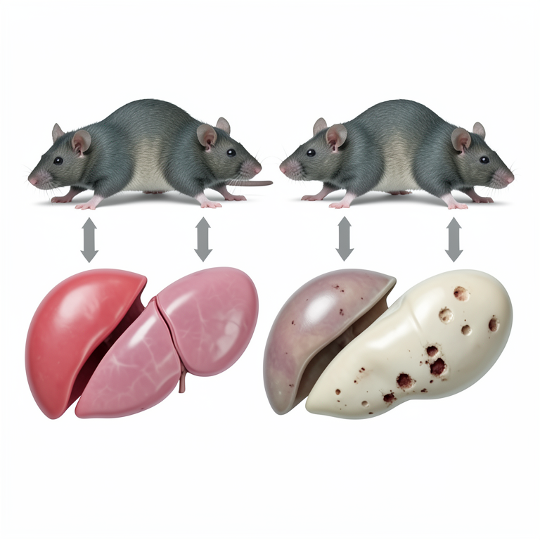
Experimental Grouping Diagram
AI-generated conceptual illustration (adult male rats and rat liver tissue). It explains the idea and does not show study data.
View source evidence ↗Figures & tables
From the original paper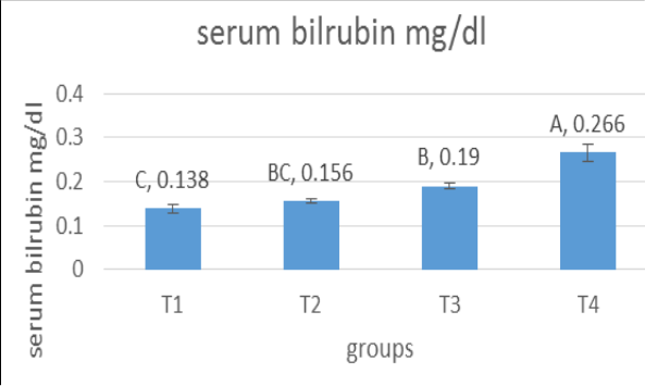 Figure 1
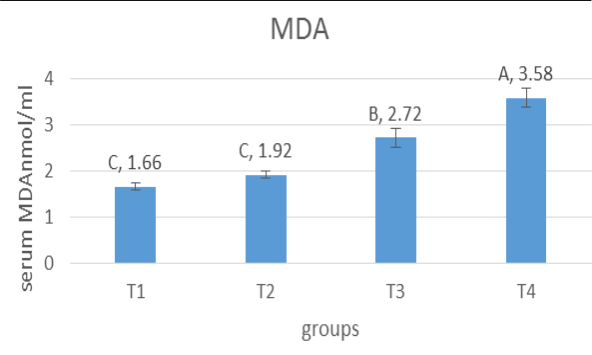 Figure 2
Introduction
The process of aging is defined by the slow accumulation of biological changes that eventually cause a person's functional abilities to deteriorate. According to estimates, the percentage of older individuals (those 60 and over) would almost double from 12% in 2015 to 22% by 2050 (Nations, 2015) [31] . In general, there are two types of aging models: those that age naturally and those that age more quickly. Because rapid aging models are easier to implement, needless study time, and have a better animal survival rate throughout the course of the experiment, they are preferred over naturally occurring aging models, which take a long time and are very expensive in vitro research uses hydroxyurea therapy and D-galactose-induced models, while in vivo studies use radiation-induced, accelerated susceptible mice, thymus-removed, and Dgalactose-induced models. The D-galactose-induced ageing model is the most extensively utilised among various ageing models because of its ease, absence of adverse effects, and better survival rate during the experiment (Cebe et al., 2014; Yanar et al., 2011) [10,42] . A reducing sugar called D-galactose is found naturally in the body and in a variety of foods, including butter, milk products, dairy products, honey, beets, and fruit like celery, plum berries, and figs in particular. (Acosta and Gross, 1995) [1] .
The maximum amount of galactose that is advised for a healthy adult to consume each day is 50 g, and the majority of it can be broken down and eliminated from the bloodstream within 8 hours of consumption. (Morava, 2014) [28] . However, galactose oxidase can catalyze its conversion to aldose and hydroperoxide at high concentrations, producing reactive oxygen forms. Significantly higher concentrations of reactive oxygen compounds (ROS) and malondialdehyde (MDA) and reduced levels of superoxide dismutase (SOD) and total antioxidant capacity were indicative of D-galactose-induced oxidative stress in the NSCs. D-galactose rats were showed considerably greater levels of pro-inflammatory cytokines, such as interleukin 1 beta (IL-1b), the interleukin 6 (IL-6), and TNF-a, compared to the normal group. Additionally, the expression of genes linked to cellular senescence, P53 and P21, was increased. These demonstrated that D-galactose may cause senescence through inflammatory pathways. (Azman and Zakaria, 2019) [8] . The onslaught of ROS is the primary cause of the progressive deterioration in liver functioning that occurs with aging. A surplus of D-galactose in the bloodstream can have a major impact on the liver because it is primarily processed there by raising NO, MDA and 8-hydroxy-2-deoxyguanosine, as well as decreasing the CAT, GPx, SOD, the NOS, the hormone GSH, and the overall antioxidant capacity of liver tissues. It has been shown that D Galactose treatment results in oxidative stress in the liver (Feng et al., 2016; Kong et al., 2018) [17,26] . Reactive oxygen species are very reactive substances that can affect the activities of different cellular components (Al-Tamemi and Al-Okaily, 2023) [2] . Advanced glycation end products are created when D-galactose combines with unbound amines that are present in proteins via nonenzymatic glycation. Additionally, long-term D-gal treatment may increase the production of ROS through glycation end products (AGEs) and D-gal oxidative metabolism (Parameshwaran et al., 2010) [33] . Regular cellular metabolism produces reactive species (ROS), which can damage lipids, proteins, and DNA. Oxidative damage contributes to ageing and cellular dysfunction over time (Andrés Juan et al., 2021) [4] . Ageing can cause cirrhosis, hepatocyte necrosis, and a malfunction in the liver, which plays a part in immunity and homeostasis (Fan et al., 2017) [16] . About 88% of D-Gal is absorbed in the gut and has the ability to buffer against liver retention when it is dissolved in drinking water and given to mice and rats (Homolak et al., 2023) [21] . D-Gal is given intraperitoneally or subcutaneously for 6-12 weeks at a dose of 50-1200 mg/kg/day to induce liver ageing (Azman, Safdar and Zakaria, 2021) [7] . Ω-3 fatty acids' ability to prevent liver and kidney damage by controlling the inflammatory NF-KB and antioxidant Nrf2 pathways (Eraky and El-Magd, 2020). Supplementing with fish oil that is high in ω-3FA may be a natural way to prevent hepatorenal damage in people who are accidentally or professionally exposed to manganese, improving both human and veterinary medical conditions (Owumi et al., 2020) [32] . Omega-3 polyunsaturated fatty acid supplementation was suggested as a therapeutic approach to lower plasma triglyceride levels by reducing hepatic lipogenesis and raising lipoprotein lipase (LPL) activity, which speeds up the breakdown of chylomicrons and VLDL (Santos et al., 2013) [36] . Alpha linolenic acid (ALA), eicosapentaenoic acid (EPA), and docosahexaenoic acid (DHA) are the three fatty acids that make up the omega-3 series. Some cereals, legumes, and vegetable oils contain ALA, while fish from deeper waters, such as salmon and sardines, have higher concentrations of the fatty acids EPA and DHA (Innes and Calder, 2018). It's possible that omega-3 fatty acids can lower oxidative stress, TG levels, and the severity of hepatic steatosis (Ratziu et al., 2019) [35] .
Materials and Methods Animal Ethical approval
Ethical approval was granted by the local committee of animal care and use (P.G. 584 date 3-12-2024), and the study's animal care and treatment were conducted at the College of Veterinary Medicine within Al-qasim Green University with strict compliance in the code of ethics for animal experiments.
Animals and experimental design
In this study, twenty-eight male rats (Rattus norvegicus), ages 4-5 months, weighing between 210-240 g, were used. The animals were housed in cages at 22-25 °C with a 12-hour light/dark cycle. During the two weeks that they were housed at the animal's house/college of veterinary medicine/Al-Qasim Green University for acclimation, the rats had complete access to water and pellets. For 75 days, the rats were divided into four equal groups at random and given daily treatments. While the animals in group T 2 received an intraperitoneal injection (I/P) of D-galactose (150 mg/kg bwt), the animals in the control group T 1 received an intraperitoneal injection of normal saline and were drenched in normal saline (Chang et al., 2017)
Tissue samples collection
The animals were subjected to regular weighing both prior to and throughout the duration of the experiment. This was done in order to accurately administer D-galactose and Omega 3 dosages in accordance with the rats' respective weights. Rats were subjected to anaesthesia with ketamine and xylazine according to (Veilleux-Lemieux et al., 2013) [38] . Liver tissues of the sacrificed specimens were preserved in a solution of natural buffer formalin (10%) in order to facilitate further histopathological examinations. Blood samples were collected at the end of the experiment to quantify the serum malondialdehyde (MDA) content using a modified method, MDA assay kit was obtained from Solarbio Life Science /china. Operation Equipment: Spectrophotometer. Serum samples were separated for measurement of alanine transaminase (ALT), alkaline phosphatase (ALP) and bilirubin, immediately after serum samples were separated, and estimated by using the clinical chemistry analyzer Cobas c 311an open reagent system, Roche diagnostic cobas c311 automated, software-controlled analyzer.
Results
Serum AST (IU)/L: The T 4 group's serum AST levels were significantly greater (p≤0.05) than those of the other groups, as shown in Figure (1-A). A significant decrease (p≤0.05) was observed in the T 1 group when compared to the T 3 and T 4 groups. Comparing T 2 to the T 1 control group, not a noticeable rise was seen. Serum ALT (IU)/L: The T 4 group's serum ALT levels were substantially greater (p≤0.05) than those of the other groups, as shown in Figure 1-B. When compared to the T 3 and T 4 groups, the T 1 group displayed a statistically significant decrease (p≤0.05). Comparing T 2 to the T 1 control group, no discernible increase was seen. Total bilirubin mg/dl According to fig. (1-C) results, the T 4 group's serum bilirubin level was statistically significantly (p≤0.05) greater than that of the other groups. The results indicated a statistically significant decrease (p≤0.05) between the T 1 group and the T 3 and T 4 groups. Likewise, T 2 displayed non-significant increases in serum malondialdehyde (MDA) concentration (mmol/ml) compared to T 1 : According to the blood MDA concentration measurement results in figure (2), the D-galactose exposed group (T 4 ) had a statistically significant (p<0.05) greater level than the negative control (T 1 ) and the other treatment groups. T 1 and T 2 were also found to not differ statistically substantially (P>0.05). But when T 1 and T 2 were contrasted with T 3 and T 4 , a significant (p<0.05) decrease in serum MDA levels. According to the results of the current study we can notice the harmful effect of 75 days of IP injection of D-galactose in T 4 rats for the above parameters, AST, ALT, total bilirubin and MDA, and we can notice the improvement effect of Omega-3 for the same parameters for group T 2 which has been drenched for 75 days as compared with those who received omega 3 for only 21 days with IP D-galactose injection 75 days values expressed as mean ± SE. N=7 rats. T 1 =Control group rats were injected with normal saline.
Discussion
Ageing makes the liver more susceptible to progressive structural and functional changes. As the liver ages, its mitochondrial count decreases, oxidative stress-related genes like cytochrome c are expressed less frequently, and hepatocyte telomere length decreases, which lowers cell proliferation and apoptosis (Azman, Safdar and Zakaria, 2021) [7] . Preservation of mitochondrial function has been found to be crucial in the restoration of organ function after severe injury. Age-related progressive organ failure, an elevated inflammatory response, and accelerated cellular senescence all contribute to the liver's diminished ability to fight injury. It has been demonstrated that aging-related mitochondrial function reduction increases the susceptibility of the heart, lungs, liver, and kidney to ischemia/reperfusion injury (Poulose and Raju, 2014) [34] . Aging decreases the ability of liver to withstand trauma (Hoare, Das and Alexander, 2010) [20] .
Receiving IP injections of D-galactose for 75 days as shown in figures (1-A, B, C) causing a significant (p≤0.05) increase in rats serum AST, ALT and bilirubin as compared with control and treated Omega-3 groups, these results were in agree with many studies,a study which has results were in agree with the present study confirmed that when compared to normal rats, D-Gal dramatically raises the liver enzyme indicators ALT, AST, TBIL, and DBIL. It has previously been documented that D-Gal-induced liver ageing causes an increase in liver aminotransferases, which are early, sensitive indicators of liver damage (Chen, Chen and Zhou, 2018) [12] . Because D-gal is a reducing sugar and an essential nutrient, It creates advancing substance related to glycosylation when it joins with amino acids, as a result, when it is injected in large quantities, it causes reactive oxygen species to be produced through the metabolism of hydrogen peroxide by galactoseoxidas. Additionally, D-Gal has been demonstrated to dramatically increase serum total and direct bilirubin in rat sera, according to another investigation that supported findings comparable to ours. Since liver function is necessary for bilirubin catabolism, increased bilirubin levels are indicative of hepatocytes destruction (Boland et al., 2014) [9] . Bilirubin is a sensitive biomarker of liver disorders and one of the three markers used to evaluate the model for the end-stage liver disease score (Kamath and Kim, 2007) [25] . Additionally, hepatic dysfunction and impaired intrahepatic excretion of conjugated bilirubin from bile duct hepatocytes are indicated by increased direct bilirubin in rats administered D-galactose (Astegiano et al., 2004) [5] . One of the mediators of cytoprotection for inflammation and cellular homeostasis balance against oxidation processes is bilirubin (Gomes et al., 2010) [19] . This may reveal the reason why bilirubin levels increased in serum of group exposed to D-Gal. and other oxidative stress stimuli. Intraperitoneal D-Gal, at 150 mg/kg for 75 days, induced significant increase in liver function markers compared to control and treated groups with omega-3. In addition, serum bilirubin levels also elevated significantly in d-galactose treated group T4 as compared with control and treated group with omega 3, significantly help in restore liver function markers to normal levels compared to D-Gal treated group. A byproduct of lipid peroxidation of polyunsaturated fatty acids, malondialdehyde (MDA) includes Moving around One of the most popular and extensively utilised indicators of oxidative stress is MDA (Al-Tamemi and Al-Okaily, 2023) [2] . Some studies found that MDA plasma levels dropped following n-3 PUFA intake (Véricel et al., 2015) [39] . It has been hypothesised that immunomodulation and a decrease in leukocyte activation may be the mechanisms by which n-3 PUFA supplementation could lessen oxidative stress which were in agree with present study according to Figure 2 showing a significant decrease in omega-3 exposed rats. Another study confirmed that omega-3 fatty acids supplementation had beneficial effects, Omega-3 fatty acids increased activity of SOD and decreased lipid peroxidation, changes in oxidative/antioxidative balance are a result of effects on lipids and enzymes of antioxidative system (Avramovic et al., 2012) [6] . (Fenton et al., 2013) [18] , through lipid synthesis and oxidation, dietary LCn-3 PUFAs impact hepatic lipid homeostasis. Research is being done on how these fatty acids interfere with nuclear processes that are not dependent on eicosanoid formation and regulate the expression of genes related to lipid metabolism (Molendi-Coste, Legry and Leclercq, 2011) [27] . When administered to animals in conjunction with DOX, omega-3 fatty acids improved the histological and biochemical changes brought on by DOX, significantly reduced the amount of MDA, and increased the activity of antioxidant enzymes. Because of its antioxidant properties, omega-3 fatty acids protected against liver damage caused by doxorubicin; they also improved the levels of AST and ALT and, because of their anti-inflammatory properties, reduced the generation of pro-inflammatory cytokines (Moussa et al., 2020) [29] .
In [14] . Studies evaluating the effect of dietary ω3 PUFA on NAFLD show that changes in hepatic PUFA impact NAFLD indicators, such as steatosis, inflammation, and fibrosis (Jump et al., 2018) [23] . Excess Dgalactose administration contributes to the increased formation of ROS, which subsequently leads to oxidative stress and hepatocyte damage (Azman, Safdar and Zakaria, 2021) [7] . When the antioxidant system's function declines or significant amounts of free radicals are produced, tissues and cells are vulnerable to damage caused by oxidation. Hepatocellular necrosis and cytoplasm vacuolar degradation in the D-gal group showed changes to the typical lobular architecture and structure of the liver cells in the control group (Xiao et al., 2018) [41] . Elevated expression amounts of oxygen-derived free radicals in mouse hepatocytes are linked to hepatic ageing and necrosis brought on by high D-gal levels (Timchenko, 2009) [37] . Free radicals are playing important role in damage a number of cellular components including DNA, proteins and lipids (Das and Vasudevan, 2007) [13] .
The benefits of administering omega-3 in reducing DOX hepatotoxicity and the efficacy of omega-3 in preventing it were demonstrated. The rat liver treated with omega-3 maintained its normal architecture and was also able to reduce congestion, hepatocyte vacuolation, and degeneration (Moussa et al., 2020) [29] . Pretreatment with omega-3 FAs and concurrent treatment with Gifosfamide reduced the liver damage caused by Gifosfamide, demonstrating its hepatoprotective properties (El Kalawy et al., 2017) [24] . The present study histopathological examination were in agree with (Ali and Rifaai, 2019), who confirmed that when liver sections from the control and omega-3 groups were examined under a microscope, the cords of hepatocytes radiating from the central vein revealed a typical histologic pattern. Hepatocytes had acidophilic cytoplasm and central vesicular nuclei, giving them a polyhedral appearance. Blood sinusoids were used to divide the cords. Hepatic artery, portal vein, and bile duct branches were found in the portal tract.
Conclusion
A current study found that Omega-3 caused an improvement in oxidative stress biomarkers and biochemical liver function biomarkers which were affected by the harmful effect of Dgalactose treatment for 75 days also results suggest that Omega 3 mitigates histological changes induced by D-galactose due to its properties as an antioxidant, antiaging, anti-inflammatory, antiapoptotic properties.
Ethical consideration
Authors has examined the manuscripts for ethical issues, including redundancy, multiple publishing and/or submission, misconduct, data fabrication and/or falsification, plagiarism, and permission to publish.
Conflicts of Interest:
Conflicts of Interest:
Acknowledgement
AcknowledgmentsThe authors express their gratitude to Al-Qasim green University, namely the Faculty of Veterinary Medicine, for granting permission to carry out the present study and for providing valuable resources and support during the research process.
References
41 in the original paper- Acosta PB, Gross KC. Hidden sources of galactose in the environment. Eur J Pediatr. 1995;154:S87-92.
- Al-Tamemi ZS, Al-Okaily BN. Effect of sodium thiosulfate and resveratrol on blood pressure and arterial health in nicotine-exposed rats. Rev Latinoam Hipertens. 2023;18(10):522-9.
- Ali FF, Rifaai RA. Preventive effect of omega-3 fatty acids in a rat model of stress-induced liver injury. J Cell Physiol. 2019;234(7):11960-119608.
- Astegiano M, et al. Laboratory evaluation of the patient with liver disease. Eur Rev Med Pharmacol Sci. 2004;8:3-10.
- Avramovic N, et al. The effects of omega 3 fatty acid supplementation on brain tissue oxidative status in aged Wistar rats. Hippokratia. 2012;16(3):241.
- Azman KF, Safdar A, Zakaria R. D-galactose-induced liver aging model: Its underlying mechanisms and potential therapeutic interventions. Exp Gerontol. 2021;150:111372.
- Azman KF, Zakaria R. D-galactose-induced accelerated aging model: An overview. Biogerontology. 2019;20(6):763-82.
- Boland BS, et al. Association of serum bilirubin with aging and mortality. J Clin Exp Hepatol. 2014;4(1):1-7.
- Cebe T, et al. Oxidation scrutiny in persuaded aging and chronological aging at systemic redox homeostasis level. Exp Gerontol. 2014;57:132-40.
- Chang YM, et al. Inhibition of cardiac hypertrophy effects in D-galactose-induced senescent hearts by alpinate oxyphyllae fructus treatment. Evid Based Complement Alternat Med. 2017;2017:2624384.
- Chen P, Chen F, Zhou B. Antioxidative, antiinflammatory and anti-apoptotic effects of ellagic acid in liver and brain of rats treated by D-galactose. Sci Rep. 2018;8(1):1465.
- Das SK, Vasudevan DM. Alcohol-induced oxidative stress. Life Sci. 2007;81(3):177-187.
- El-Baz FK, et al. Astaxanthin-rich Haematococcus pluvialis algal hepatic modulation in D-galactose-induced aging in rats: Role of Nrf2. Adv Pharm Bull. 2018;8(3):523.
- Eraky SM, El-Magd NFA. Omega-3 fatty acids protect against acetaminophen-induced hepatic and renal toxicity in rats through HO-1-Nrf2-BACH1 pathway. Arch Biochem Biophys. 2020;687:108387.
- Fan J, et al. Spermidine coupled with exercise rescues skeletal muscle atrophy from D-gal-induced aging rats through enhanced autophagy and reduced apoptosis via AMPK-FOXO3a signal pathway. Oncotarget. 2017;8(11):17475.
- Feng Y, et al. Chlorogenic acid protects D-galactoseinduced liver and kidney injury via antioxidation and anti-inflammation effects in mice. Pharm Biol. 2016;54(6):1027-34.
- Fenton JI, et al. Long chain omega-3 fatty acid immunomodulation and the potential for adverse health outcomes. Prostaglandins Leukot Essent Fatty Acids. 2013;89(6):379.
- Gomes AS, et al. Gastroprotective effect of hemeoxygenase 1/biliverdin/CO pathway in ethanol-induced gastric damage in mice. Eur J Pharmacol. 2010;642(13):140-145.
- Hoare M, Das T, Alexander G. Ageing, telomeres, senescence, and liver injury. J Hepatol. 2010;53(5):950961.
- Homolak J, et al. D-galactose might protect against ionizing radiation by stimulating oxidative metabolism and modulating redox homeostasis. J Radiat Res. 2023;64(4):743-745.
- Innes JK, Calder PC. The differential effects of eicosapentaenoic acid and docosahexaenoic acid on cardiometabolic risk factors: A systematic review. Int J Mol Sci. 2018;19(2):532.
- Jump DB, et al. Omega-3 polyunsaturated fatty acids as a treatment strategy for nonalcoholic fatty liver disease. Pharmacol Ther. 2018;181:108-125.
- El Kalawy S, et al. Histological and immunohistochemical study of the effect of omega-3 fatty acids on ifosfamide-induced liver injury in adult male albino rat. J Med Histol. 2017;1(2):134-145.
- Kamath PS, Kim WR. The model for end-stage liver disease (MELD). Hepatology. 2007;45(3):797-805.
- Kong S-Z, et al. Anti-aging effect of chitosan oligosaccharide on D-galactose-induced subacute aging in mice. Mar Drugs. 2018;16(6):181.
- Molendi-Coste O, Legry V, Leclercq IA. Why and how meet n-3 PUFA dietary recommendations? Gastroenterol Res Pract. 2011;2011:364040.
- Morava E. Galactose supplementation in phosphoglucomutase-1 deficiency; review and outlook for a novel treatable CDG. Mol Genet Metab. 2014;112(4):275-279.
- Moussa FI, et al. Protective effect of omega-3 on doxorubicin-induced hepatotoxicity in male albino rats. J Biosci Appl Res. 2020;6(4):207-219.
- Mu X, et al. Angelica Sinensis polysaccharide prevents hematopoietic stem cells senescence in D-galactoseinduced aging mouse model. Stem Cells Int. 2017;2017:3508907.
- Nations U. Department of Economic and Social Affairs, population division. Int Migr Rep [Preprint]. 2015.
- Owumi SE, et al. Gallic acid and omega-3 fatty acids decrease inflammatory and oxidative stress in manganese-treated rats. Exp Biol Med. 2020;245(9):83544.
- Parameshwaran K, et al. D-galactose effectiveness in modeling aging and therapeutic antioxidant treatment in mice. Rejuvenation Res. 2010;13(6):729-735.
- Poulose N, Raju R. Aging and injury: alterations in cellular energetics and organ function. Aging Dis. 2014;5(2):101.
- Ratziu V, et al. Recommendations for management and treatment of nonalcoholic steatohepatitis. Transplantation. 2019;103(1):28-38.
- Santos RD, et al. I Diretriz sobre o consumo de gorduras e saúde cardiovascular. Arq Bras Cardiol. 2013;100:1-40.
- Timchenko NA. Aging and liver regeneration. Trends Endocrinol Metab. 2009;20(4):171-176.
- Veilleux-Lemieux D, et al. Pharmacokinetics of ketamine and xylazine in young and old Sprague-Dawley rats. J Am Assoc Lab Anim Sci. 2013;52(5):567-570.
- Véricel E, et al. Moderate oral supplementation with docosahexaenoic acid improves platelet function and oxidative stress in type 2 diabetic patients. Thromb Haemost. 2015;114(08):289-296.
- Wu D, et al. Purple sweet potato color repairs Dgalactose-induced spatial learning and memory impairment by regulating the expression of synaptic proteins. Neurobiol Learn Mem. 2008;90(1):19-27.
- Xiao M, et al. Ginsenoside Rg1 attenuates liver injury induced by D-galactose in mice. Exp Ther Med. 2018;16(5):4100-4106.
- Yanar K, et al. Protein and DNA oxidation in different anatomic regions of rat brain in a mimetic ageing model. Basic Clin Pharmacol Toxicol. 2011;109(6):423-433.