
Research article · 2024 · Full text
International Journal of Veterinary Sciences and Animal HusbandrySurgical management of bilateral lateral luxation of patella in a Doberman dog: A case report
Volume 9(6S) · Pages 181–183 · DOI: 10.22271/veterinary.2024.v9.i6sc.1938 ↗
Research areasVeterinary Orthopedics and NeurologyTendon Structure and TreatmentVeterinary Equine Medical Research
Abstract
Author-writtenA one-year-old male Doberman was presented to Small Animal Surgery Unit of Veterinary Clinical Complex, Veterinary College and Research Institute, Orathanadu with the history of non–weight bearing lameness in both the hind limbs since birth. Physical examination and radiographic evaluation revealed lateral luxation of patella in the both the hind limbs. The case was confirmed as congenital bilateral lateral luxation of patella and surgical correction through trochlear wedge recession sulcoplasty with tibial tuberosity repositioning was performed in both the legs under general anaesthesia as staged procedure. Post operative radiography revealed patella in normal position. Recovery was good and improvement in gait noticed on 14th post operative day and the animal made an uneventful recovery.
Summary
Source-checkedPlain-language reading of the full paper, prepared with AI and checked against the text. Each point links to the authors' own words.
Why it matters
- Patellar luxation is a frequent cause of lameness in dogs, affecting both small and large breeds. View evidence ↗
- Lateral luxation of the patella is less common than medial luxation and typically occurs in giant or large breed dogs. View evidence ↗
- The condition is caused by joint abnormalities that shift the pull of the quadriceps muscle, pulling the patella out of its groove. View evidence ↗
The question
- The study aims to describe the surgical management of a specific case of bilateral lateral patellar luxation in a dog. View evidence ↗
What they did
- The subject was a one-year-old male Doberman with non-weight bearing lameness in both hind limbs since birth. View evidence ↗
- Diagnosis was confirmed as Grade IV congenital lateral luxation of the patella using physical examination and skyline and lateral radiographs. View evidence ↗
- The surgical correction involved a staged procedure using trochlear wedge recession sulcoplasty and tibial tuberosity repositioning under general anesthesia. View evidence ↗
- The procedure included cutting a wedge of bone from the trochlear groove and reinserting it in an inverted position to deepen the groove. View evidence ↗
- A Kirschner wire was used to fix the repositioned tibial tuberosity into its normal anatomical position. View evidence ↗
What they found
- Post-operative radiographs confirmed that the patella had been returned to its normal position. View evidence ↗
- An improvement in the dog's gait was observed on the 14th day after the surgery. View evidence ↗
- The dog experienced an uneventful recovery following the surgical procedures. View evidence ↗
What it means
- The authors conclude that congenital bilateral lateral luxation of the patella can be successfully corrected using trochlear wedge resection sulcoplasty and tibial tuberosity repositioning. View evidence ↗
Visual summary
1 visualsIllustrations explain the paper; they do not show study data.
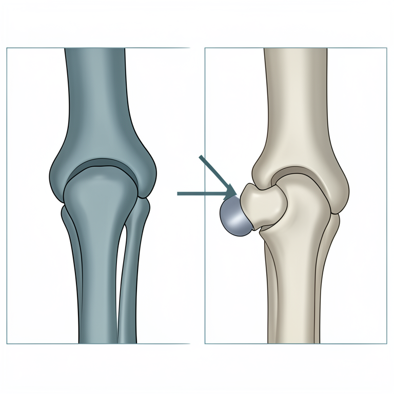
Mechanism of Patellar Luxation
AI-generated conceptual illustration (Doberman dog stifle joint). It explains the idea and does not show study data.
View source evidence ↗Figures & tables
From the original paper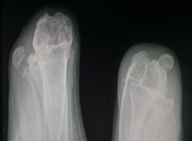 Figure 1
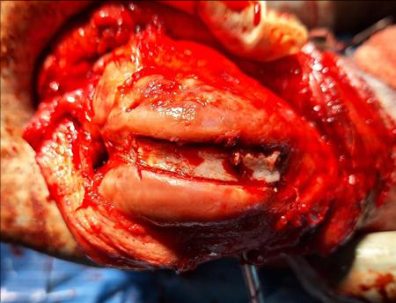 Figure 2
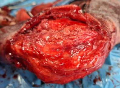 Figure 3
Introduction
Patellar luxation is one of the most common causes of lameness in the dog. Both large and small breed dogs may be affected; the disease may be seen in cats as well. (Nunamaker DM, 1985) [11] . The luxation can be medial, lateral or bidirectional (Ness et al. 1996) [10] . Patellar luxation is an intermittent or permanent displacement of patella from the trochlear sulcus. Most luxation are termed 'congenital' because they occur in early life and not associated with trauma (DeAngelis M, 1996) [3] . Lateral luxation occurs less frequently and is commonly diagnosed in large or giant breed dogs (Linney et al. 2011) [9] . The reported incidence of medial patellar luxation in small breed dogs is 12 times higher compared with large breed dogs (Roush JK, 1993) [12] . The patellar luxation is related to the anteversion or coxa valga of the coxo-femoral joint, which shifts the line of force produced by the pull of quadriceps lateral to the longitudinal axis of the trochlear groove. This abnormally directed force pulls the patella from the trochlear sulcus (Bosio, F. 2017) [2] . Tibial deformities seen with patellar luxation were the result of abnormal forces acting on the proximal and distal physis of tibia. These include lateral displacement of tibial tuberosity and lateral torsion of distal tibia (Hayes AG et al., 1994) [6] . A system of classifying patellar luxation into four grades has been developed. In grade I, spontaneous luxation of patella during normal joint motion occurs but the patella reduces when the pressure is released. In grade II, angular and torsional deformities of femur occur to mild degree and the patella remains luxated until it is manually reduced. In grade III, patella is permanently luxated and can be reduced manually. In grade IV, patella is permanently luxated permanently and it cannot be reduced manually. Abnormalities of supporting soft tissues of stifle joint and femur are notable. Clinical signs may vary from animal to animal. Lameness may be intermittent or continuous, mild to moderate weight bearing lameness in grade III, non-weight bearing lameness and crouched position of rear quarters in grade IV (LaFond E. et al., 1994) [8] . Radiography reveals laterally displaced patella from the trochlear groove. Surgical treatment is Trochlear Wedge Recession Sulcoplasty (TWRS) with Tibial tuberosity transposition (LaFond E. et al. 2002) [8] . This case report describes about a clinical case of a dog with bilateral lateral patellar luxation and its surgical management.
Case history
A one-year-old male Doberman was presented to Small Animal Surgery Unit of Veterinary Clinical Complex, Veterinary College and Research Institute, Orathanadu with the history of non-weight bearing lameness in both the hind limbs since birth.
Diagnosis
Physical examination revealed the patella was luxated laterally away from the trochlear groove and cannot be reduced. Skyline and lateral radiographs of both the patella confirmed the case as Grade IV congenital lateral luxation of patella (Figure 1). The haematological and biochemical values were normal.
Materials and Methods
The food and water were withdrawal for 24 and 12 hours respectively before the day of surgery. The animal was premedicated using Inj. Atropine sulphate @ 0.02mg/kg s.c. and Inj. Xylazine @ 1mg/kg i.m. and Inj. Butorphanol @ 0.2mg/kg i.m. After 10 minutes, the anaesthesia was induced using Inj. Ketamine @ 5mg/kg i.v. and Inj. Diazepam @ 0.5mg/kg i.v. and the anaesthesia was maintained using 2% Isoflurane with variable vaporizer setting and fresh gas flow of oxygen @ 20 ml/kg/minute. Animal was placed in left lateral recumbency and the right stifle was aseptically prepared using 2.5 per cent povidone iodine, 70 per cent ethyl alcohol and 2 per cent chlorhexidine. A 10 cm long skin incision was made on the cranial aspect of stifle joint starting from the distal femur to proximal tibia to expose the retinaculum. The retinaculum was loosened from its attachment and the trochlea was exposed. Using an osteotome, partial osteotomy was performed on tibial tuberosity for trans positioning of tibial tuberosity to its normal position. A wedge-shaped bone was cut from the trochlear groove and it is placed into the sulcus in an inverted manner after deepening the trochlear groove towards the lateral side (Figure 2). The trans positioned tibial tuberosity was fixed in normal position using a Kirschner wire. Retinaculum was sutured towards medially using PGA (2-0) by Ford interlocking pattern and skin using polyamide (2-0) by cross mattress suture pattern (Figure 3). Post operative radiography showed patella was in position. Post operative antibiotics and analgesics were followed for 2 weeks. The cutaneous sutures were removed on tenth post operative day and the animal made an uneventful recovery.
Results and Discussion
The cause includes congenital, developmental and traumatic. Most of the prevalence studies identified that females are more predisposed than males. Neuter status of the animal seems to be a risk factor, with neutered dogs having about 3 times odds of developing this condition (L'Eplattenier HF and Montavon P., 2002) [7] Occasionally, patellar luxation may occur as a complication secondary to treatment of cranial cruciate ligament disease or fractures involving femur or tibia. Because there is evidence that this condition is genetic, dogs diagnosed with patellar luxation should not be bred. Dogs with Grade I patellar luxation are generally asymptomatic. Occasionally "skipping" type lameness typical in dogs with Grade II patellar luxation. Grade III and Grade IV patellar luxation are characterized by persistent lameness and abnormal posture (Di Dona F et al. 2016) [4] . In bilateral lateral dislocation, the stifle appears to be close to each other while the distal extremities are abducted. Large breed dogs may have "knocked-in-knee" appearance. Diagnosis is based on clinical signs, orthopedic examinations and radiography (lateral and skyline view). Medical management may be indicated in dogs with Grade I patellar luxation with nonsteroidal anti-inflammatory drugs, weight control and physical rehabilitation exercises. Surgical correction of patellar luxation include oft tissue reconstruction procedures (Overlap of medial retinaculum, patellar and tibial antirotational suture ligaments, desmotomy and partial capsulectomy and quadriceps release) and bone reconstruction procedures (Trochleoplasty-trochlear chondroplasty, trochlear wedge or block recession sulcoplasty), Tibial tuberosity transposition, patellectomy and femoral osteotomy (Harasen G 2006) [5] . Post operative care includes soft padded bandage for 10-14 days, pain medications and physiotherapy. Prognosis is favorable. Recurrence may occur but always of a lesser grade than preoperative grade. The most common complication includes patellar relaxation, loose or broken implants or fracture of proximal tibia (Anna E Dunlap, 2016) [1] .
Conclusion
The case of congenital bilateral lateral luxation of patella was successfully corrected through trochlear wedge resection sulcoplasty with tibial tuberosity reposition. The animal made uneventful recovery with normal gait.
Acknowledgement
AcknowledgementThe authors are thankful to the Dean, Veterinary College and Research Institute, Orathanadu and the Professor and Head, Veterinary Clinical Complex, Veterinary College and Research Institute, Orathanadu for the technical support rendered during this study.
References
12 in the original paper- Dunlap AE. Outcomes and complications following surgical correction of grade IV medial patellar luxation in dogs. Available from: https://avmajournals.avma.org. Accessed: 22 Apr 2024.
- Bosio F, Bufalari A, Peirone B, Petazzoni M, Vezzoni A. Prevalence, treatment and outcome of patellar luxation in dogs in Italy: A retrospective multicentric study (20092014). Vet Comp Orthop Traumatol. 2017;30(5):364-70.
- DeAngelis M. Patellar luxation in dogs. Vet Clin North Am Small Anim Pract. 1996;1:403-15.
- Di Dona F, Della Valle G, Balestriere C. Lateral patellar luxation in nine small breed dogs. Open Vet J. 2016;6(3):255-8.
- Harasen G. Patellar luxation: pathogenesis and surgical correction. Can Vet J. 2006;47(10):1037-9.
- Hayes AG, Boudrieau RJ, Hungerford LL. Frequency and distribution of medial and lateral patellar luxation in dogs: 124 cases (1982-1992). J Am Vet Med Assoc. 1994;205(5):716-20.
- L'Eplattenier HF, Montavon P. Patellar luxation in dogs and cats: management and prevention. Comp Cont Edu Pract Vet. 2002;24:292-300.
- LaFond E, Breur GJ, Austin CC. Breed susceptibility for developmental orthopedic diseases in dogs. J Am Anim Hosp Assoc. 2002;38(5):467-77.
- Linney WR, Hammer DL, Shott S. Surgical treatment of medial patellar luxation without femoral trochlear groove deepening procedures in dogs: 91 cases (1998-2009). J Am Vet Med Assoc. 2011;238(9):1168-72.
- Ness MG, Abercromby RH, May C, Turner BM, Carmichael S. A survey of orthopaedic conditions in small animal veterinary practice in Britain. Vet Comp Orthop Traumatol. 1996;9(2):43-52.
- Nunamaker DM. Patellar luxation. In: Newton CD, Nunamaker DM, editors. Textbook of small animal orthopaedics. Philadelphia: Lippincott; 1985. p. 941-7.
- Roush JK. Canine patellar luxation. Vet Clin North Am Small Anim Pract. 1993;23(4):855-68.