
Research article · 2024 · Full text
International Journal of Veterinary Sciences and Animal HusbandryThe effect of aging on skin cells in domestic rabbits, a histological study using a special dye
Volume 9(6) · Pages 587–592 · DOI: 10.22271/veterinary.2024.v9.i6i.1937 ↗
Research areasRabbits: Nutrition, Reproduction, Health
Abstract
Author-writtenThe study provides a comprehensive analysis of the histological changes in the skin of domestic rabbits with age using Verhoff for collagen stain, which is considered a special dye. A group of histological staining techniques was used to examine skin samples taken from rabbits at different age stages. We were able to analyze and identify changes in the structure of the skin, including the dermis, epidermis, and subcutaneous fatty tissue. The results showed that aging leads to clear changes in the structure of the skin, including an increase in the thickness of the dermis and epidermis, a change in the distribution of collagen and elastic fibers, and the appearance of fatty aggregates. An increase in inflammatory cell activity in the skin has also been observed with age, indicating an effect of age on the skin inflammatory response. In addition, the study revealed changes in the composition of blood vessels in the skin, as an increase in the size and number of blood vessels was observed in aging skin tissue. These changes reflect the effects of aging on skin circulation, which may affect skin tissue nutrition and its ability to regenerate. This study provides a comprehensive understanding of the histological and functional changes in the skin of domestic rabbits with age. The results indicate that aging significantly affects skin structure and function, reinforcing the need to develop new therapeutic strategies to maintain skin health and delay signs of aging.
Summary
Source-checkedPlain-language reading of the full paper, prepared with AI and checked against the text. Each point links to the authors' own words.
Why it matters
- The skin is the body's largest organ and is essential for temperature regulation, sensation, and protection against environmental hazards. View evidence ↗
- Aging leads to a decline in the skin's regenerative capacity and the emergence of physical signs like wrinkles and reduced elasticity. View evidence ↗
- Verhoff's collagen stain is a histological tool used to identify and evaluate collagen and elastic fibers in tissues. View evidence ↗
The question
- The research aims to understand how aging affects the histological structure and overall function of the skin in domestic rabbits. View evidence ↗
What they did
- The study used 40 local rabbits across various age stages, including early, medium, and long-lived individuals. View evidence ↗
- Rabbits were collected between August 2023 and February 2024 and divided into seven distinct groups. View evidence ↗
- Skin samples were harvested from the neck area and washed four times in 0.75% normal saline before fixation. View evidence ↗
- Data were analyzed using the SAS system, employing one-way ANOVA to compare arithmetic averages and determine standard errors between groups. View evidence ↗
What they found
- Aging in rabbits is associated with a significant increase in the thickness of both the epidermis and the dermis. View evidence ↗
- Older rabbits exhibit degenerated epithelial cells in the epidermis and a higher frequency of surface folds and grooves. View evidence ↗
- As rabbits age, the dermis shows a more pronounced dense accumulation of collagen fibers and an increase in vertically oriented hair follicles. View evidence ↗
- The subdermal layer in older rabbits contains a mixture of dense connective tissue and adipose tissue, with evidence of fat cell degeneration. View evidence ↗
- Aging triggers increased inflammatory cell activity in the skin, which the authors link to a reduced capacity for tissue repair and regeneration. View evidence ↗
- Aging skin shows an increase in the size and number of blood vessels, potentially impacting circulation and tissue nutrition. View evidence ↗
- Changes in the density and distribution of collagen and elastic fibers result in increased rigidity and reduced elasticity in aging skin. View evidence ↗
What it means
- The authors conclude that aging significantly impacts rabbit skin structure and function, suggesting that rabbits are a useful model for studying human skin aging. View evidence ↗
- The authors recommend developing therapies to delay aging signs by managing inflammation, improving elasticity, and enhancing collagen production. View evidence ↗
Visual summary
2 visualsIllustrations explain the paper; charts are replotted from the paper's own table values.
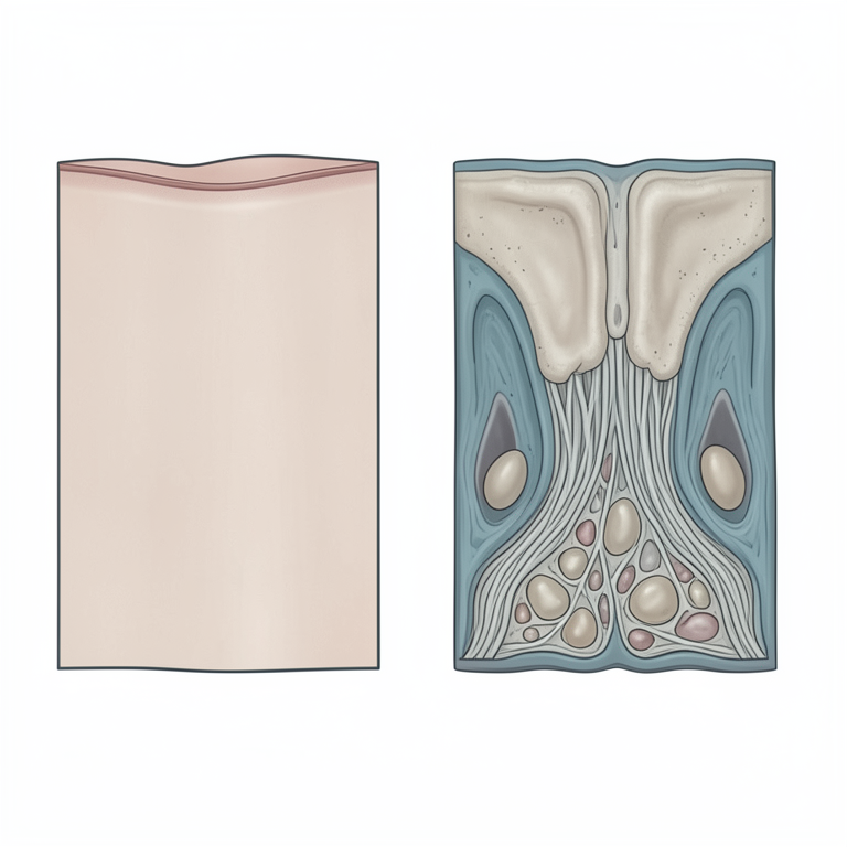
Comparison of Young vs. Old Rabbit Skin Layers
AI-generated conceptual illustration (domestic rabbit skin tissue). It explains the idea and does not show study data.
View source evidence ↗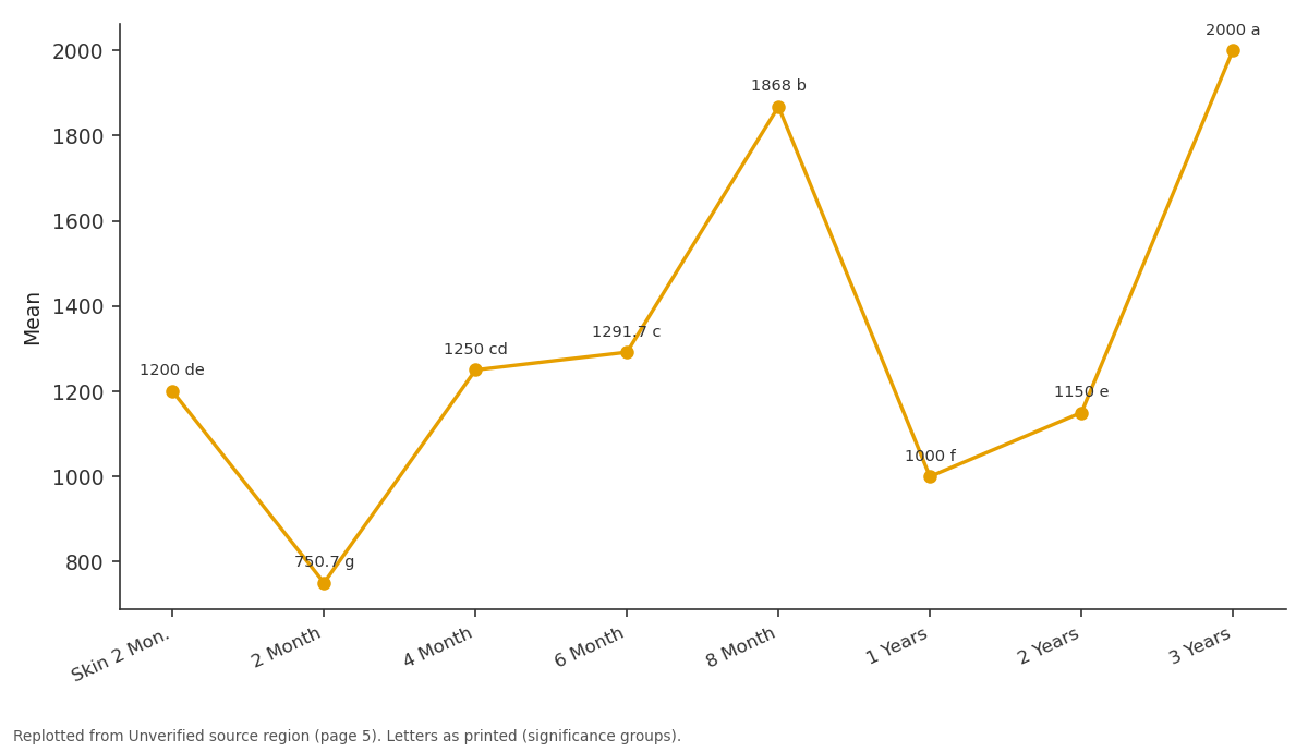
Mean: Skin 2 Mon. vs 2 Month vs 4 Month vs 6 Month vs 8 Month
Replotted from a table (page 5) of the original paper. Every bar is the paper's own table value.
View source evidence ↗Figures & tables
From the original paper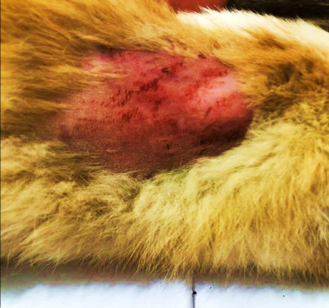 Figure 1
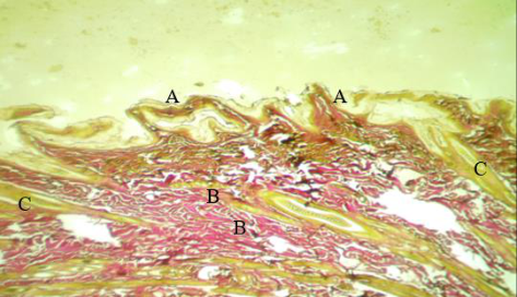 Figure 2
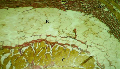 Figure 3
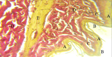 Figure 4
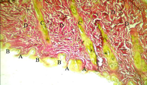 Figure 5
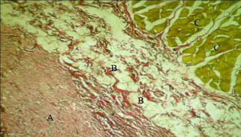 Figure 6
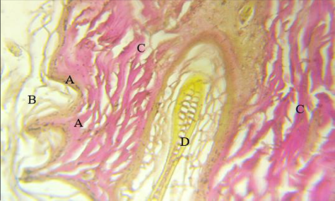 Figure 7
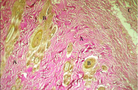 Figure 8
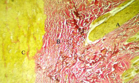 Figure 9
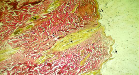 Figure 10
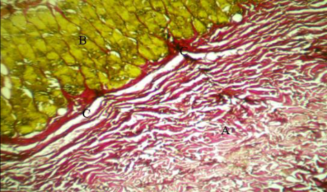 Figure 11
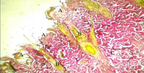 Figure 12
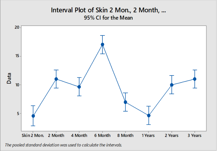 Figure 13
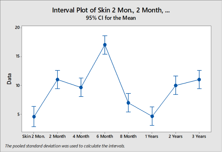 Figure 14
Introduction
The skin is the largest organ in the body, and plays a vital role in protecting the body from harmful environmental factors, as well as regulating temperature and sensation. Skin accounts for approximately 15% of the total body weight and is characterized by its ability to regenerate and repair (Burgess, 2015). The skin consists of three main layers: the epidermis, the dermis, and the subcutaneous tissue, and each layer plays a specific role in maintaining the integrity and functions of the skin (Kolarsick et al., 2011) [1] . With age, all skin functions undergo significant changes. The skin's ability to regenerate is diminished, and signs of aging such as wrinkles and loss of elasticity appear (Farage et al., 2008) [2] . The aging process affects the outer layer of the skin, making it more susceptible to damage and dryness (Makrantonaki & Zouboulis, 2007) [3] . Skin changes resulting from aging are not only the result of internal influences, but also chronic exposure to ultraviolet rays from the sun. Exposure to ultraviolet radiation accelerates the appearance of signs of aging, such as pigmentation and dark spots, which is known as photoaging (Krutmann, 2000) [4] . Oxidative stress from exposure to UV radiation and free radicals is thought to play a key role in this process, causing damage to DNA, proteins and lipids in skin cells (Fisher et al., 2002) [5] . Understanding the biological changes that occur in the skin with aging is essential for developing effective strategies to maintain healthy skin. Recent research highlights the importance of prevention through the use of sun protection and daily skin care (Rigel, 2010) [6] . Furthermore, aging is associated with slower wound healing, which increases the importance of careful care for aging skin (Wong & Pomerantz, 2008) [7] .
Recent studies confirm that skin changes associated with aging are caused by a combination of genetic and environmental factors. Therefore, strategies focusing on the prevention of UV damage and oxidative stress, in addition to proper skin care, can be effective in reducing the effects of aging on the skin (Vierkötter & Krutmann, 2012) [8] . Verhoff's collagen stain is a staining technique commonly used in histology to detect elastic fibers and collagen in tissues. This stain is an essential tool for identifying and evaluating ultrastructural changes in collagen-containing tissues, making it useful in the diagnosis of a variety of diseases and pathological conditions (Verhoeff, 1908) [9] . Verhoff's collagen stain works by using a mixture of iron hematoxylin, ferrichloride, and thiourin. These compounds react with elastic fibers to produce a characteristic black color, while collagen is stained red to pink using another dye such as Van Gieson (Gordon & Sweet, 1936) [10] . This technique allows a clear distinction between elastic and collagen fibers in tissues, which helps in the evaluation of structural and pathological changes. Verhoff's collagen stain is particularly used in the diagnosis of diseases affecting connective tissue, such as atherosclerosis, cirrhosis, and fibrotic lung disease. For example, they can be used to determine the extent of damage to elastic fibers in arterial walls in cases of atherosclerosis, helping doctors assess the progress of the disease (Lie, 1959) [11] . Furthermore, this dye is used in research studies to understand the changes that occur in connective tissue with age or as a result of exposure to certain environmental factors. Understanding these changes can help in the development of new treatments for diseases affecting connective tissues (Gamble & St. Mary, 1952) [12] . The study aims to provide a comprehensive understanding of the histological and functional changes that occur in the skin of domestic rabbits with age and the extent to which aging significantly affects the structure and functions of the skin, which in turn reinforces the need to develop new therapeutic strategies to maintain skin health and delay signs of aging. It also aims to open the door to more research for a deeper understanding of the biological mechanisms that control the aging process and its effects on living tissues.
Materials and Working Methods 2.1 Collecting rabbits
The animals being investigated, namely local rabbits, were acquired with a total of 40 rabbits at various stages of age, encompassing early, medium, and long-lived individuals. The rabbits were gathered between August 2023 and February 2024, and were separated into seven groups. Young and adult rabbits were acquired through the rearing and breeding of rabbits within the animal facility affiliated with the College of Veterinary Medicine/Tikrit University. Mature rabbits were sourced from reputable rabbit breeders, ensuring their age and disease-free status, and subsequently housed in dedicated enclosures for a period of time. Their health was regularly evaluated to assure their safety, considering laboratory circumstances such as ventilation, temperature ranging from 28 to 25 degrees Celsius, and a lighting schedule of 12 hours of light followed by 12 hours of darkness. The cages were lined with sawdust on the floor, and it was changed twice a week to maintain safety. The rabbits underwent a thorough examination by a veterinarian to verify their physical condition and appropriateness for the ongoing research.
Laboratory work
After the rabbits were acclimated into their seven groups, they were transferred to the surgical laboratory of the animal house at the College of Veterinary Medicine at Tikrit University for the purpose of dissection. Initially, the weights of the rabbits were recorded using a scale designed for small animals. The rabbits were then anesthetized with chloroform in preparation for dissection. Hair was removed from the neck area, then the outer skin and inner membrane were opened. Next, the skin samples were transferred to Petri dishes containing normal saline (0.75%) for washing. As shown in Figure1, the washing process was repeated four times, alternating the solution each time to maintain its concentration. After obtaining clean samples free of blood residue, they were placed directly into sealed plastic vials containing a fixative solution to avoid damage and decomposition of the samples. Bottles are labeled with member name, lot number, and sample number to ensure accurate documentation.
Microscopic Examination and Measurements
The rabbit's skin histological sections were analyzed using a Miotic light microscope at various magnification levels. Subsequently, the necessary portions of the histological sections were measured using an ocular micrometer.
Statistical analysis
Statistical analysis was conducted using the ready-made statistical program SAS system to calculate the significant differences between groups of study animals, by extracting the arithmetic averages for each group to compare with each other, then using one-way analysis of variance (ANOVA), as well as knowing the standard error (SE).
Results and Discussion
Results and Discussion
Laboratory results 2 months belly (A), ver
The epidermis contained a multi-folded paired squamous epithelium and the dermis contained a dense connective tissue composed of colloidal fiber bundles surrounding numerous hair follicles each containing a cortex and a medulla (Figure 2).
yellow color
The epidermis contains paired squamous epithelium, the epidermis has outer folds, and the inner bandages with the dermis, forming human papillae -the dermis is composed of dense colloidal fiber bundles and has hair follicles extending toward the epidermis (Figure 4)
2 years Dorsal (A) ver
The skin tissue contained degenerated epithelium, and the epidermis appeared with several pointed folds, with grooves emerging around those folds. The dermis contained hair follicles in large numbers, oriented vertically towards the epidermis, surrounded by dense connective tissue (Figure 5(.
2 years Dorsal (B) ver
The subdermis layer contains dense connective tissue adjacent to the adipose tissue, which contains degenerated fat cells, and under the skin, skeletal muscle fibers are found surrounded by fascia of colloidal fibers (Figure 6).
3 year Leg (A), ver
The epidermis of the skin is surrounded by multi-rowed strands of keratin, and the epidermis contains degenerated, dark-colored epithelium. The dermis contains bundles of colloidal fibers. Some hair follicles are also found, consisting of a cortex with indistinguishable cells and a medulla that contains numerous follicles and vacuoles devoid of pigment (Figure 7)
3 year Leg (A), ver
The dermis of the skin appeared wide, composed of bundles of colloidal fibers surrounding hair follicles spread throughout the entire dermis area, and the subdermis layer contained loose loose connective tissue fibers (Figure 8).
8 months Dorsal (A) ver
The dermis of the skin contained some hair follicles extending vertically towards the epidermis, surrounded by dense connective tissue with the presence of muscle tissue composed of skeletal muscle fibers interconnected from the subcutaneous layer (Figure 9).
8 months Dorsal (B) ver
The skin tissue contained a very narrow epidermis that appeared in the form of a thin strip, and hair follicles protruded from the dermis towards the epidermis and outside it through extensions, which are hair shafts, and around them from the dermis, bundles of colloidal fibers emerged (Figure 10) The subdermis layer contained connective tissue composed of colloidal fibers separated from the subcutaneous layer by muscle fascia with dense connective tissue, and the subcutaneous layer contained rows of compact skeletal muscle fibers (Figure 11). The epidermis of the skin is disintegrated and some of its parts have been shed, with the spread of many hair follicles in the dermis emerging from the epidermis in the hair shaft and connected to the sebaceous glands. The bundles of colloidal fibers in the dermis appeared moderately oriented and wavy (Figure 12).
Discussion and Results
Discussion and Results
Epidermal and Dermal Changes
The study shows that aging leads to significant changes in the thickness and structure of the epidermis and dermis in rabbits. The thickness of these layers increases with age. In older rabbits, the epidermis contains degenerated epithelial cells, and there is an increase in the number of folds and grooves in the skin surface.
The dermis shows a dense accumulation of collagen fibers, which becomes more pronounced with age. Hair follicles also become more numerous and oriented vertically towards the epidermis.
Subdermal Changes
The subdermis in older rabbits shows a mix of dense connective tissue and adipose tissue, with signs of degeneration in the fat cells.
Skeletal muscle fibers under the skin become more prominent and are surrounded by connective tissue, indicating structural changes in the subcutaneous layers.
Inflammatory Response and Vascular Changes
Aging appears to trigger an increase in inflammatory cell activity within the skin, which could be linked to a decreased ability to repair and regenerate skin tissues. The study notes changes in blood vessel composition, including an increase in both size and number of vessels, which might affect skin circulation and, consequently, tissue nutrition and regeneration.
Collagen and Elastic Fibers
The use of Verhoff's collagen stain highlighted changes in collagen and elastic fibers in the skin as rabbits age. The density and distribution of these fibers are altered, reflecting the reduced elasticity and increased rigidity of aging skin.
Conclusion Aging Effects:
The study provides clear evidence that aging has a significant impact on the structural integrity and functionality of rabbit skin. These changes are similar to those observed in human skin, making rabbits a potentially useful model for studying skin aging. Therapeutic Implications: The findings suggest the need for developing therapeutic strategies aimed at maintaining skin health and delaying aging signs. This could include approaches to enhance collagen production, improve skin elasticity, and manage inflammation. Further Research: The study opens avenues for further research into the biological mechanisms behind skin aging, potentially leading to new treatments for agerelated skin conditions.
Figures and tables from the paper
References
12 in the original paper- Kolarsick PA, Kolarsick MA, Goodwin C. Anatomy and physiology of the skin. J Dermatol Nurses' Assoc. 2011;3(4):203-213.
- Farage MA, Miller KW, Elsner P, Maibach HI. Intrinsic and extrinsic factors in skin ageing: a review. Int J Cosmet Sci. 2008;30(2):87-95.
- Makrantonaki E, Zouboulis CC. Molecular mechanisms of skin aging: state of the art. Ann N Y Acad Sci. 2007;1119(1):40-50.
- Krutmann J. Ultraviolet A radiation-induced biological effects in human skin: relevance for photoaging and photodermatosis. J Dermatol Sci. 2000;23:S22-6.
- Fisher GJ, Wang ZQ, Datta SC, Varani J, Kang S, Voorhees JJ. Pathophysiology of premature skin aging induced by ultraviolet light. N Engl J Med. 2002;337(20):1419-1428.
- Rigel DS. Cutaneous ultraviolet exposure and its relationship to the development of skin cancer. J Am Acad Dermatol. 2010;58(5):S129-32.
- Wong VW, Pomerantz H. Skin wound healing. N Engl J Med. 2008;359(6):670-673.
- Vierkötter A, Krutmann J. Environmental influences on skin aging and ethnic-specific manifestations. Dermatoendocrinol. 2012;4(3):227-231.
- Verhoeff FH. Some new methods of preparing sections of the eye for study by the microscope. Trans Am Ophthalmol Soc. 1908;10:568-571.
- Gordon H, Sweet HH. A simple method for the silver impregnation of reticulin. Am J Pathol. 1936;12(4):545551.
- Lie JT. Verhoeff's elastic stain: a practical method for the demonstration of elastic tissue. Arch Pathol. 1959;67(1):138-142.
- Gamble JS, St. Mary D. The elastic tissue of the lungs: its development and its regression in old age and emphysema. J Pathol Bacteriol. 1952;64(1):43-54.
Cited by 1
From OpenAlex, OpenCitations and reference lists read by Scinovex
- Morphological and histometrical analysis of skin in albino rabbits (Oryctolagus cuniculus) during postnatal period
Shahad Salih Abd-Almahde, Ekhlas Abid Hamza Al-Alwany · International Journal of Veterinary Sciences and Animal Husbandry · 2026
Background: The skin is a dynamic organ, which is subject to significant structural modification during postnatal life, and is an important organ in comparative anatomy as well as experimental studies. Aim of study: The present study was devised to understand the changes in the skin of albino rabbit (Oryctolagus cuniculus) of the factors related to morphology and histometrics after parturition regarding the regional difference between dorsal neck and dorsal abdomen. Methods: Fifteen apparently healthy rabbits were used and divided into three postnatal age groups, 7 days, 1 month, and 3 months