
Research article · 2024 · Full text
International Journal of Veterinary Sciences and Animal HusbandryConventional blood smear diagnosis and clinico-pathological findings of canine monocytic Ehrlichia canis in a rottweiler dog
Volume 9(6) · Pages 521–523 · DOI: 10.22271/veterinary.2024.v9.i6h.1926 ↗
Research areasVector-borne infectious diseasesYersinia bacterium, plague, ectoparasites researchViral Infections and Vectors
Abstract
Author-writtenA four year old, male, Rottweiler dog was brought to small animal medicine outpatient ward unit at Veterinary Clinical Complex, Tirunelveli, Tamil Nadu, India with the history of anorexia, panting and nasal discharge. On physical examination, the prescapular lymph node was swollen and mucous membrane congested. Pinpoint petechial hemorrhages were present on ventral abdomen. For hematological and biochemical analysis, the blood was collected and examined under automated hematology and biochemical analyzers. Blood smears were prepared from peripheral site and stained with leishman’s - Giemsa stain. Hematological examination revealed anemia, leukocytosis along with monocytosis and thrombocytopenia. Blood pictures revealed presence of hypochromasia and anisocytosis. The blood smears showed presence of morulae of Ehrlichia canis. It was present in both monocytes and lymphocytes. Biochemical examination showed elevation of blood urea nitrogen (BUN), alkaline phosphatase (ALP) and reduced albumin level. The case was diagnosed as an Ehrlichia canis based on presence of initial bodies and morulae within lymphocytes and monocytes. The case was treated successfully with Doxycycline @ 10 mg/kg/B/Wt for 14 days.
Summary
Source-checkedPlain-language reading of the full paper, prepared with AI and checked against the text. Each point links to the authors' own words.
Why it matters
- Canine monocytic ehrlichiosis (CME) is a disease in dogs caused by the intracellular rickettsia Ehrlichia canis. View evidence ↗
- The disease is transmitted via the bite of the brown dog tick, Rhipicephalus sanguineus, leading to serious clinical symptoms. View evidence ↗
- Identifying morulae on a blood smear provides a rapid diagnostic approach for ehrlichiosis. View evidence ↗
The question
- The study aimed to report the cytological diagnosis, clinico-pathological changes, and treatment of canine monocytic ehrlichiosis in a single Rottweiler dog. View evidence ↗
What they did
- The subject was a four-year-old male Rottweiler dog presented at a veterinary clinic in Tirunelveli, Tamil Nadu, India. View evidence ↗
- Blood samples were analyzed using automated machines for both hematological and biochemical examinations. View evidence ↗
- Peripheral blood smears from the ear were fixed with methanol and stained using Leishman-Giemsa for cytological examination. View evidence ↗
- The dog was treated with Doxycycline at a dose of 10 mg/kg body weight for 14 days. View evidence ↗
What they found
- Physical examination revealed anorexia, a fever of 102.5°C, congested conjunctival mucous membranes, and a swollen prescapular lymph node. View evidence ↗
- Multifocal petechial hemorrhages were observed on the ventral abdomen of the dog. View evidence ↗
- Hematological results showed anemia (Hemoglobin 5.7 g/dL), thrombocytopenia (Platelets 55,000/ul), and monocytosis (Monocytes 18%). View evidence ↗
- Biochemical analysis showed elevated blood urea nitrogen (351.45 mg/dl) and alkaline phosphatase (1217 IU/dl), and reduced albumin (1.9 g/dl). View evidence ↗
- Blood smears confirmed the presence of E. canis morulae and initial bodies within both monocytes and lymphocytes. View evidence ↗
- The dog showed clinical improvement in feeding and activity after one week, and hematobiochemical values returned to normal after 14 days of therapy. View evidence ↗
What it means
- The authors concluded that the presence of morulae and initial bodies in blood smears confirmed Canine Ehrlichiosis, and the dog achieved complete recovery within 14 days of treatment. View evidence ↗
Visual summary
1 visualsIllustrations explain the paper; they do not show study data.
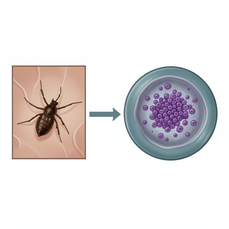
Transmission and Cellular Infection Cycle
AI-generated conceptual illustration (Rhipicephalus sanguineus tick and canine monocytes). It explains the idea and does not show study data.
View source evidence ↗Figures & tables
From the original paper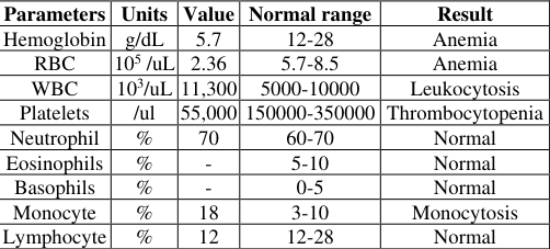 Table 1
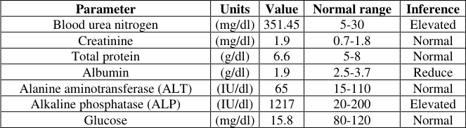 Table 2
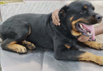 Fig1
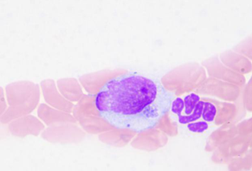 Figure 2
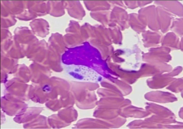 Figure 3
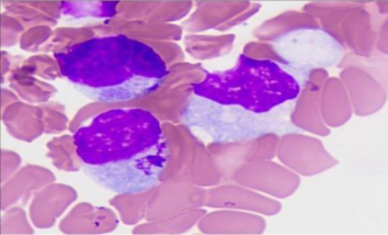 Figure 3
Introduction
Canine monocytic ehrlichiosis (CME) is a disease of dogs and caused by obligatory intracellular pleomorphic rickettsia Ehrlichia canis (Huxsoll et al., 1970) [7] . The brown dog tick, Rhipicephalus sanguineus, spreads through its bite, which causes serious clinical signs in affected dogs (Aziz et al., 2022) [3] . The infection is more prevalent in tropical and subtropical regions where R. sanguineus serves as a primary vector. Subsequent incubation period, three typical stages of the disease may develop like acute, chronic, and subclinical (Davis et al., 2013) [5] . Intracytoplasmic inclusion bodies (morulae) in circulating monocytes and lymphocytes are suggestive of E. canis infection. The clinical symptoms of canine ehrlichiosis viz, fever, weakness, loss of appetite, epistaxis, enlarged lymph nodes, tick infestation and changes in the eyes colour (Huxsoll et al., 1970) [7] . A few reports of E. canis infection are recorded with typical clinical sign like epistaxis (Venkatesakumar et al., 2018) [11] . The definitive diagnosis of ehrlichiosis based on clinical sigs, haemato biochemical changes and solely presence of organism within the cytoplasm of circulating monocytes. But current challenges, as natural infections may manifest with diverse clinical signs that vary across geographical regions and stray presence of the organism in advance stages of infection. Presence of morulae on blood smear examination is snap shot approach for diagnosis of ehrlichiosis (Sheikh et al., 2017) [10] . The present study reported the cytological diagnosis, clinico pathological alterations and therapeutic management of canine monocytic Ehrlichiosis in a Rottweiler Dog.
Materials and Methods
A four year old, male, Rottweiler dog (Fig. 1) was presented at Veterinary Clinical Complex, Veterinary College and Research Institute, Tirunelveli with history of not taking food for past one week, difficulty in breathing, panting along and nasal discharge. For further investigation blood was collected from cephalic vein in the forelimb. With the use of 5 ml syringe and the blood was pour into EDTA tube and clot activator tube. Collected blood was analyzed in complete automated blood cell count machine. Biochemical examination was carried in fully automated biochemical analyzer. Blood smears were prepared from peripheral vein of ear. Smears were fixed with methanol and stained with Leishman -Giemsa as per standard procedure. Stained blood smears were washed with distilled water after 20 minutes for cytological examination.
Results and Discussion
The physical examination was conducted. It revealed general appearance of dog was normal. The dog had history of anorexia it correlated with finding of (Asmita et al., 2016) [2] and pyrexia (Temperature: 102.5 C ), congested conjunctival mucous membrane along with swollen prescapular lymph node as same clinical findings reported by (Senthil et al., 2020) [10] . Presence of multifocal petechial hemorrhage in the ventral aspect of abdomen with it mentioned in (Azize et al., 2016) [3] could be due to thrombocytopenia. The results were obtained from hematology and biochemical examinations are presented in Table 1 and2. Hematology indicated that dog had anemia, thrombocytopenia, monocytosis as same as (Bulla et al., 2004) [4] . In present study, epistaxis does not happened though thrombocytopenia exists. Biochemical analysis of our study showed elevation of blood urea nitrogen, alkaline phosphatase and reduction of albumin. These findings were contradicted with previous report of ( Asmita et al., 2016) [2] and concurred with (Azize et al., 2016) [3] . Blood smear examination revealed presences of both morulae (Fig. 2) and initial bodies (Fig. 3) of E. canis in monocytes and also within lymphocytes (Fig. 4). These results of presence of morula of Ehrlichia canis in lymphocytes and monocytes it is correlated with (Elias et al., 1991) [6] . Animal was treated with doxycycline 10mg/kg/Bwt as a drug of choice in many clinical studies (Amyx et al., 1971) [1] : (Asmita et al., 2016) [2] : (Komal et al., 2020) [8] . On the first day, the dog was treated with Inj. doxycycline @10 mg/kg/Bwt and to correct the electrolyte deficit infused ringer's lactate with dose rate based on dehydration, dexamethasone was given at dose rate of 2 mg/kg body weight. Then owner was advised to have Tab -Doxycycline -10 mg/kg body weight for 14 days. The dog feeding habits and other activities improved clinically one week after treatment. The haematobiochemical values were within the normal range after 14 days of therapy.
| Parameters | Units | Value | Normal range | Result |
|---|---|---|---|---|
| Hemoglobin | g/dL | 5.7 | 12-28 | Anemia |
| RBC | 105 /uL | 2.36 | 5.7-8.5 | Anemia |
| WBC | 103/uL | 11,300 | 5000-10000 | Leukocytosis |
| Platelets | /ul | 55,000 | 150000-350000 | Thrombocytopenia |
| Neutrophil | % | 70 | 60-70 | Normal |
| Eosinophils | % | - | 5-10 | Normal |
| Basophils | % | - | 0-5 | Normal |
| Monocyte | % | 18 | 3-10 | Monocytosis |
| Lymphocyte | % | 12 | 12-28 | Normal |
Table text extracted from the original PDF; superscript letters are shown inline. The original table image is below.
View original table image
Conclusion
Based on conventional blood smear examination the case was diagnosed, recorded the clinical pathological parameters and treated with doxycycline and supportive therapy for 14 days. Morulae and initial bodies presence within the circulating monocytes and lymphocytes of the dog blood smear confirm the Canine Ehrlichiosis. The dog recovered completely within 14 days of post treatment.
Figures and tables from the paper
| Parameter | Units | Value | Normal range | Inference |
|---|---|---|---|---|
| Blood urea nitrogen | (mg/dl) | 351.45 | 5-30 | Elevated |
| Creatinine | (mg/dl) | 1.9 | 0.7-1.8 | Normal |
| Total protein | (g/dl) | 6.6 | 5-8 | Normal |
| Albumin | (g/dl) | 1.9 | 2.5-3.7 | Reduce |
| Alanine aminotransferase (ALT) | (IU/dl) | 65 | 15-110 | Normal |
| Alkaline phosphatase (ALP) | (IU/dl) | 1217 | 20-200 | Elevated |
| Glucose | (mg/dl) | 15.8 | 80-120 | Normal |
Table text extracted from the original PDF; superscript letters are shown inline. The original table image is below.
View original table image
References
11 in the original paper- Amyx HL, Huxsoll DL, Zeiler DC, Hildebrandt PK. Therapeutic and prophylactic value of tetracycline in dogs infected with the agent of tropical canine pancytopenia. J Am Vet Med Assoc. 1971;159:14281432.
- Asmita S, Rockson K. Ehrlichiosis in a dog: A case study. Int J Vet Sci Anim Husbandry. 2016;1(5):20-23.
- Aziz MU, Hussain S, Song B, Ghauri HN, Zeb J, Sparagano OA. Ehrlichiosis in dogs: A comprehensive review about the pathogen and its vectors with emphasis on South and East Asian countries. Vet Sci. 2022 Dec 29;10(1):21.
- Bulla C, Kiomi TR, Pessoa AJJ, Trinca LA, Souza LR, Wiedmeyer CE. The relationship between the degree of thrombocytopenia and infection with Ehrlichia canis in an endemic area. Vet Res. 2004;35(1):141-146.
- Davis H, Jensen T, Johnson A, et al. American Animal Hospital Association/American Association of Feline Practitioners fluid therapy guidelines for dogs and cats. J Am Anim Hosp Assoc. 2013;49(3):149-159.
- Elias E. Diagnosis of Ehrlichiosis from the presence of inclusion bodies of morulae of Ehrlichia canis. J Small Anim Pract. 1991;33:540-543.
- Huxsoll DL, Hildebrandt PK, Nims RM, Walker JS. Tropical canine pancytopenia. J Am Vet Med Assoc. 1970;157(11):1627-1632.
- Komal, Sweety, Vaishali, Jain T, Singh M. Ehrlichiosis in a dog: A case report. J Entomol Zool Stud. 2020;8(1):349-350.
- Senthil NR, Chakravarthi R, Vairamuthu S. Epidemiology of canine ehrlichiosis from 2010-2019 in Chennai, India. The Pharma Innovation J. 2020;SP9(8):1-3.
- Sheikh TI, Mohd YG, Singh AK, Gupta T, Sharma S, Noor S. Ehrlichiosis in a Labrador dog: A case study. The Pharma Innovation J. 2017;6(11):690-692.
- Venkatesakumar E, Kumar V, Ramprabhu R. Diagnosis and management of concurrent Ehrlichiosis and Babesiosis in a dog. Intas Polivet. 2018;19(II):267-268.