
Research article · 2024 · Full text
International Journal of Veterinary Sciences and Animal HusbandryA clinical study on clinical evaluation of eggshell derived nano hydroxyapatite on augmenting healing of long bone fractures in dogs
Volume 9(6) · Pages 446–455 · DOI: 10.22271/veterinary.2024.v9.i6g.1913 ↗
Research areasBone fractures and treatmentsBone Tissue Engineering Materials
Abstract
Author-writtenAmong 158, 126 were long bone fractures accounting for 3.78% of total cases and 79.74% of fracture cases. The highest incidence was recorded in Mongrel dogs in the juvenile age group, male dogs, right hind limb and femur bone. The most frequent cause of fractures in the present study was automobile accidents. Out of 12 cases, fractures of radius ulna (N=6), tibio-fibula (N=5) and femur (N=1) were selected and were randomly divided in to two groups of six each. In group I, repair of fractures was done by DCP and in group II, along with bone plating, eHA was applied at the fracture site under general anaesthesia. Cranio-medial approach for the radius and ulna cranio-lateral approach for the femur and cranio-medial approach for the tibio-fibula were adopted. Lower values of ALP were recorded during the preoperative and immediate postoperative days which then increased significantly. Lower values of serum calcium and phosphorus were recorded initially in group I and higher values observed in group II. Lower values of serum vitamin D were recorded that increased significantly (p
Summary
Source-checkedPlain-language reading of the full paper, prepared with AI and checked against the text. Each point links to the authors' own words.
Why it matters
- The development of biomaterials has been more critical to the advancement of modern veterinary orthopedics than any other clinical specialty. View evidence ↗
- Bioactive coatings are currently a key area of focus for expanding the capabilities of orthopedic biomaterials. View evidence ↗
The question
- The study aimed to evaluate whether eggshell-derived nano hydroxyapatite (eHA) could augment the healing of long bone fractures in dogs. View evidence ↗
What they did
- The research involved 12 dogs with long bone fractures treated at the NTR College of Veterinary Science between March and November 2024. View evidence ↗
- The 12 cases, consisting of radius ulna (6), tibio-fibula (5), and femur (1) fractures, were randomly split into two groups of six dogs each. View evidence ↗
- Radiographic healing was measured using a scoring system, with results recorded up to the 90th postoperative day. View evidence ↗
What they found
- By the 90th postoperative day, Group II had a lower mean radiographic healing score (0.17±0.16) compared to Group I (0.50±0.22), indicating better healing. View evidence ↗
- Group II showed higher serum alkaline phosphatase (ALP) levels than Group I on the 7th, 15th, and 30th postoperative days, suggesting increased osteoblastic activity. View evidence ↗
- Group II exhibited significantly higher serum phosphorus levels between the 7th and 30th postoperative days compared to Group I, which the authors associate with an augmented healing process. View evidence ↗
- Serum vitamin D levels increased significantly from the 7th to 60th postoperative day in Group II, whereas the same trend was non-significant in Group I. View evidence ↗
- Dogs in Group II demonstrated better lameness scores on the 7th, 15th, and 30th postoperative days, suggesting earlier clinical union. View evidence ↗
- By the 90th postoperative day, a majority of dogs in Group II showed bone remodeling with corticomedullary continuity compared to those in Group I. View evidence ↗
- At the study's conclusion, five dogs in Group II and four dogs in Group I achieved excellent usage of the affected limb. View evidence ↗
What it means
- The authors conclude that eggshell-derived nano-hydroxyapatite is an effective biomaterial for inducing osteoblastic activity when used alongside dynamic compression plating. View evidence ↗
- The authors state that supplementing Vitamin D after long bone fracture repair can influence the restoration of the animal's ability to walk. View evidence ↗
Visual summary
1 visualsIllustrations explain the paper; they do not show study data.
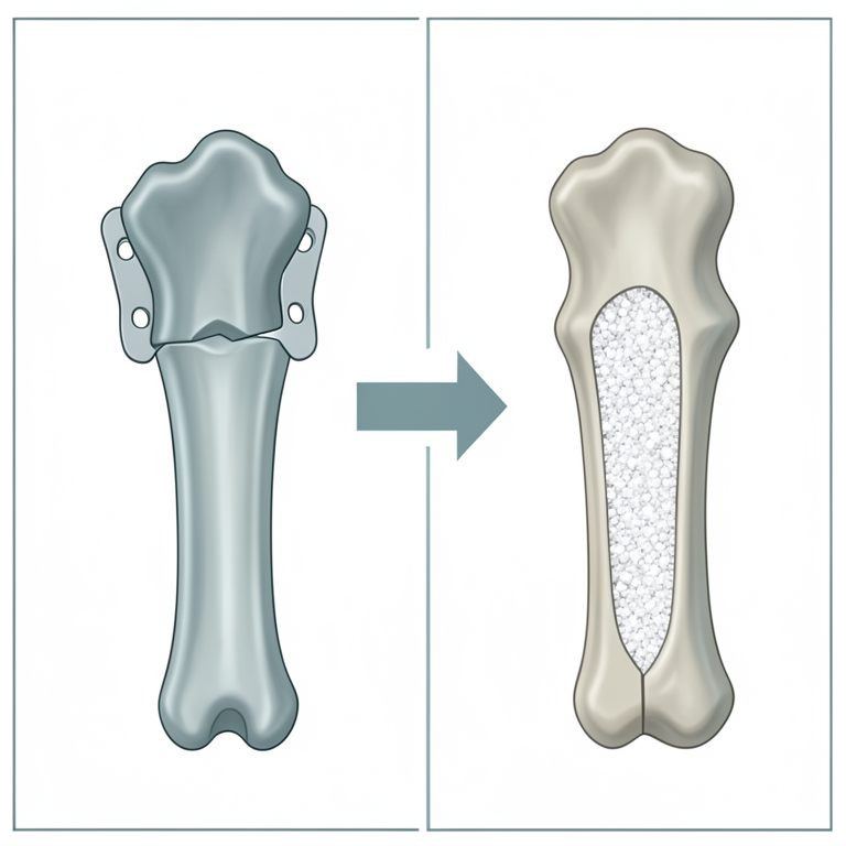
Study overview
AI-generated conceptual illustration (canine long bone fractures). It explains the idea and does not show study data.
View source evidence ↗Introduction
Orthopedics owes its present status of modern care to the development of biomaterials more than any other veterinary clinical specialty. In the realm of modern veterinary orthopaedics, the integration of biomaterials led to enhanced care of patients, refined techniques, and unprecedented therapeutic possibilities.
The insatiable search for better clinical outcome propels the development of many biomaterials in the field of orthopaedics. The vista of bioactive coatings emerges as a beacon guiding the expansion of orthopaedic biomaterials. Hence the present study was undertaken to test the efficacy of egg shell derived nano hydroxy apatite in augmenting the healing of the fractured bones in dogs and the results are discussed as following.
Materials and Methods
The present study was carried out on 12 clinical cases of long bone fractures in dogs that were brought to the Department of Veterinary Surgery and Radiology, NTR CVSc, Gannavaram, from March to November of 2024 with symptoms of nonweight bearing lameness of the pectoral and pelvic limbs. Thorough clinical, orthopedic, neurological, and radiographic examination was conducted for the dogs included in the present study. The fracture cases were so selected that both the fragments could accommodate at least two screws each. When an animal refused to co-operate for a radiographic examination, butorphanol tartarate 1 (0.2 mg/kg body weight) was injected IM for sedation. The selected cases with long bone fractures were categorized into two group of six dogs each and were treated as following. Group-I (N=6): The fracture was repaired by dynamic compression plating. Group-II (N=6): The fracture was repaired by dynamic compression plating and a mixture of eggshell derived Nano hydroxyapatite 2 was doped at the fracture site. The latter was procured from the Department of Clinical Research, SIBAR Institute of Dental Sciences and Guntur.
Anesthesia
All the dogs were premedicated with Butorphanol tartarate @ 0.2mg/Kg body weight IM, atropine sulphate [8] @ 0.04mg/Kg body weight SC and thirty minutes prior to induction of anesthesia. After cannulation of cephalic vein, general anesthesia was induced by intravenous injection of Ketamine hydrochloride [9] @ 5mg/Kg body weight and Midazolam 10 @ 0.2mg/Kg body weight (till effect) and was maintained by inhalation anesthesia using a mixture of Isoflurane 11 and oxygen.
Patient positioning
The dogs were controlled in lateral recumbency with the fractured limb touching the operating table, exposing the medial aspect while treating radio-ulnar and tibial fractures, however for the repair of femoral fractures, lateral surface of thigh was positioned upper to the face of surgeon.
Instrumentation and implants
In the present study used a general instrumentation set and a conventional orthopedic set comprising specific equipment for the fixation of bone plates.
Material used for bone grafting
Egg shell derived Nano hydroxyapatite powder.
Surgical approaches to the long bones Surgical approach to the radius
In order to access the fracture fragments and fix the bone plate, a large, linear skin incision was made on the craniomedial aspect of the radius. On the same line as the skin, the subcutaneous fascia was also incised with the distal part of the incision paralleling the extensor carpi radialis muscle. The deep antebrachial fascia was cut between the pronator teres and extensor carpi radialis muscles proximally. The supinator muscle was visible upon retraction of lateral extensor muscle.
For increased exposure of the radial shaft, the deep digital flexor and flexor carpi radialis muscles were raised caudally. Throughout the process, cephalic vein was shielded carefully.
Surgical approach to the Femoral Diaphysis
Along the craniolateral surface of the femur, beginning from the trochanter major to the lateral condyle of the femur the skin incision was made. The muscle tensor fascia lata was made visible by cutting the subcutaneous tissue. The tensor fascia lata was cut at the point where it inserted to the cranial border of the biceps femoris muscle. The vastus lateralis muscle was made visible by the caudal retraction of the biceps femoris muscle. The shaft of the femur was made visible by the separation and retraction of these two muscles.
Following the repair of fracture, the incision on the tensor fascia lata was closed using No. 2/0 polyglactin 910 in a straightforward continuous pattern and subcuticular sutures was placed using 2-0 polyglactin 910. Using no-1 polyamide, the edges of incision were approximated with a row of cruciate mattress or simple interrupted sutures.
Surgical approach to the tibial diaphysis
A curved incision was made to prevent the skin incision from being directly over a bone plate. The incision was made on the skin proximally over the medial tibial condyle, curving cranially to the midline of tibia at its midshaft, and then caudally in the direction of the medial malleolus. With caution to preserve the saphenous nerve and saphenous artery that traverse the middle to distal third of the tibial diaphysis, the subcutaneous fascia was dissected in the same direction as the skin incision. Depending on the location of the bone, the saphenous nerve and arteries were drawn back either proximally or distally. The fragmented bone became visible after the deep crural fascia on the shaft of tibia was cut (Fossum, 2013) [4] . The subcutaneous fascia was sealed with continuous sutures with of No. 2-0 polyglactin 910 after the fracture was fixed. The skin incision was closed using No. 2-0 polyamide, with a row of horizontal mattress sutures or with simple interrupted sutures.
Procedure for dynamic compression plating
Once the fracture site was reached, the broken fragments of the bone were visible and their ends were restored using bone hooks. Using bone-holding forceps to provide gentle traction and counter traction, fracture reduction was achieved with caution to avoid damaging the thin cortex and periosteum. Soft tissue damage was avoided by gentle dissection to preserve the blood supply at fracture site. Following reduction of the fracture fragments, dynamic compression plates (DCP) of the proper size were used for stability. Once the right size plate was chosen, the plate benders were used to precisely shape it to fit the counter of the bone. Bone and plate holding forceps was used to properly apply and secure the plate over the cranial surface of the fractured radius ulna. Using the proper size drill bit (e.g. 2.7mm for 3.5mm plate), the screw holes were drilled away from the fracture line, and the second hole was drilled eccentrically in the opposite fragment. To avoid heat necrosis of the bone, normal saline was irrigated at the drilling site. The remaining screws were inserted in a different order such that each fragment had at least two threaded screws that purchased four cortices installed. Repair of fracture of long bones in dogs of group I was done with SS DCP; while in group II in addition to SS DCP, eggshell derived Nano hydroxyapatite powder was also applied at the fracture site.
The antebrachial fascia was sutured with 2-0 polyglactin 910 by simple interrupted sutures followed by subcuticular sutures with 2-0 polyglactin 910. The skin edges were approximated with 2-0 polyamide using simple interrupted sutures.
Postoperative care
To evaluate the degree of fracture fixation, the operated limb's immediate post-operative radiographs were obtained. After surgery, the operated limb was supported by reinforced and modified Robert Jones bandages that were changed every alternate day along with the antiseptic dressing of the surgical wound. In certain situations that required more support, a stronger, reinforced Robert Jones bandage was also used.
Following surgery, all of the dogs were given IM injections of ceftriaxone sodium (25-50 mg/kg) twice daily for seven days, meloxicam (0.2 mg/kg) once daily for three days, and oral calcium syrup (10mL/day) orally once daily for eight weeks.
In those dogs that were deficient in vitamin D were orally administered on fasting stomach at weekly intervals. After 10-12 days of surgery, the skin sutures were removed.
In order to maintain normal joint movement following two weeks of surgery, physiotherapy including passive exercises was recommended for ten minutes three times a day.
Result and Discussion
This procedure of DCP fixation provided good results for fracture healing in present investigation. Dynamic compression plating was described as a versatile and stable repair procedure that enabled rapid weight bearing for transverse fractures of the radius (Milovancev and Ralphs, 2004 and Mukherjee et al., 2015) [5,6] , as it provided rigid stabilization by neutralizing rotational, bending, compression, and shearing stresses on the bone acting at the fracture sites and producing osteosynthesis without causing any complications (Silbernagel, et al., 2004 and Stiffler, 2004) [7,8] . DCP permitted early ambulation and immediate movement of neighboring joints (Uhthoff et al., 2006) [9] . This procedure was very well recommended for the repair of transverse overriding or oblique or comminuted fractures of radius and ulna by Claes and Heigele (1999) [10] and Denny and Butterworth (2000) [11] and tibial fractures by Conzemius and Swainson (1999) [12] . The favorable results after the usage of DCP in the present study might be due to the restoration of vascularity at the fracture site as the blood supply to the cortex beneath a conventional DCP was found to be significantly higher than that of the cortex opposite the plate. Furthermore, the blood supply to both cortices was found to be significantly higher than that of the contralateral normal radius bone as explained by Protopsaltis et al. (2002) [13] . The DCP was considered superior to other bone plates (Kotwal, 2008) [14] and the success of the technique largely depended on the selection of suitable plate length (Niemeyer and Sudkamp, 2006) [15] . The successful outcome as reported in the present study, was also documented by Aikawa et al. (2018) [16] who considered that DCP produced a normal functional outcome without developing non-union. Ramirez and Macias (2016) [17] and Parent et al. (2017) [18] reported excellent functional outcomes after treating distal radial and ulnar fractures in toy breed dogs.
Antibiotics and non-steroidal anti-inflammatory drugs administered prevented the postoperative sepsis and provided good analgesia. During the initial postoperative period, application of reinforced Robert Jones bandages helped to limit joint motion, gave the fractured limb more support, and provided good adjunct support to the internal fixation. The physiotherapy and passive exercises that were advised oneweek post-surgery helped in improving the range of motion of the joints proximal and distal to the affected bone. Similar postoperative care was also exercised by Das et al. (2012) [19] , Reddy et al. (2020) [20] and Das et al. (2021) [21] following internal fixation of fractures in dogs. [22][23][24][25][26] . Serum levels of bone markers, such as ALP, are higher in relation with osteoblastic activity at the fracture site (Komnenou et al., 2005) [23] . This enzyme facilitates the transfer of calcium from bone fragments to plasma when the pH is acidic as the pH shifts to alkaline, the formation of the major bone callus occurs (Chhatrola et al., 2022) [27] .
In the current study, lower values of ALP were recorded during the preoperative and immediate postoperative days which then increased significantly from the 7th postoperative day to 60 th postoperative day with the peak values on the 30 th day and then decreased significantly (p<0.05) by the 90 th postoperative day in both the groups. The higher ALP values could be attributed to the damaged periosteum that happens at the time of the fracture and at the time of the bone plating procedure. Based on these results, it appeared that callus development was evident on the 30 th postoperative day, when osteoblastic activity peaked which was in agreement with that of Chaudhari et al. (2000) [28] , Kulkarni (2000) [29] and Chaurasia et al. (2019) [30] . A similar trend in the levels of ALP i.e. higher values during the early postoperative days followed by a gradual decrease to later stages of fracture healing process were also reported by Johnson and Watson (2000) [31] , Rani and Ganesh (2003), Reddy et al. (2020) [20] and Bhavani et al. (2022) [26] and Mehdi et al. (2024) [33] . However, Suryawanshi et al. (2001) [34] recorded higher values immediate postoperatively that remained elevated till the end. The values of ALP were found elevated in Group-II than group I on the 7 th , 15t h and 30 th postoperative days indicating more osteoblastic activity at the site of fracture in these dogs. The use of egg shell derived nano-hydroxy apatite at the fracture site might have encouraged this augmented activity as opined by Chaurasia et al. (2019) [30] . The normal values of ALP in Groups I & II on the 90 th postoperative day can be thought to represent the complete healing. In the current study, serum calcium levels were lower during the preoperative and immediate postoperative days that increased significantly from the 7th postoperative day and decreased gradually up to the 90th postoperative day. These observations were in correlation with those of Vasantha (1991) [35] , Suryawanshi et al. (2001) [34] , Hegde et al. (2007) [36] , Patil et al. (2017) [37] , Bhavani et al. (2022) [26] , Chhatrola et al. (2022) [27] and Mehdi et al. (2024) [33] . The deposition of excess calcium at the fracture site might have led to decreased serum calcium levels during the early stages and bone remodeling might have contributed to a subsequent increase in the levels during the later stages of fracture healing as explained by Chaurasia et al. (2019) [30] .
In the current study, lower values of serum phosphorus were recorded during the preoperative and immediate postoperative days that increased significantly from the 7th to 30 th postoperative days and eventually decreased significantly by the 90 th postoperative day. There was a significant difference in the values of serum phosphorus between the groups at 7 th to 30 th postoperative days. The increased levels of inorganic phosphorus in group-II can be thought to indicate augmented healing process. A similar trend i.e. higher values during the early postoperative days followed by a gradual decline in later stages of fracture healing was also observed by Hegde et al. (2007) [36] . Suryawanshi et al. (2001) [34] and Reddy et al. (2020) [20] observed a gradual increase in the values from preoperative day to 60 th postoperative day. The high levels of extracellular concentrations of phosphorus resulted in the deposition of calcium at the site of fracture, thereby facilitating the ossification of the callus (Komnenou et al., 2005) [23] . Hazewinkel and Tryfonidou (2002) [38] opined that that vitamin D was necessary for the systematic growth and mineralization of cartilage in the growth plate in young dogs. Lower values of serum vitamin D were recorded during the preoperative and immediate postoperative day that increased significantly from 7 th to 60 th postoperative day and decreased gradually up to the 90th postoperative day in animals of group II, while same trends were observed non-significantly in group I. There was significant difference in the values of serum vitamin D between the groups at 7 th to 30 th postoperative days. The difference in the total mean serum vitamin D concentration between the groups was significant. Similar to these observations, Ettehad (2014) [39] also reported decreased vitamin D levels in patients with femoral and tibial fractures and the magnitude of decline was found more pronounced in the first week, when compared to that in subsequent week. Aslan (2006) [40] and Sharma et al. (2017) [42] administered vitamin D injection to rats that had a fracture and observed better biomechanical characteristics and higher bone content of the damaged bones indicating better healing. A clinical lameness scoring system of 0-5-point scale was used to evaluate the usage of limb as suggested by Cook et al. (1999) [43] . In both the groups, partial usage of the affected limb was observed between the 1 st and 4 th postoperative days. The partial weight bearing was observed early as one day (Abhishek et al., 2022) [44] , third day (Singh and Coutinho, 2012) and between 7 th to 10 th days (Al-Harby et al, 1996) [46] following dynamic compression plating. Better lameness scores on 7th, 15th and 30th postoperative days were observed in group II which could be attributed to the better and early clinical union in that group, suggesting the efficacy of eggshell derived nano hydroxyapatite on augmenting healing of bone fractures. Radiographic evaluation of fracture healing was done by obtaining follow-up radiographs that were taken on the immediate postoperative then on the 30th, 60th and 90th postoperative days. Based on the development of a callus and/or the disappearance of the radiolucent fracture line, fracture healing scores (0-3) were assigned as recommended by Cook et al. (1999) [43] . On immediate postoperative radiographs, an excellent anatomical reduction, well apposed fracture ends, and without any gap between the fracture fragments were observed in animals of both the groups. Disappearance of the fracture line in group II could be attributed to radiopaque biomaterial i.e. eggshell derived nano hydroxyapatite which was applied at the fracture site whereas fracture line was still observable in all cases of group I. Radiographs which were taken on the 15 th day depicted the initiation of callus in all the cases and this finding is in agreement with that of Dakhane et al. (2021) [47] . The formation of soft bridging callus was observed between10 to 12 postoperative days (Hobbs, 2003) [48] , the second postoperative week and at the fourth postoperative week (Umarani et al., 2007) [50] following bone plating. The present study defined radiographic healing as the presence of bridging callus and disappearance of radiolucent fracture line. Radiographs obtained after 30 days in Group-II suggested early healing of fractures as the mean fracture healing score was close to 0 with evidence of periosteal callus with a small fracture gap, with an appropriate progression towards healing. Similar to this observation, Doijode et al. (2018) [51] and Abhishek et al. (2022) [44] also observed a bridging callus around the fracture site with the fracture fragments in proper alignment, on the 30 th postoperative day. On the 60th postoperative day, the mean fracture healing scores of animals of group-II were lower than that of group-I indicating the superiority of the treatment group in terms of fracture healing. Follow up radiographs of two cases in group II showed cortical and medullary continuity with fracture remodeling on the 60 th postoperative day itself as also observed by Doijode et al. (2018) [51] . Quite parallel to this finding, Patil and Dilip Kumar (2018) [52] , Reddy et al. (2020) [20] and Abhishek et al. (2022) [44] , noted completely filled fracture gap with moderate callus by the 60 th postoperative day. In contrast to the present study, Vardhan et al. (2017) [53] recorded resorption of exuberant callus at the fractured site as early as the 60 th postoperative day following repair of diaphyseal fractures in dogs. However, the radiolucent region surrounding bone screws as observed by Feng et al. (2019) [54] that was ascribed it to bone loss carried due to implants was not recorded in the present study. Bone remodeling with corticomedullary continuity was recorded in majority of group II dogs when compared to those of group I by the end of 90 th postoperative day. In the present study, all the cases displayed complete radiographic healing by the 90th postoperative day with the exception one dog of group I, which could be attributed to the rigid fracture fixation provided by SS plates that restricted micro motion between the fracture fragments and minimal periosteal callus at the fracture site. At various periodic intervals of the present study, group II had better mean fracture healing scores than group I, suggesting the impact of eggshell derived nano hydroxyapatite on augmenting healing of long bone fractures in dogs. The hydroxyapatite with its newly discovered osteo-inductive properties is widely known for its ability to promote bone growth by conduction or by serving as a scaffold to fill in defects and it holds a great promise for stimulating the formation of new bone (Kattimani et al., 2016) [55] . It was considered as a suitable alternative for autogenous bone grafting in periodontal tissue, since it possessed several merits including biocompatibility and lack of toxicity (Bayani et al., 2017) [56] . Very recently, Mohamed et al. (2024) [1] recommended xeno-hydroxyapatite as a good choice for bone reconstruction as it reduced the healing time in area of fracture line. At the end of the study, an excellent usage of the affected limb was recorded in five cases of group-II and in four cases of group-I. One case from group II and two cases from group I showed exercise induced lameness, and the outcome was graded as "good". Similar findings were also observed by Patil and Dilip Kumar (2018) [52] who recorded complete weight bearing following dynamic compression plating in dogs. The most prominent side effects of fractures as osteomyelitis, nonunion, delayed union, malunion, early physeal closure, sarcoma related with the fracture, synostosis, implant failure etc. which were reported by a few authors including Denny and Butterworth (2000) [11] , Jackson and Pacchiana (2004) [57] , Wheeler et al. (2004) [58] , Chandy et al. (2007) [59] , Kallianpur et al. (2018) [60] , Deveci et al. (2022) [61] were not observed in the present study. Pedal edema observed in case 5 of group I in the immediate postoperative period was attributed to application of tight bandage. Similar complication in animals treated with DCP were also reported by Baltzer et al. (2015) [62] and Ramirez and Macias (2016) [17] , who ascribed them to pressure imposed by the MRJ bandage. Group-II dogs showed better outcome than those of group-I which can be attributed to the application of eggshell derived nano hydroxyapatite at the fracture gap after fracture fixation with dynamic compression plates. Egg shell derived nano-hydroxy apatite is a superior osteo-inductive growth factor and osteogenic cell carrier, which increases its usefulness as a bioactive delivery vehicle in the fractures. Hydroxyapatite had an unstable crystalline structure which allowed exchange of ions, highly porous nature that acted as a scaffold and induced fibrovascular ingrowth, inward migration of bone producing cells, and osteoid deposition onto the well-developed surface area as explained by Bhatt and Rozental, 2012) [63] .
Conclusion
Dynamic compression plating is a versatile and stable repair procedure that enables rapid weight bearing for transverse fractures of the long bones by providing rigid stabilization by neutralizing rotational, bending, compression, and shearing stresses on the bone. Egg shell derived nano-hydroxy apatite is an affective biomaterial which can induce the osteoblastic activity when used as an adjunct at the site of fracture following dynamic compression plating. Vitamin D supplementation following repair of long bone fractures can have a bearing on the restoration of ambulation following bone plating. Regular and periodic radiographic evaluation of fracture healing can help in minimizing the complications related to the implant failure and monitor the progress of radiographic union of the fracture fragments.
Assessment of bone specific biomarkers like alkaline phosphatase can be used to predict the non-union or delayed union at an early date
Conflict of Interest Not available
Conflict of Interest Not available
Financial Support
Financial Support
References
62 in the original paper- Mohamed ISS, Metwally E, Ahmed IH, Elkorash SAH, Ghanem WA, Hamed MS. Effect of XenoHydroxyapatite Bone Graft on the Healing of Unfavorable Mandibular Fractures in Dogs. Dent Sci Updates. 2024;5(1):141-149.
- Johnson KA. Piermattei's Atlas of Surgical Approaches to the Bones and Joints of the Dog and Cat. 5th ed. St Louis, MO: Elsevier, 2013, p. 244-92.
- Piermattei DL, Johnson KA. Approach to the shaft of the radius through a medial incision. In: Piermattei DL, Johnson KA, editors. An Atlas of Surgical Approaches to the Bones and Joints of the Dog and Cat. 3 rd Ed, 2004, p. 196-9.
- Fossum TW, Dewey CW, Horn CV, Johnson AL, MacPhail CM, Radlinsky MG, et al. Small Animal Surgery. 4th ed. Mosby Elsevier, 2013, p. 1033-1199.
- Milovancev M, Ralphs SC. Radius/ulna fracture repair. Clin Tech Small Anim Pract. 2004;19(3):128-33.
- Silbernagel JT, Johnson AL, Pijanowski GJ, Ehrhart N, Schaeffer DJ. A mechanical comparison of 4.5 mm narrow and 3.5 mm broad plating systems for stabilization of gapped fracture models. Vet Surg. 2004;33(2):173-179.
- Stiffler KS. Internal fracture fixation. Clin Tech Small Anim Pract. 2004;19:105-113.
- Uhthoff HK, Poitras P, Backman DS. Internal plate fixation of fractures: short history and recent developments. J Orthop Sci. 2006;11(2):118-126.
- Claes LE, Heigele CA. Magnitudes of local stress and strain along bony surfaces predict the course and type of fracture healing. J Biomech. 1999;32(3):255-266.
- Denny HR, Butterworth SJ. The radius and ulna. A Guide to Canine and Feline Orthopaedic Surgery. 4th ed. Blackwell Science, UK, 2000, p. 389-408.
- Conzemius M, Swainson S. Fracture fixation with screws and bone plates. Vet Clin Small Anim Pract. 1999;29(5):1117-1133.
- Protopsaltis TS, Raia FJ, Lane K, Rosenwasser MP. The effect of the dynamic compression plate on cortical vascularity after fracture healing: A study in the Beagle dog. Osteosynth Trauma Care. 2002;10(02):101-105.
- Kotwal PP. Fractures of the radius and ulna. In: Kulkarni GS, editor. Textbook of Orthopaedics and Trauma. 2 nd Ed. New Delhi: Jaypee, 2008, p. 1967.
- Niemeyer P, Sudkamp NP. Principles and clinical application of the locking compression plate (LCP). Acta Chir Orthop Traumatol Cech. 2006;73(4):221-8.
- Aikawa T, Miyazaki Y, Shimatsu T, Iizuka K, Nishimura M. Clinical outcomes and complications after open reduction and internal fixation utilizing conventional plates in 65 distal radial and ulnar fractures of miniatureand toy-breed dogs. Vet Comp Orthop Traumatol. 2018;31(03):214-217.
- Ramirez JM, Macias C. Conventional bone plate fixation of distal radius and ulna fractures in toy breed dogs. Aust Vet J. 2016;94(3):76-80.
- Parent RDA, Benamou J, Gatineau M, Clerfond P, Plante J. Open reduction and cranial bone plate fixation of fractures involving the distal aspect of the radius and ulna in miniature-and toy-breed dogs: 102 cases (2008-2015). J Am Vet Med Assoc. 2017;250(12):1419-1426.
- Das BC, Thilagar S, Ayyappan S, William JB, Mohd Shafiuzama, Prasad AA. Surgical management of unstable diaphyseal tibial fracture with conventional dynamic compression plating (DCP) in dogs. Int Res J Appl Life Sci. 2012;1(2):36-48.
- Reddy KJM, Kumar DD, Sekhar CEL, Kulkarni S, Kumar VM, Dhoolappa M. Clinical study on the use of Titanium Dynamic Compression Plate (Ti-DCP) for repair of femur fractures in dogs. The Pharma Innovation J. 2020;9(12S):8-18.
- Das BC, Bristi SZT, Paul T, Sarker D, Yadav SK, Sutradhar BC. Evaluation of tibial fracture management by using dynamic compression plating with two proximal and two distal cortical screws: A case report in a dog. Bangladesh J Vet Anim Sci. 2021;9(1):87-92.
- Allen MJ, Hoffmann WE, Richardson DC, Breur GJ. Serum markers of bone metabolism in dogs. Am J Vet Res. 1998;59:250-254.
- Komnenou A, Karayannopoulou M, Polizopoulou ZS, Constantinidis TC, Dessiris A. Correlation of serum alkaline phosphatase activity with the healing process of long bone fractures in dogs. Vet Clin Pathol. 2005;34(1):35-38.
- Sousa C, Abreu H, Viegas C, Azevedo J, Reis R, Gomes M, Dias I. Serum total and bone alkaline phosphatase and tartrate-resistant acid phosphatase activities for the assessment of bone fracture healing in dogs. Arq Bras Med Vet Zootec. 2011;63(4):1007-1011.
- Joshi S, Venugopal SK, Nair SS, NS S, KD JM. Management of fracture of long bones using String of Pearls (SOP) plating technique in dogs. Int J Sci Res. 2021;10(10):1086-1091.
- Bhavani DS, Krishna NH, Sreenu M, Venkata G. Evaluation of biochemical parameters for assessment of long bone fracture healing in young dogs subjected to bone plating. The Pharma Innovation J. 2022;11(7S):642644.
- Chhatrola M, Marvania N, Fefar D. Evaluation of minimally invasive plate osteosynthesis for long bone fractures in dogs. The Pharma Innovation J. 2022;11(11):938-941.
- Chaudhari MM, Ganesh TN, David WPA. Biochemical and pathological studies during compound tibial fracture management using Ilizarov's circular external skeletal fixator in dogs. Indian J Vet Surg. 2000;77:313-316.
- Kulkarni SD. Studies on treatment of fracture of femur in canines with horn pegs and Kuntscher's nail. M.V.Sc. Thesis, Marathwada Agricultural University, Parbhani, 2000.
- Chaurasia A, Jawre S, Singh R, Shahi A, Pathak R, Das B, et al. Evaluation of haemato-biochemical parameters using different biomaterials in fracture healing of dogs. Int J Curr Microbiol Appl Sci. 2019;8(05):2265-2271.
- Johnson KA, Watson ADJ. Skeletal diseases. In: Ettinger SJ, Feldman EC, Editors. Textbook of Veterinary Internal Medicine. Diseases of the Dog and Cat. 5th ed. Saunders, Philadelphia, PA, 2000, p. 1887-916.
- Rani RU, Ganesh TN. Study of serum calcium, phosphorus and alkaline phosphatase during fracture healing of femur in goats. Indian Vet J. 2003;80(4):377378.
- Mehdi G, Bhagavantappa B, Shivaprakash S, Kumar D, Venkatgiri V, Kumar M, et al. Biomarkers for assessment of fracture healing in dogs with tibial fracture. Ind J Vet Sci Biotech. 2024;20(4):143-145.
- Suryawanshi SB, Maiti, Singh GR. A comparative study of two different fixation techniques in management of tibial metaphyseal fracture in dogs. Appl Anim Res. 2001;20:89-98.
- Vasantha MS. Studies on the effect of ultrasonic therapy and shortwave diathermy on femoral fracture healing in canines. Doctoral dissertation, College of Veterinary Science, 1991.
- Hegde Y, Dilipkumar D, Usturge S. Comparative evaluation of biochemical parameters during fracture healing in dogs. Karnataka J Agric Sci. 2007;20(3):694695.
- Patil M, Dilipkumar D, Shivaprakash BV, Kasaralikar VR, Tikare VP, Ramesh BK. Physiological and haematobiochemical changes during repair of femur fracture in dogs. The Pharma Innovation J. 2017;6(8):381.
- Hazewinkel M, Tryfonidou M. Vitamin D3 metabolism in dogs. Mol Cell Endocrinol. 2002;197:23-33.
- Ettehad H, Mirbolook A, Mohammadi F, Mousavi M, Ebrahimi H, Shirangi A. Changes in the serum level of vitamin D during healing of tibial and femoral shaft fractures. Trauma Mon. 2014;19(1):e10946.
- Aslan B, Aydıner MDK, Murat MD, Atik MDE, Yanat MDAN, Taşçi MDA. Effects of Vitamin D3 and Calcium on Fracture Healing in Rats. Turkiye Klinikleri J Med Sci. 2006;26:507-513.
- Hong HA, Yen CHA, Hong AA, Chou T. Association of vitamin D3 with alveolar bone regeneration in dogs. J Cell Mol Med. 2015;19(6):1208-1217.
- Sharma N, Singh R, Mohindroo J, Sharma AK. Evaluation of Vitamin D3-Calcium Therapy in Rachitic Dogs. J Anim Res. 2017;7(3):515-521.
- Cook JL, Tomlinson JL, Reed AL. Fluoroscopically guided closed reduction and internal fixation of fractures of the lateral portion of the humeral condyle: prospective clinical study of the technique and results in ten dogs. Vet Surg. 1999;28(5):315-321.
- Abhishek B, Reddy JMK, Chandra SEL, Radha KRJ, Rajendranath N. Clinical study on repair of tibial fractures using intramedullary interlocking nailing in dogs. The Pharma Innovation J. 2022;SP-11(11):17551761.
- Singh V, Coutinho N. Application of Dynamic Compression Plate (DCP) for the management of distal diaphyseal tibial fracture in a dog. Intas Polivet. 2012;13(2):456-459.
- Al-Harby SW, Samy MT, El Naggar MI, Al-Damegh SA, Nooreldin MA. Delayed healing of experimental fractures in the denervated limbs of dogs: clinical and radiological study. Bahrain Med Bull. 1996;18(1):16-19.
- Dakhane PS, Lokhande DU, Khandekar GS, Yadav GU, Tripathi SD, Ingole SD, et al. Radiographic evaluation of long bone fractures by using antibiotic loaded bone cement (ALBC) and biosynthetic bone graft in dogs. Int J Curr Microbiol Appl Sci. 2021;10(6):2319-7706.
- Hobbs SL. Biology and radiological assessment of fracture healing. In Pract. 2003;25(1):26-35.
- Aithal HP, Singh GR, Bisht GS. Fractures in dogs: A survey of 402 cases. Indian J Vet Surg. 1999;20(1):15-21.
- Umarani R, Rajendran N, Vairavasamy K. Immobilisation and treatment of femoral diaphyseal oblique fractures in dogs using double intramedullary pinning and cerclage wiring: a study in twelve patients. Intas Polivet. 2012;13(2):411-5.
- Doijode V, Desai D, Bhari S. Comparative evaluation of veterinary cuttable plate and polypropylene mesh impregnated PMMA plate for fracture repair of tibia bone in goats. Int J Livestock Res. 2018;8(6):160-169.
- Patil M, Dilipkumar D. Dynamic compression plating for repair of diaphyseal femoral fractures in dogs. Intas Polivet. 2018;19(1):156-157.
- Vardhan K, Devi Prasad V, Sreenu M. Histopathological evaluation of polymethyl methacrylate and hydroxyapatite implants for fracture healing in rabbits. Res Rev: J Vet Sci Technol. 2017;6(3):3-6.
- Feng X, Lin G, Fang CX, Lu WW, Chen B, Leung FK. Bone resorption triggered by high radial stress: the mechanism of screw loosening in plate fixation of long bone fractures. J Orthop Res. 2019;37(7):1498-1507.
- Kattimani VS, Kondaka S, Lingamaneni KP. Hydroxyapatite: past, present, and future in bone regeneration. Bone Tissue Regener Insights. 2016;7:S36138.
- Bayani M, Torabi S, Shahnaz A, Mohammad P. Main properties of nanocrystalline hydroxyapatite as a bone graft material in treatment of periodontal defects: a review of literature. Biotechnol Biotechnol Equip. 2017;31(3):457-468.
- Jackson LC, Pacchiana PD. Common complications of fracture repair. Clin Tech Small Anim Pract. 2004;19(3):168-179.
- Wheeler JL, Lewis D, Cross AR, Stubbs WP, Parker RB. Intramedullary interlocking nail fixation in dogs and cats: clinical applications. Compend. 2004;26(7).
- Chandy G. Clinical evaluation of stainless steel and acrylic external skeletal fixators as adjuncts to intramedullary pinning for fracture of femur in dogs. M.V.Sc. thesis, Tamil Nadu Veterinary and Animal Sciences University, Chennai, 2000, p. 75.
- Kallianpur N, Singh K, Gopinathan A, Sarangom SB, John C, Sowbharenya C, Sharma P. Investigation on relation between factors affecting occurrence and outcome of repair of long bone fractures in 216 dogs. Int J Livestock Res. 2018;8(2):225-34.
- Deveci MZY, İşler CT, Altuğ ME, Kırgız Ö, Yurtal Z, Alakuş H, et al. Treatment of radius and ulna fractures in toy and miniature breed dogs (22 cases): radius and ulna fractures in small breed dogs. Int J Vet Anim Res. 2022;5(2):66-72.
- Baltzer WI, Cooley S, Warnock JJ, Nemanic S, Vanegas SSM. Augmentation of diaphyseal fractures of the radius and ulna in toy breed dogs using a free autogenous omental graft and bone plating. Vet Comp Orthop Traumatol. 2015;28(02):131-139.
- Bhatt RA, Rozental TD. Bone graft substitutes. Hand Clin. 2012;28(4):457-468.