
Research article · 2024 · Full text
International Journal of Veterinary Sciences and Animal HusbandrySuccessful therapeutic management of canine ehrlichiosis with aid of blood transfusion: A case report
Volume 9(6) · Pages 380–382 · DOI: 10.22271/veterinary.2024.v9.i6f.1902 ↗
Research areasVector-borne infectious diseasesDermatological diseases and infestationsBartonella species infections research
Abstract
Author-writtenEhrlichiosis is a globally distributed canine vector- borne disease transmitted by the brown dog tick, Rhipicephalus sanguineus. A 45- day- old male husky dog weighing 2 kg is presented with the history of tick infestation, inappetence, fever, lethargy, anemia and melena. Clinical examination revealed elevated rectal temperature of 104.60 F, increased respiratory rate and pale mucous membrane. Ehrlichiosis was diagnosed in a dog from blood smear examination with Giemsa staining, Ehrlichia morulae were noticed in lymphocytes and also confirmed by rapid test kit. Complete blood picture revealed a decrease in red blood cell count, platelet count and low hemoglobin concentration compared to the normal range. The dog was treated with doxycycline at a dose rate of 10 mg/kg body weight intravenously with DNS slowly, Imidocarb was given in two doses with an interval of 14 days at a dose rate of 6.6 mg/kg body weight subcutaneously, followed by supportive drugs. Blood transfusion was done on the first day of treatment. After 21 days of therapy, the dog was completely recovered from the ehrlichiosis.
Summary
Source-checkedPlain-language reading of the full paper, prepared with AI and checked against the text. Each point links to the authors' own words.
Why it matters
- Canine ehrlichiosis is a disease caused by the gram-negative bacterium Ehrlichia canis and is spread by the brown dog tick, Rhipicephalus sanguineus. View evidence ↗
- The chronic phase of the disease can lead to high mortality rates due to severe bleeding or septicemia, often characterized by bone marrow aplasia and pancytopenia. View evidence ↗
- Thrombocytopenia is considered the most consistent hematological abnormality in dogs infected with E. canis. View evidence ↗
The question
- The study reports on the successful therapeutic management of a dog with ehrlichiosis using a combination of medication and blood transfusion. View evidence ↗
What they did
- The subject was a 2 kg, 45-day-old male husky dog presenting with fever, anemia, lethargy, inappetence, melena, and a history of tick infestation. View evidence ↗
- Diagnosis involved screening thin blood smears stained with Giemsa under a light microscope to look for hemoprotozoa. View evidence ↗
- An antibody rapid test kit for E. canis, Anaplasma, and Babesia was used to aid in the diagnosis. View evidence ↗
- The dog received intravenous doxycycline (10 mg/kg) and clindamycin (11 mg/kg) for 3 days, followed by oral administration of both drugs twice daily for 21 days. View evidence ↗
- Imidocarb was administered subcutaneously at 6.6 mg/kg in two doses spaced 14 days apart. View evidence ↗
- A blood transfusion of 60 ml was administered on the first day of the treatment protocol. View evidence ↗
What they found
- Initial clinical examination showed a high rectal temperature of 104.60 F, pale mucous membranes, and an increased respiratory rate. View evidence ↗
- The dog's hemoglobin level was severely low at 2.9 g/dl, compared to the reference range of 12-18 g/dl. View evidence ↗
- The platelet count was 121,000 cells/cumm, which is below the normal reference range of 150,000-350,000 cells/cumm. View evidence ↗
- Microscopic examination of the Giemsa-stained blood smear revealed light blue-stained morulae within the lymphocytes. View evidence ↗
- The rapid antibody test was positive for E. canis antibodies, confirming the diagnosis of canine ehrlichiosis. View evidence ↗
What it means
- The authors conclude that the combination of blood transfusion, doxycycline, and supportive medications significantly aided the animal's recovery. View evidence ↗
- The authors recommend year-round tick control via manual removal or acaricides as the most critical preventive measure against E. canis infection. View evidence ↗
- The authors note that dogs do not develop permanent immunity after recovering from the infection and remain susceptible to re-infection. View evidence ↗
Visual summary
1 visualsIllustrations explain the paper; they do not show study data.
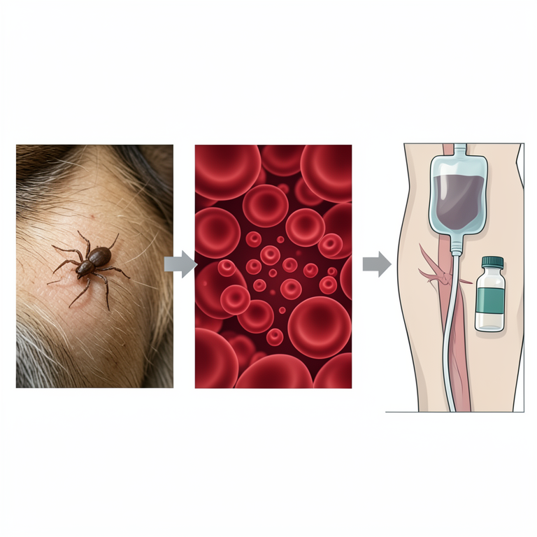
Transmission and Treatment Cycle
AI-generated conceptual illustration (a dog and a brown dog tick). It explains the idea and does not show study data.
View source evidence ↗Figures & tables
From the original paper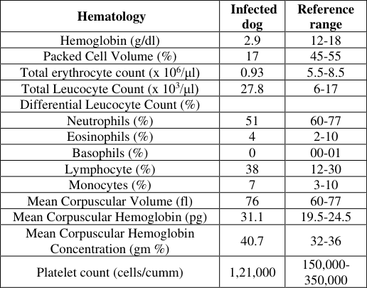 Table 1
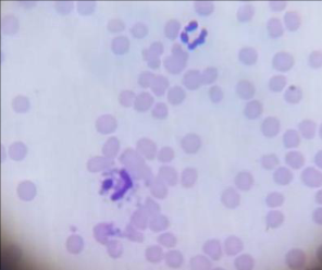 Figure 1
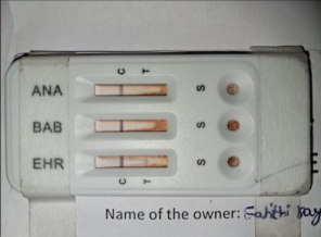 Figure 2
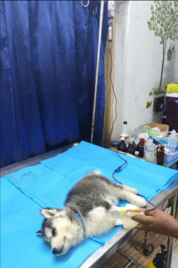 Figure 3
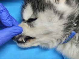 Figure 4
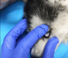 Figure 4
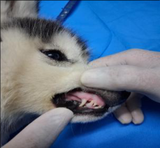 Figure 5
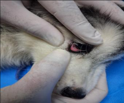 Figure 5
Introduction
Ehrlichiosis is a tick-borne disease caused by Ehrlichia canis, a gram-negative obligate intracellular bacterium, and transmitted by the brown dog tick Rhipicephalus sanguineus. Canine ehrlichiosis is caused by three different Ehrlichia species namely E. canis, E. chaffeensis and E. ewingii. The disease is also known as canine typhus, canine hemorrhagic fever, tracker dog disease, idiopathic hemorrhagic syndrome and tropical canine pancytopenia (Ristic and Holland, 1993) [1] . The incidence of canine ehrlichiosis in India was reported for the first time by Mudaliar in a Spaniel dog of eight months old. Ehrlichia canis is naturally spread by the tick Rhipicephalus sanguineous both intrastadially and transstadially, but not transovarially (Sainz et al., 2015) [2] . After an incubation period of 8-20 days, the course of E. canis infection, can be sequentially divided into acute, subclinical and chronic phases (Syaputra et al., 2020) [3] . Immunocompetent dogs may eliminate the infection during the acute or subclinical phases (Breitschwerdt et al., 1998; Harrus et al., 1998) [4,5] , but some will eventually develop the chronic phase, characterized by bone marrow aplasia, peripheral blood pancytopenia and high mortality due to septicemia and/or severe bleeding (Mylonakis et al., 2004) [6] . There are many clinical signs of the disease, but the most prevalent ones are depression, lethargy, weight loss, anorexia, pyrexia, lymph adenomegaly, splenomegaly, and bleeding tendencies. The most common and consistent hematological abnormality of dogs infected with E. canis naturally or experimentally is thought to be thrombocytopenia (Waner et al., 1999) [7] . Mechanisms assumed to be involved in the pathogenesis of thrombocytopenia in the acute phase of the disease include increased platelet consumption due to inflammatory changes in blood vessel endothelium, increased splenic sequestration of platelets, and immunologic destruction or injury resulting in a significantly decreased platelet life span (Troy and Forrester, 1990) [8] . Ehrlichia spp. infect primarily leukocytes, forming intracytoplasmic, membrane-bound bacterial aggregates, called morulae (Sainz et al., 2015) [2] . Demonstration of Ehrlichia spp. morulae in monocytes, macrophages and lymphocytes in Romanowsky type stained smears from buffy coat and less frequently lymph node, BM, spleen, liver and cerebrospinal fluid smears is helpful in establishing a definitive diagnosis of acute CΜE (Harrus et al., 2012; Harrus and Waner, 2011) [9,10] . Though tetracycline and oxytetracycline have been considered to be the initial drugs of choice, doxycycline and minocycline were also used as drugs of choice (Chandran et al., 2021) [11] . For up to five months following engorgement, the disease can be spread by ticks. The infection can also be transmitted through blood transfusions. The acute cases occur mostly in summer because of the greatest activity of the tick vector during that period (Sharma et al., 2010) [12] .
Case History and Observations
A 45-day-old male husky dog weighing 2 kg is presented to the veterinary clinic, Vijayawada with the history of tick infestation, inappetence, fever, lethargy, anemia and melena. Clinical examination revealed elevated rectal temperature of 104.6 0 F, increased respiratory rate and pale mucous membranes (Figure 4). superficial lymph nodes especially the popliteal lymph node were found to be enlarged in size.
Materials and Methods
The blood was collected in an EDTA vial for studying the complete blood picture (CBP). Giemsa stain was used in the thin blood smear method to examine the blood sample (Bhatia et al., 2010) [13] . The stained blood smear was screened for hemoprotozoa under the light microscope. Hematological analysis was carried out by using automatic hematological analyzer. Fecal sample is also examined for parasitic ova under the light microscope. E. canis/Anaplasma/Babesia Ab Test Kit was used for diagnosis. One drop of whole blood is placed in each well followed by three drops of diluent in the same wells. The presence of two color bands ("T" and "C") within the result window, indicates a positive result. Based on history, clinical and laboratory findings it was diagnosed as a case of canine ehrlichiosis.
Results and Discussion
Hematological analysis revealed anemia with decreased levels of hemoglobin and red blood cell count, thrombocytopenia and lymphocytosis (Table 1). These results were in accordance with the findings of Waner et al., 1999 [7] ; Meneses, 1997 [14] . Upon Giemsa staining and microscopic examination of blood smear, a light blue-stained morulae was observed in the lymphocytes (Figure 1) (Harrus and Waner, 2011) [10] . The E. canis/Anaplasma/ Babesia antibody rapid test showed positive results for E. canis antibody (Figure 2), so the dog was diagnosed with canine ehrlichiosis (Syaputra et al., 2020) [3] . Fecal sample is negative for parasitic ova. Dog was treated with doxycycline at a dose rate 10 mg/kg and clindamycin @ 11 mg/kg body weight intravenously with DNS slowly for 3 days, from the 4 th day onwards both were given orally twice daily for 21 days. Similar results were observed by Shrestha & Karmacharya, 2016 [15] ; Qurollo et al., 2013 [16] ; Sainz et al., 2015 [2] and Neer et al., 2002 [17] . Metronidazole 15 mg/kg body weight was administered intravenously for 3 days, and then it was given orally for 21 days, Imidocarb was given in two doses with an interval of 14 days at a dose rate of 6.6 mg/kg body weight subcutaneously (Chandran et al., 2021) [11] , Advaplat 5 ml orally 3 times a day, aRBC 1ml orally twice daily, Livoferol 2 ml orally twice a day. Blood transfusion (60 ml) was done on the first day of treatment (Figure 3). After 3 weeks of treatment, the dog showed marked improvement in clinical condition (Figure 5). Haematological values were found to be within a normal range and peripheral blood smear was negative for E. canis on the 21 st day of post-treatment. Blood transfusion, use of Doxycycline, along with supportive drugs had a significant impact in recovery of the animal. Dogs that have recovered and cleared of the infection do not acquire permanent immunity and may become re-infected. Therefore, tick control with careful manual removal or by applying appropriate acaricides on a year-round basis, is the single most important measure for the prevention of E. canis infection.
| Hematology | Infected dog | Reference range |
|---|---|---|
| Hemoglobin (g/dl) | 2.9 | 12-18 |
| Packed Cell Volume (%) | 17 | 45-55 |
| Total erythrocyte count (x 106/μl) | 0.93 | 5.5-8.5 |
| Total Leucocyte Count (x 103/μl) | 27.8 | 6-17 |
| Differential Leucocyte Count (%) | ||
| Neutrophils (%) | 51 | 60-77 |
| Eosinophils (%) | 4 | 2-10 |
| Basophils (%) | 0 | 00-01 |
| Lymphocyte (%) | 38 | 12-30 |
| Monocytes (%) | 7 | 3-10 |
| Mean Corpuscular Volume (fl) | 76 | 60-77 |
| Mean Corpuscular Hemoglobin (pg) | 31.1 | 19.5-24.5 |
| Mean Corpuscular Hemoglobin Concentration (gm %) | 40.7 | 32-36 |
| Platelet count (cells/cumm) | 1,21,000 | 150,000- 350,000 |
Table text extracted from the original PDF; superscript letters are shown inline. The original table image is below.
View original table image
References
16 in the original paper- Ristic M, Holland CJ. Canine ehrlichiosis. In: Woldehiwet Z, Ristic M, editors. Rickettsial and chlamydial diseases of domestic animals. Oxford: Pergamon Press; 1993. p. 169-186.
- Sainz A, Roura X, Miro G, Estrada-Pena A, Kohn B, Harrus S, Solano-Gallego L. Guidelines for veterinary practitioners on canine ehrlichiosis and anaplasmosis in Europe. Parasit Vectors. 2015;8:75.
- Syaputra JGE, Sajuthi CK, Sajuthi TP, Permata FS, Purwatiningsih W. Case study of canine monocytic ehrlichiosis (CME) in a Pomeranian dog at PDHB drh. Cucu K. Sajuthi. J Phys Conf Ser. 2020;1430(1):012009.
- Breitschwerdt EB, Hegarty BC, Hancock SI. Sequential evaluation of dogs naturally infected with Ehrlichia canis, Ehrlichia chaffeensis, Ehrlichia equi, Ehrlichia ewingii, or Bartonella vinsonii. J Clin Microbiol. 1998;36:2645-2651.
- Harrus S, Waner T, Aizenberg I, Bark H. Therapeutic effect of doxycycline in experimental subclinical canine monocytic ehrlichiosis: evaluation of a 6-week course. J Clin Microbiol. 1998;36:2140-2142.
- Mylonakis ME, Koutinas AF, Breitscwerdt EB, Hegarty BC, Billinis CD, Leontides LS, et al. Chronic canine ehrlichiosis (Ehrlichia canis): a retrospective study of 19 natural cases. J Am Anim Hosp Assoc. 2004;40:174-184.
- Waner T, Keysary A, Bark H, Sharabani E, Harrus S. Canine monocytic ehrlichiosis -an overview. Israel J Vet Med. 1999;54:103-107.
- Troy GC, Forrester SD. Canine Ehrlichiosis. In: Greene CE, editor. Infectious Diseases of the Dog and Cat. Philadelphia: W.B. Saunders; 1990. p. 48-59.
- Harrus S, Waner T, Neer M. Ehrlichia canis infection. In: Greene CE, editor. Infectious Diseases of the Dog and Cat. St. Louis, Missouri: Elsevier Saunders; 2012. p. 227238.
- Harrus S, Waner T. Diagnosis of canine monocytotropic ehrlichiosis (Ehrlichia canis): an overview. Vet J. 2011;187:292-296.
- Chandran D, Edana GG, Varghese S, Kumar AS. A case study of assessment of haemato-biochemical profile, clinical diagnosis and therapeutic management of canine monocytic ehrlichiosis (Tropical Canine Pancytopenia) in a male Doberman dog. Int J Sci Res Sci Technol. 2021;8(4):426-434.
- Bhatia BB, Pathak KML, Juyal PD. Textbook of veterinary parasitology. J Vet Parasitol. 2010;24(2):211.
- Meneses A. Diagnosis of canine ehrlichiosis by detecting inclusion bodies and morulae in blood smears. Ciencias Vet Heredia. 1997;20:57-63.
- Shrestha A, Karmacharya R. Ehrlichiosis in a dog: A case study. Int J Vet Sci Anim Husbandry. 2016;1(5):20-23.
- Qurollo BA, Davenport AC, Sherbert BM, Grindem CB, Birkenheuer AJ, Breitschwerdt EB. Infection with Panola Mountain Ehrlichia sp. in a dog with atypical lymphocytes and clonal T-cell expansion. J Vet Intern Med. 2013;27:1251-1255.
- Neer TM, Breitschwerdt EB, Greene RT, Lappin MR. Consensus statement on Ehrlichial disease of small animals from the infectious disease study group of the ACVIM. J Vet Intern Med. 2002;16:309-315.