
Research article · 2024 · Full text
International Journal of Veterinary Sciences and Animal HusbandryCanine trypanosomiasis in a non-descript dog: A case report
Volume 9(6S) · Pages 155–157 · DOI: 10.22271/veterinary.2024.v9.i6sc.1897 ↗
Research areasTrypanosoma species research and implicationsResearch on Leishmaniasis StudiesVector-borne infectious diseases
Abstract
Author-writtenA six-year-old male mongrel dog, weighing 26 kg was presented to Veterinary Clinical Complex of NTR College of Veterinary Science, Gannavaram with the history of inappetence, pyrexia, bilateral blindness accompanied by hyphema and mild corneal opacity, as well as significant weight loss for the past four days. Clinical examination revealed elevated rectal temperature (103.9oF), pallor mucous membranes, enlarged lymph nodes, tachycardia and lack of menace reflex in both the eyes. Haemato-biochemical examination indicated anemia, neutrophilia, hypoglycemia and slightly elevated total bilirubin. Microscopic evaluation of the wet blood film revealed motile Trypanosomes, while the Giemsa-stained blood smear showed a massive infection of Trypanosomes. Based on the diagnostic evaluation the case was confirmed as canine trypanosomiasis. The animal was successfully treated with a single intramuscular dosage of diminazene aceturate at a rate of 3.5 mg/kg body weight, along with supportive care. The dog showed recovery following a week-long course of treatment.
Summary
Source-checkedPlain-language reading of the full paper, prepared with AI and checked against the text. Each point links to the authors' own words.
Why it matters
- Trypanosomiasis is a disease caused by extracellular parasites of the Trypanosoma genus that can infect both wild and domestic animals. View evidence ↗
- In the Indian subcontinent, the canine form of this disease is mainly caused by Trypanosoma evansi. View evidence ↗
- While T. evansi causes chronic disease in horses and camels, it is typically acute and fatal in dogs, often leading to death within 2 to 4 weeks without treatment. View evidence ↗
The question
- The authors aim to document a specific case of canine trypanosomiasis that included ophthalmic complications. View evidence ↗
What they did
- The subject was a 26 kg, six-year-old male mongrel dog treated at the Veterinary Clinical Complex of NTR College of Veterinary Science. View evidence ↗
- Diagnosis was achieved through microscopic evaluation of wet blood films and Giemsa-stained blood smears to detect Trypanosomes. View evidence ↗
- Treatment consisted of a single intramuscular dose of Diminazine aceturate (3.5 mg/kg) along with supportive medications including DNS, Oxytetracycline, Meloxicam, and Chlorpheniramine maleate. View evidence ↗
What they found
- Initial clinical signs included a fever of 103.9°F, tachycardia, enlarged lymph nodes, pale mucous membranes, and a lack of menace reflex in both eyes. View evidence ↗
- The dog presented with a four-day history of weight loss, inappetence, fever, and bilateral blindness with hyphema and mild corneal opacity. View evidence ↗
- The dog's hemoglobin increased from 7.2 g/dL to 8.4 g/dL and packed cell volume rose from 19% to 22% following treatment. View evidence ↗
- Blood glucose levels improved from a pre-treatment value of 57 mg/dL to 72 mg/dL post-treatment. View evidence ↗
- Recovery progressed rapidly: temperature normalized to 102.1°F by day two, appetite returned by day three, full recovery occurred by day five, and corneal clarity was restored by day seven. View evidence ↗
- By the end of the treatment period, blood smears no longer showed the presence of Trypanosoma species. View evidence ↗
What it means
- The authors conclude that a single dose of diminazene aceturate, combined with supportive care, is effective for treating canine trypanosomiasis. View evidence ↗
- The authors note that distinguishing between Trypanosoma species is difficult and requires serological or molecular testing for accurate identification. View evidence ↗
Visual summary
3 visualsIllustrations explain the paper; charts are replotted from the paper's own table values.
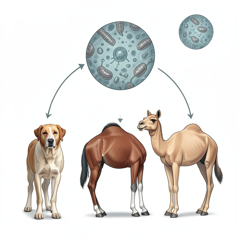
Pathogen Overview
AI-generated conceptual illustration (Trypanosoma evansi parasites, a dog, a horse, and a camel). It explains the idea and does not show study data.
View source evidence ↗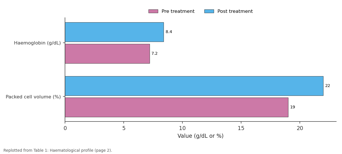
Pre treatment and Post treatment: Haemoglobin (g/dL) vs Packed cell volume (%)
Replotted from Table 1 (page 2) of the original paper. Every bar is the paper's own table value.
View source evidence ↗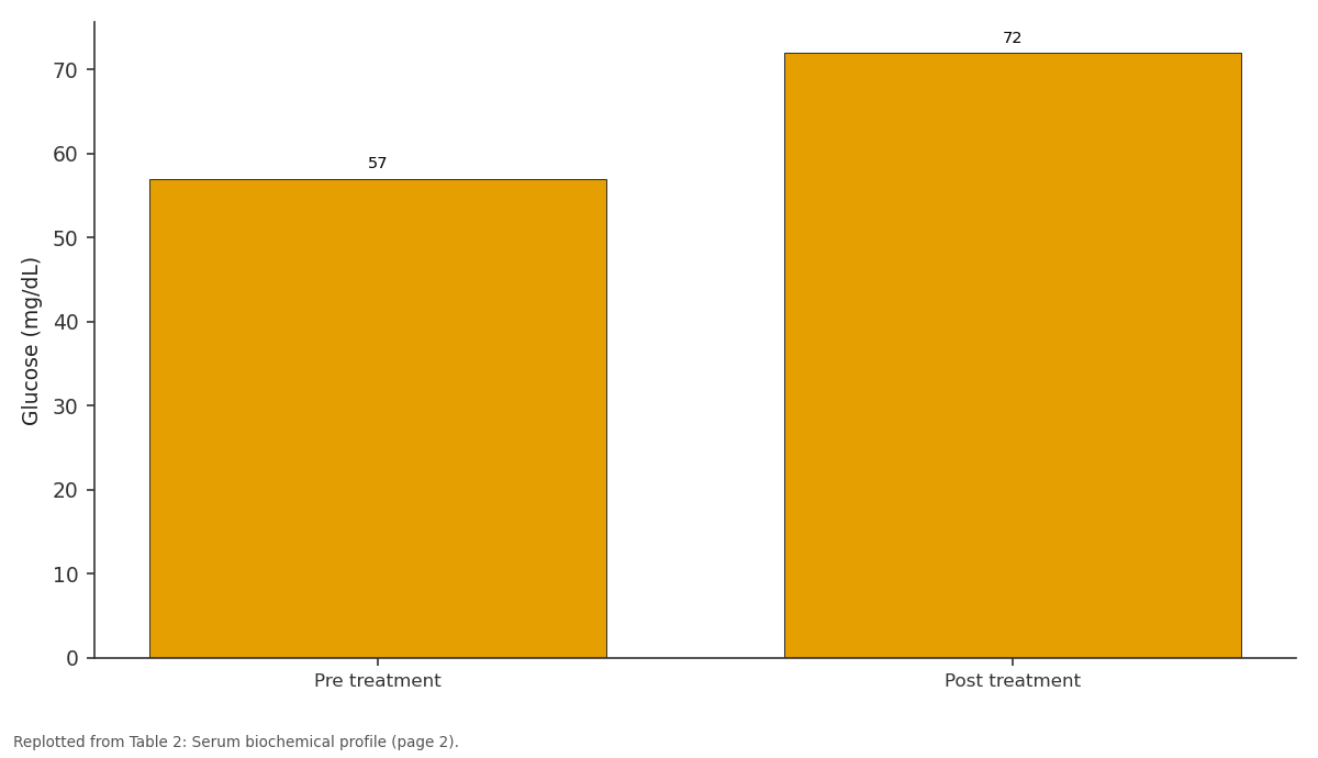
Glucose (mg/dL): Pre treatment vs Post treatment
Replotted from Table 2 (page 2) of the original paper. Every bar is the paper's own table value.
View source evidence ↗Figures & tables
From the original paper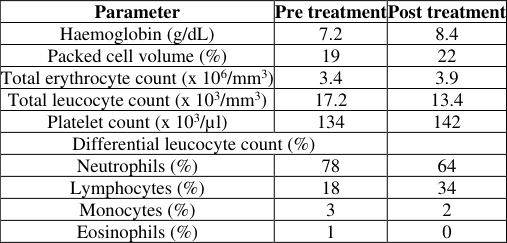 Table 1
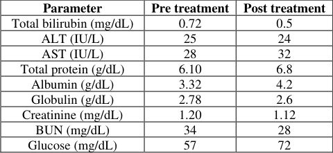 Table 2
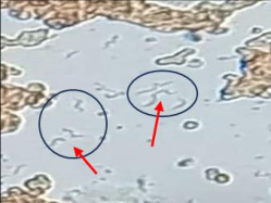 Figure 1
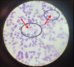 Figure 2
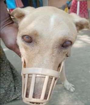 Figure 3
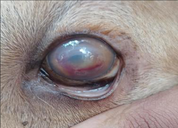 Figure 4A
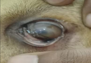 Figure 4B
Introduction
Trypanosomiasis is a haemoprotozoan disease caused by various species of Trypanosoma, an extracellular parasite that affects both domestic and wild animals (Agrawal et al., 2020) [1] . Canine trypanosomiasis is classified into two main forms: the American form (Chagas disease), caused by Trypanosoma cruzi infection, and the African form (sleeping sickness or surra), caused by Trypanosoma evansi (Haritha et al., 2024) [7] . However, in the Indian subcontinent, canine trypanosomiasis is primarily caused by Trypanosoma evansi (Behera et al., 2018) [4] and has been recorded the prevalence of T. evansi in dogs has been recorded in various regions, including Madhya Pradesh (7.69%), Andhra Pradesh (2.40%) and Kerala (2.7%) (Rangaswamy et al., 2024) [10] . Trypanosoma evansi causes chronic disease (Surra) in camels and horses, but in dogs, it is usually acute and fatal, causing death within 2 to 4 weeks if untreated (Haritha et al., 2024) [7] . The disease is primarily transmitted by various biting flies, including Tsetse, Tabanus, Stomoxys, and Culicoides (Agrawal et al, 2020) [1] . During its infectious life cycle, the Trypomastigote form of the parasite immediately enters host cells, proliferates sub-clinically, evades the immune system (Ahmed, 2018), and spreads throughout the host body, primarily within macrophages. Symptomatic parasitemia typically develops within 3-5 days post-infection (Khan et al., 2022) [8] . The severity of canine trypanosomiasis ranges from acute and subacute to chronic forms. Clinical signs of Trypanosomiasis are characterized by weight loss, progressive weakness, anorexia, anemia, intermittent fever, conjunctivitis, swelling of the limbs, enlarged superficial lymph nodes, and corneal opacity, which are typical findings in chronic Trypanosomiasis (Thirunavukkarasu et al., 2004) [12] . Several effective trypanosomacidal agents are available for dogs, including suramin, quinapyramine, and diminazene. However, a single dose of diminazene aceturate has been shown to be effective in eliminating natural Trypanosomiasis infection in canines (Rani and Suresh, 2007) [11] . The present report places on record a case of canine trypanosomiasis with ophthalmic involvement.
Case history and observations
A six year old male mongrel dog weighing 26 kg was presented to Veterinary Clinical Complex, NTR College of Veterinary Science, Gannavaram, with the history of inappetence, pyrexia, bilateral blindness accompanied by hyphema and mild corneal opacity, as well as significant weight loss for the past four days. Clinical examination revealed elevated rectal temperature (103.9 o F), pallor mucous membranes, enlarged lymph nodes, tachycardia and lack of menace reflex in both the eyes. Haematobiochemical examination indicated anemia, neutrophilia, hypoglycemia and slightly elevated total bilirubin. Microscopic evaluation of the wet blood film revealed motile Trypanosomes, while the Giemsa-stained blood smear showed a massive infection of Trypanosomes. Stomatocytes are abnormal red blood cells with a slit-like central pallor surrounded by a dense zone, giving them the appearance of a human mouth. These cells arise from defects in the red blood cell membrane, commonly associated with haemolytic diseases. Based on the microscopic examination, the case was confirmed as canine trypanosomiasis. [4] , Khan et al. (2022) [8] and Dhakane et al. (2024), who also noted anorexia, corneal opacity, high temperature, gradual weight loss and lymph node enlargement. Chandrasekar et al. (2016) reported that corneal opacity results from parasites descending along the optic artery, leading to immune complex formation in the aqueous humor, which creates granular deposits that cloud the cornea and impair vision. Aref et al.
(2013) also reported bilateral blindness accompanied by hyphema. Microscopic examination of Giemsa-stained blood smears and wet film examinations are diagnostic tools used for detecting Trypanosoma infections. However, distinguishing between Trypanosoma species can be challenging, highlighting the need for serological and molecular tests to accurately identify the species (Haritha et al., 2024) [7] . Anemia in the present case report was in agreement with earlier reports of Haritha et al. (2024) [7] and Rangaswamy et al. (2024) [10] who opined that anemia might be caused by haemolysis due to erythrophagocytosis, haemodilution, and suppression of erythropoiesis. Neutrophilia in the present case report might be due to secondary bacterial infections. Hypoglycemia and elevated bilirubin values were also reported by Khan et al. (2022) [8] . Hypoglycemia occurs due to the utilization of blood glucose by circulating parasites. Most of the literature reports the single use of Diminazine aceturate at a dose of 3.5 mg/kg body weight administered intramuscularly (Rani and Suresh, 2007 and Agrawal et al., 2020) [11,1] . Uneventful clinical recovery was observed after a week of therapy. Similar clinical recovery within one week was reported by Agrawal et al. (2020) [1] and Haritha et al. (2024) [7] in Trypanosoma positive dogs.
Figures and tables from the paper
| Parameter | Pre treatment | Post treatment |
|---|---|---|
| Haemoglobin (g/dL) | 7.2 | 8.4 |
| Packed cell volume (%) | 19 | 22 |
| Total erythrocyte count (x 106/mm3) | 3.4 | 3.9 |
| Total leucocyte count (x 103/mm3) | 17.2 | 13.4 |
| Platelet count (x 103/µl) | 134 | 142 |
| Differential leucocyte count (%) | ||
| Neutrophils (%) | 78 | 64 |
| Lymphocytes (%) | 18 | 34 |
| Monocytes (%) | 3 | 2 |
| Eosinophils (%) | 1 | 0 |
Table text extracted from the original PDF; superscript letters are shown inline. The original table image is below.
View original table image
| Parameter | Pre treatment | Post treatment |
|---|---|---|
| Total bilirubin (mg/dL) | 0.72 | 0.5 |
| ALT (IU/L) | 25 | 24 |
| AST (IU/L) | 28 | 32 |
| Total protein (g/dL) | 6.10 | 6.8 |
| Albumin (g/dL) | 3.32 | 4.2 |
| Globulin (g/dL) | 2.78 | 2.6 |
| Creatinine (mg/dL) | 1.20 | 1.12 |
| BUN (mg/dL) | 34 | 28 |
| Glucose (mg/dL) | 57 | 72 |
Table text extracted from the original PDF; superscript letters are shown inline. The original table image is below.
View original table image
References
12 in the original paper- Agrawal H, Jaiswal M, Tripathi AK. Successful management of trypanosomiasis in a dog. Indian J Vet Med. 2020;40(2):35-36.
- Ahmed W, Hafeez MA, Ahmad R, Mahmood S. CRISPR-Cas system in regulation of immunity and virulence of bacterial pathogens. Gene Reports. 2018;13:151-157.
- Aref M, Yasin SM, Bahear W, Ghulam Z, Hastie L, Dennison T, et al. Canine Trypanosoma evansi infection in Afghanistan. Vet Parasitol. 2013;197(3-4):638-641.
- Behera SK, Sarma K, Behera Parthasarathi, Ali MA. Therapeutic management of trypanosomosis with ophthalmic involvement in a dog. J Parasit Dis. 2018;42(2):329.
- Chandrasekar M, Senthilkumar G, Prathaban S. Management of Trypanosomiasis and Associated Corneal Opacity in a Dog. Intas Polivet. 2017;18(1):178-179.
- Dakhane PS, Suryawanshi AA, Thorat GC, Gimmvanekar SS. Therapeutic management of Canine Trypanosomiasis: A case report. Int J Vet Sci Anim Husbandry. 2024;9(3):277-278.
- Haritha GS, Ramesh P, Hemanth I, Devi RP. Trypanosomiasis in a dog-A case report. J Krishi Vigyan. 2024;12(1):173-177.
- Khan AA. A clinical note on canine trypanosomiasis. Pak J Sci. 2022;74(2):128-131.
- Prasad KL, Kondaiah PM, Rayulu VC, Srilatha C. Prevalence of canine trypanosomiasis in certain areas of Andhra Pradesh. J Parasit Dis. 2015;39:238-240.
- Rangasamy V, Annamalai L, Kanniappan VM, Mani S, Natarajan BP, Chinnaswamy PP, et al. Occurrence of Trypanosoma evansi infection in Rottweiler dog from Cauvery Delta region of Tamil Nadu, India: a case report. Iranian J Parasitol. 2024;19(3):370.
- Rani NL, Suresh K. Canine trypanosomiasis. India Vet J. 2007;84:186-187.
- Thirunavukkarasu PS, Rao VV, Srinivasan SR, Nambi AP, Dhanapalan P. Haematobiochemical findings in case of trypanosomiasis in dog: a clinical study. Ind J Vet Med. 2004;24:117.