
Research article · 2024 · Full text
International Journal of Biology SciencesEvaluating the extent of prematurity-related retinopathy (ROP): Frequency and availability regarding risky individuals
Volume 6(2) · Pages 233–238 · DOI: 10.33545/26649926.2024.v6.i2d.258 ↗
Research areasRetinopathy of Prematurity StudiesNeonatal and fetal brain pathologyNeonatal Respiratory Health Research
Abstract
Author-writtenThe current investigation aimed to clarify the previous findings about the prevalence and incidence of retinopathy of prematurity (ROP) in children delivered before 34 weeks of gestation or in those with birth weights (BW) ≤ 2000 g and gestational ages between 34 and 36 weeks. After receiving ethical clearance, 160 newborns participated in a prospective, hospital-based observational study over a four-month period. Neonates younger than 34 weeks gestational age and those with a body weight (BW) under 2000 gm and a gestational age between 34 and 36 weeks comprised the study population. Number and percentage were used to express categorical/nominal variables, which were then evaluated using the Fischer exact test or the Chi square test. The mean and standard deviation were used to represent continuous variables. All statistical analyses were conducted using appropriate statistical software, such as SPSS (Statistical Sciences Package for Social Sciences). After screening all 160 newborns that were part of the current study, 30 of them had ROP, representing an 18.8% prevalence. The majority of neonates evaluated for ROP were delivered between 33 and 34 weeks gestational age (56.9%), then between 30 and 32 weeks (28.8%); 16 (10%) neonates were born before 30 weeks, and seven (4.4%) after 34 weeks. Ten (33.3%) of the thirty neonates with ROP were born at gestational age less than 32 weeks, and twenty (66.7%) at gestational age more than 32 weeks. There is a noteworthy correlation (p=0.044) between the incidence of ROP and gestational age less than 32 weeks. Of the thirty neonates with ROP, twenty-four (80%) weighed less than 1500 g, while six (20%) weighed more than 1500 g. There was a strong correlation (p
Summary
Source-checkedPlain-language reading of the full paper, prepared with AI and checked against the text. Each point links to the authors' own words.
Why it matters
- Retinopathy of Prematurity (ROP) is a vaso-proliferative syndrome caused by oxygen therapy in premature children and is a leading cause of preventable cataracts affecting approximately 50,000 children globally. View evidence ↗
- In India, ROP is estimated to cause blindness in about 500 children annually. View evidence ↗
- The condition results from abnormal blood vessel growth in the retina, triggered by inadequate hyperoxic blood circulation that suppresses VEGF and destroys endothelial cells. View evidence ↗
The question
- The study aimed to clarify the prevalence and incidence of ROP in infants born before 34 weeks of gestation or those with birth weights ≤ 2000 g and gestational ages between 34 and 36 weeks. View evidence ↗
What they did
- The research was a prospective, hospital-based observational study involving 160 newborns over a four-month period. View evidence ↗
- The study population included newborns under 34 weeks of gestation and infants weighing over 2000 g with gestational ages between 34 and 36 weeks from the NICU. View evidence ↗
- Screening for ROP was performed by a skilled ophthalmologist using a RETCAM computerized neonatal retinal examination instrument. View evidence ↗
- Categorical variables were analyzed using the Chi square or Fischer exact tests, while continuous variables were represented by mean and standard deviation. View evidence ↗
What they found
- Out of 160 screened newborns, 30 were diagnosed with ROP, resulting in a prevalence of 18.8%. View evidence ↗
- A significant correlation (p=0.044) was found between the incidence of ROP and a gestational age of less than 32 weeks. View evidence ↗
- Birth weight under 1500 g was strongly correlated with ROP (p<0.001), with 80% of ROP-positive newborns weighing less than 1500 g. View evidence ↗
What it means
- The authors conclude that there is a need for standardized screening, early treatment, and improved access to neonatal care to prevent vision loss. View evidence ↗
- The authors recommend improving healthcare infrastructure and addressing regional disparities to reduce the global burden of ROP. View evidence ↗
Visual summary
3 visualsIllustrations explain the paper; charts are replotted from the paper's own table values.
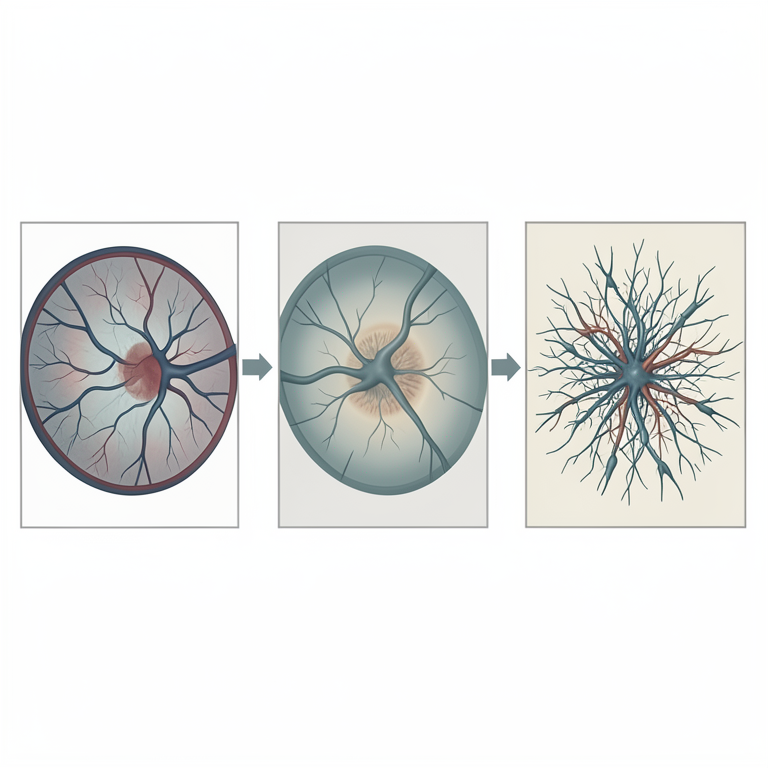
Pathophysiology of ROP
AI-generated conceptual illustration (premature infant retina). It explains the idea and does not show study data.
View source evidence ↗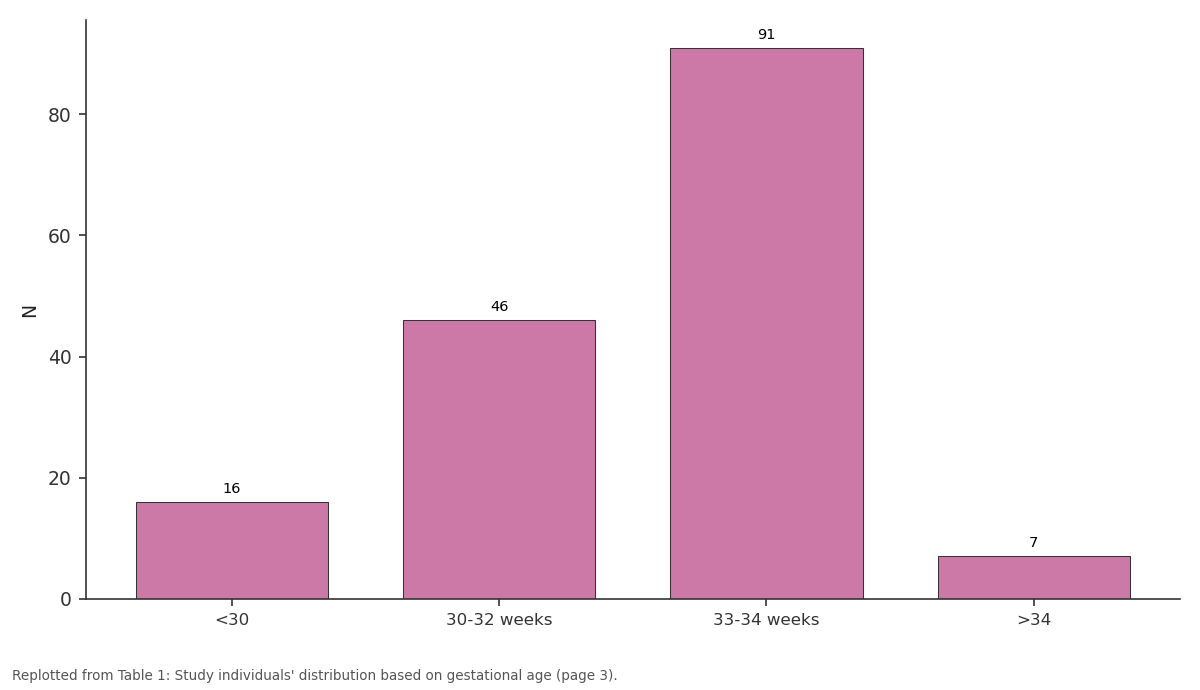
N: <30 vs 30-32 weeks vs 33-34 weeks vs >34
Replotted from Table 1 (page 3) of the original paper. Every bar is the paper's own table value.
View source evidence ↗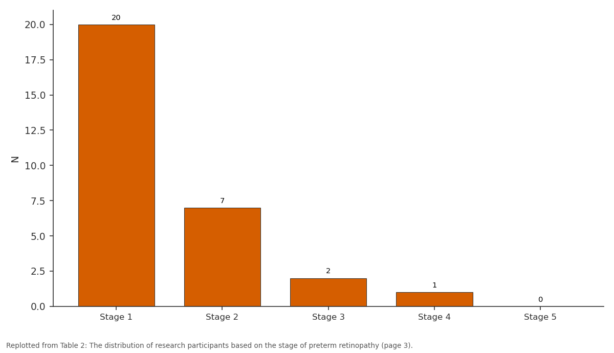
N: Stage 1 vs Stage 2 vs Stage 3 vs Stage 4 vs Stage 5
Replotted from Table 2 (page 3) of the original paper. Every bar is the paper's own table value.
View source evidence ↗Figures & tables
From the original paper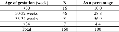 Table 1
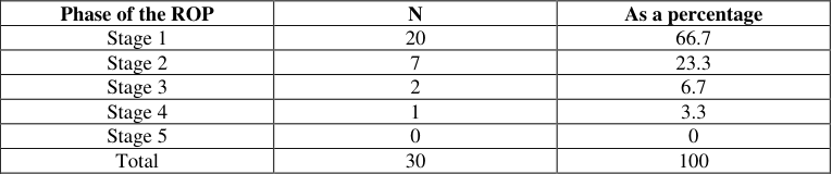 Table 2
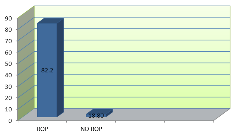 Figure 1
 Table 3
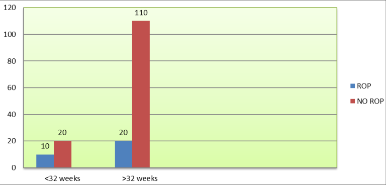 Figure 2
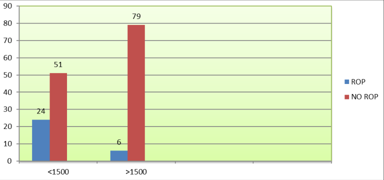 Figure 3
Introduction
Children born prematurely may typically affected by prematurity-related ophthalmology (ROP), a vaso-proliferative syndrome brought on by oxygen therapy. That constitutes the primary contributor to preventable cataracts that affects about 50,000 children worldwide and is present in both developed and developing countries. ROP was discovered to have an increasing incidence (second epidemic), despite improvements in monitoring methods for oxygen supplementation and an increase in premature newborn survival in the following decades. Over the past ten years, ROP blindness has become more common in low-income countries. It is estimated that 500 youngsters in India lose their sight due to ROP every year. The disorder has garnered significant global investigation due to the elevated survival rates among preterm newborns with The people particularly vulnerable of having ROP are individuals from exceptionally low gestation measurements, particularly those weighing less than 1500 g. These greater numbers might be the result of improved prenatal care. The disorder is brought on by aberrant vasculature multiplication that impacts the retina's development which is the outcome of inadequate hyperoxic blood circulation within the visual organs, which suppresses VEGF and kills endothelial cells. The closure of growing capillaries results in ischaemia and hypoxia of the developing retinal tissue.
The overexpression of VEGF in this phase leads to neovascularisation. A elevated chance of disability as a result of detached retinal tissue is associated with a severe form of retinalopathy of prematurity. Possible further issues include macular folds, strabismic amblyopia, and refractive amblyopia. A good examination plan is a particularly crucial element to a successful ROP control approach. In accordance with the most recent ROP guidelines, screening should be done for all newborns weighing less than 2000 g BW or for all children born between thirty-four weeks in addition to if the GA is established, 34 several months with warning signs. After being discharged from the infant's medical centre (NICU) or 30 days after the baby was delivered, a monitoring is done, whenever comes earlier.
The subsequent circumstances heighten the danger of ROP: premature delivery, high foetal age, additional oxygen, protracted breathing assistance, the Apgar score, breathing issues, anaemia, for instance intraventricular bleeding (IVH), necrotising intestinal inflammation, and pneumonia, some of which can lead to the development of severe ROP. The following stages of ROP illustrate the degree of vascular alterations: Phase 1: Dividing boundary dividing the angiogenic and without blood vessels regions of the cornea Phase 2: Boundary line expanding to create a ridge Phase 3: Ridge-like fibrovascular supplementary ocular growth Phase 4: Subtotal separation of the retina Phase 5: Complete separation of the retina Furthermore sickness is marked by fragility and swelling of the anterior pole arteries is a gauge of the severity of the disease. More posterior pole vascular dilatation and tortuosity than normal but less than plus disease is known as pre-plus disease. The characteristics of aggressive posterior ROP include extremely narrow intraretinal space, significant degenerative illnesses, along with forward placement neovascularisation. It is an uncommon and rapidly advancing type of ROP. It can quickly advance severe cataracts with phase 5 of ROP if treatment is delayed. The present investigation aimed to clarify the earlier findings about ROP in children born prior to 34 weeks of gestation or in infants weighing smaller than two hundred grammes at approximately 34 to 36 weeks of the course of pregnancy, including a focus on the condition's frequency and prevalence.
Significance of this study
The condition known as of infanticide (ROP) is an important worldwide factor of avoidable cataracts in youngsters, particularly infants who are premature, hence determining the extent of ROP is crucial. The frequency and severity of ROP are affected by a variety of factors, including advancements in parenting, geographic disparities regarding healthcare accessibility, and various diagnostic methods. This study is important because it sheds light on the prevalence of ROP in various communities, demonstrating the variation in incidence rates associated with variables including birth weight, gestational age, and availability of neonatal care. Comprehending these characteristics is crucial in discerning at-risk cohorts and customizing strategies to avert or alleviate the severe consequences linked to ROP. The study also looks at screening and treatment options available in various localities, highlighting any inadequacies in the healthcare system that might be a factor in some areas having higher incidence of ROP. The study's mapping of these discrepancies can help guide public health initiatives that aim to enhance prompt intervention and early identification, both of which are critical to lowering the burden of ROP.
To sum up, this study not only adds to our understanding of the epidemiology of ROP, but it also emphasizes how critical equitable access to healthcare is. It has the potential to act as a trigger for resource allocation and policy reforms that will enhance outcomes for vulnerable neonatal populations and ultimately lessen the condition's crippling effects on a global scale.
The procedures of this study
After receiving ethical clearance, 160 neonates participated in this four-month prospective observational study at a hospital. The subject matter demographic consisted of newborns under thirty-four weeks of pregnant and infants over 2000 g after arrival with gestation periods from 34 and 36 weeks of pregnancy which were taken within the NICU during the investigation timeframe. A proforma that was semi-structured was used to gather data. After obtaining their signed informed consent, parents' pertinent medical histories were collected in order to gather information from the subjects. Those newborns (male and female) delivered within 35 and 36 weeks of pregnancy, as well as infants hospitalized to The intensive care unit for neonatal patients, or NICU who weighed a maximum of 2000 grammes and who met the following criteria for risk: hyperglycemia or hypoglycemia; oxygen therapy administered for any length of time via a mechanical ventilator, CPAP, HFNC, Nasal Prongs/HOOD, multiple pregnancies; The blood element blood transfusions, pneumonia, preterm apnoea, respiratory distress syndrome, ventilator shock/inotrope usage, open duct artrieosus, anaemia in either the mom or the kid, and neonatal haemophilia. The current study did not include infants with inherited retinal defects, those who were not ensuing until complete vascularization of the retina, or those with ocular problems that impede fundus inspection. Before being included in the study, the parent or guardian gave their written, informed consent. Every newborn enrolled in the research had the following procedures: 1) A predesigned proforma was used to record the following information: a thorough medical history of the mother; a breakdown of the intrauterine foetal wellbeing evaluation; the baby's weight and gender; the baby's the technique, length, as well as the number 2 received oxygen treatment period; the years of the infant at birth; as well as any additional associated risks. 2) After a comprehensive neurological and clinical assessment, newborns were tested for the existence of retinopathy of prematurity by an ophthalmologist. 3) To determine the ROP variables of risk, investigations such as peripheral smears, CRP, blood cultures, and complete blood counts were carried out in the central lab.
4) They underwent follow-up until 40 weeks after the initiation of treatment, the completion of retinal maturation, or both. 5) Should the child have been born beforehand thirty-eight weeks of pregnancy, the first screening exam was performed after three weeks, and if the baby was born beyond 28 weeks, the exam was performed after four weeks, whichever came first.
The ophthalmologic examination procedure These pupils were dilation using a combination of 2.5% and tropicamide, which 0.5%. Four times, every ten to fifteen minutes, starting an hour before the examination time, a single drop of tropicamide was injected. One drop of phenylephrine was then injected right before the examination. Phenylephrine injections were not given frequently due to concern about hypertension.
A skilled ophthalmologist used a computerized neonatal retinal examination instrument with an expansive scope (RETCAM) to screen for ROP.
An examination of statistics
The study's original data were all entered into Performa, cleaned, and checked before being transferred to an MS Excel sheet. The collected information were analysed using either the suitable Pearson accurate analysis or Chi square test. Generic and qualitative variables were represented as quantities and proportions. The Fischer accurate analysis was applied whenever one of the cells located in a 2 x 2 dependency table had a predicted value below five both of the samples were compared and variables that were continuous, which The results were evaluated employing the indistinguishable individual t test and reported as the median and deviation from the mean (SD).
A statistically A meaningful P value of less than 0.05 was used. Every calculation of statistics was carried out using statistical software such as SPSS (Statistical Sciences Package for Social Sciences).
Findings of this study
The majority of neonates that were evaluated for ROP were born between 33 and 34 weeks gestational age (56.9%), then between 30 and 32 weeks (28.8%); 16 neonates (10%) were born before 30 weeks, and seven (4.4%) were born beyond 34 weeks. 77 (48.1%) and 83 (51.9%) of the 160 newborns that underwent ROP screening were female. There were 1.08 males for every female (Table 1). ROP was discovered in 30 of the 160 newborns who underwent screening, yielding an 18.8% prevalence (Figure 1). Table 2 shows that of the 30 newborns diagnosed with Twenty (66.7%) of the participants had the initial phase ROP, seven two people (6.7%) had stage 3 ROP, one person (3.3%) had the fourth stage of ROP, and 23.3% had stage 2 ROP. Neither of the patients exhibited stage 5 ROP found in themselves. 2). Table 3 shows that out of the thirty newborns with ROP, five (16.7%) developed Aggressive Posterior Retinopathy of Prematurity (APROP). Of the 30 newborns with ROP, 24 (80%) weighed less than 1500 g, and 6 (20%) weighed more than 1500 g. In contrast, 51 (39.2%) and 79 (60.8%) of the 130 neonates without ROP had BWs of less than or equal to 1500 g. A noteworthy correlation was discovered between the BW of less than 1500 g and the incidence of ROP (p<0.001).
| Retinopathy of Prematurity (ROP) | N | As a percentage |
|---|---|---|
| Phases of ROP | 25 | 83.3 |
| APROP | 5 | 16.7 |
| TOTAL | 30 | 100 |
Table text extracted from the original PDF; superscript letters are shown inline. The original table image is below.
View original table image
| Phase of the ROP | N | As a percentage |
|---|---|---|
| Stage 1 | 20 | 66.7 |
| Stage 2 | 7 | 23.3 |
| Stage 3 | 2 | 6.7 |
| Stage 4 | 1 | 3.3 |
| Stage 5 | 0 | 0 |
| Total | 30 | 100 |
Table text extracted from the original PDF; superscript letters are shown inline. The original table image is below.
View original table image
| Age of gestation (week) | N | As a percentage |
|---|---|---|
| <30 | 16 | 10.0 |
| 30-32 weeks | 46 | 28.8 |
| 33-34 weeks | 91 | 56.9 |
| >34 | 7 | 4.4 |
| Total | 160 | 100 |
Table text extracted from the original PDF; superscript letters are shown inline. The original table image is below.
View original table image
Discussion
According to the current study, the majority of neonates evaluated for ROP were born between 33 and 34 weeks gestational age (56.9%), then between 30 and 32 weeks (28.8%). In a 2009 study by Chaudhari et al., (2009) [13] the mean age at delivery was 31.4±2. [15] Chang-Yo Yang et al., (2011) [16] conducted a review of the literature on 252 Very-Low-Birth-Weight (VLBW) babies in 2011. About 99 (40%) of them had been determined to have ROP. Of the 704 babies that Kumar and colleagues evaluated, 84 (11.9%) and 33 (4.7%) had mild or severe ROP, respectively. Gloria Isaza et al. noted a rate that was roughly 40% higher. Out of 259 newborns, 142 (54.8%) developed ROP in various phases, according to research by Saravanan Jothi et al., (2016) [18] According to Crystal Le et al., (2016) [19] 2.3% (n = 66) of the individuals had ROP.
According to our research, of the 30 newborns with ROP, 20 (66.7%) had stage 1 ROP, seven (23.3%) had stage 2, two (6.7%) had stage 3, and one (3.3%) had stage 4. No baby had stage 5 ROP. According to VA Shah et al., (2005) [15] , [20] likewise discovered comparable amounts. According to Eduardo Gonçalves et al., (2014) [21] among the neonates under study, stage I ROP had the highest incidence, followed by stage III ROP and stage II ROP. Additionally, Saravanan Jothi et al., (2016) [18] discovered that stage 1 ROP affected 66.9% of newborns with ROP. The context of the present investigation, of the 30 newborns with ROP, 10 (33.3%) were born before 32 weeks of pregnancy, and 20 (66.7%) after 32 weeks; in comparison, only 20 (15.4%) of the 130 newborns without ROP were born before 32 weeks of pregnancy, and most of them (84.6%) after 32 weeks of pregnancy. There was a noteworthy association (p=0.044) found among the incidence of ROP and pregnancy duration less than 32 weeks. The findings of Chaudhari et al., (2009) [13] indicated that the frequency of ROP rose as pregnancy weeks decreased (P=0.003). The one-way analysis of variance showed a strong correlation between ROP and prenatal age <32 weeks (OR=2.750). Low gestational age was also linked to ROP, according to Chang-Yo Yang et al., (2011) [16] Abdel H. A. A. Hakeem (2012) [20] and colleagues identified a substantial correlation between the occurrence of reproductive organ problems and gestational age. Comparable significant associations were also found by Kumar et al., (2011) [17] with gestational ages ≤ 30 a few days. Neonatals' maximum age at delivery with ROP was found to be approximately 26 weeks (23-31 weeks), while that of newborns without ROP was 28.5 weeks (24-36 weeks). This information was observed by Gloria Isaza et al. More logistic regression analysis revealed that among Reduced age at conception was also found to be substantially correlated with ROP by Rao KA et al., (2013) [22] Using multivariate logistic regression analysis, Saravanan Jothi et al., (2016) [18] revealed that gestational age was an independent risk factor for the development of ROP. According to Rania M.R. Bassiouny et al., (2017) [23] the mean gestational age of premature newborns evaluated for ROP was 31.5±2.3 weeks, between 24 and 36 weeks. Dhaivat Vasavada et al., (2017) [24] reported that the typical gender at childbirth was 30.6 + 1.97 weeks, with a range of 26 to 35 weeks. During the research we conducted, 24 (80%) of the 30 newborns with ROP weighed less than 1500 g, and 6 (20%) weighed more than 1500 g. On the other hand, 79 (60.8%) and 51 (39.2%) of the 130 newborns without ROP had a BW of greater than 1500 g. There was a strong correlation (p<0.001) between the incidence of ROP and a BW of less than 1500 g. According to Using univariate testing, a substantial association was found among ROP and BW <1500 g (OR=6.196). Following a comparable direction, Chaudhari et al., (2009) [13] noted that the patients' mean body weight was 1306±267 g. According to Kumar et al., (2011) [17] there is a substantial correlation between Birth Weight (BW) of 1000 g and severe ROP, with newborns with ROP having mean birth weights of 438 g, or 1113±. The median average Body Weight (BW) of babies with ROP was 840 g (500 to 1,890 g), considerably smaller than the mean BW of neonates without ROP, which was 1,191 g (470 to 2,460 g), according to Gloria Isaza et al. Also, mathematical modelling revealed that BW had an odds ratio of 1.90 and an uncertainty interval of 95% ranging from 1.34 to 2.69 was among the most significant adjusted risk factors for ROP. Low BW was also found to be substantially correlated with ROP by Rao KA et al., (2013) [22] Using multivariate logistic regression analysis, Low BW was found to be a separate risk indicator for the possibility of ROP by Saravanan Jothi et al., (2016) [18] Rania M.R. Basiouny et al., (2017) [23] calculated the median BW to be 1514±391 g (Range: 600-2860 g).
Conclusion
The prevalence and incidence of in people who are at risk Retinopathy of Prematurity (ROP) groups must be addressed, and this study emphasizes the vital need for focused treatment. Significant regional differences exist in the prevalence of ROP as well as in the accessibility of screening and treatment options. The research highlights these gaps and supports standardized screening techniques, early treatment to prevent vision loss, and better access to neonatal care. To lessen the ROP load worldwide and protect the vision of susceptible newborns, improving healthcare infrastructure and addressing these disparities are ultimately crucial initiatives. Thirty (18.8%) of the 160 babies who were checked for ROP had violent backward Twenty (66.7%) of the participants had initial ROP, seven (23.3%) had stage 2 ROP, two (6.7%) had stage 3 ROP, and one (3.3%) had stage 4 ROP. A total of 160 newborns had their ROP levels examined. There was a significant correlation seen between ROP and birth weights less than 1500 g (p<0.001) and the time of conception less than 32 weeks (p=0.044).
Figures and tables from the paper
References
23 in the original paper- Gilbert C. Retinopathy of prematurity: a global perspective of the epidemics, population of babies at risk, and implications for control. Early Human Development 2008;84:77-82.
- Zin AA, Moreira ME, Bunce C, et al. Retinopathy of prematurity in 7 neonatal units in Rio de Janeiro: screening criteria and workload implications. Pediatrics 2010;126:e410-e417.
- Gibson DL, Sheps SB, Schechter MT, et al. Retinopathy of prematurity: A new epidemic? Pediatrics 1989;83:486-492.
- Beharry KD, Valencia GB, Lazzaro DR, Aranda JV. Pharmacologic interventions for the prevention and treatment of retinopathy of prematurity. Seminars in Perinatology 2016;40:189-202.
- Celebi AR, Petricli IS, Hekimoglu E, et al. The incidence and risk factors of severe retinopathy of prematurity in extremely low birth weight infants in Turkey. Medical Science Monitor 2014;20:1647-1653.
- Bremer DL, Palmer EA, Fellows RR, et al. Strabismus in premature infants in the first year of life. Archives of Ophthalmology 1998;116:329-333.
- Repka MX, Summers CG, Palmer EA, et al. The incidence of ophthalmologic interventions in children with birth weights less than 1251 grams: results through 5½ years. Ophthalmology 1998;105:1621-1627.
- Quinn GE, Dobson V, Kivlin J, et al. Prevalence of myopia between 3 months and 5½ years in preterm infants with and without retinopathy of prematurity. Ophthalmology 1998;105:1292-1300.
- Fielder AR. The natural ocular outcome of premature birth and retinopathy: status at 1 year. Archives of Ophthalmology 1995;113:850-851.
- Cryotherapy for Retinopathy of Prematurity Cooperative Group. Multicenter trial of cryotherapy for retinopathy of prematurity: natural history ROP: ocular outcome at 5½ years in premature infants with birth weights less than 1251 g. Archives of Ophthalmology 2002;120:595-599.
- Vedantham V. Retinopathy of prematurity screening in the Indian population: it's time to set our own guidelines. Indian Journal of Ophthalmology 2007;55:329-330.
- Chaudhari S, Patwardhan V, Vaidya U, et al. Retinopathy of prematurity in a tertiary care centerincidence, risk factors, and outcome. Indian Pediatrics 2009;46:219-222.
- Yau GSK, Lee JWY, Tam VTY, et al. Risk factors for retinopathy of prematurity in extremely preterm Chinese infants. Medicine 2014;93:1-6.
- Shah VA, Yeo CL, Ling YLF, Ho LY. Incidence and risk factors of retinopathy of prematurity among very low birth weight infants in Singapore. Annals of the Academy of Medicine, Singapore 2005;34:169-178.
- Yang CY, Lien R, Peng H, et al. Analysis of incidence and risk factors of retinopathy of prematurity among very-low-birth-weight infants in North Taiwan. Pediatrics and Neonatology 2011;52:321-326.
- Kumar P, Sankar MJ, Deorari A, et al. Risk factors for severe retinopathy of prematurity in preterm low birth weight neonates. Indian Journal of Pediatrics 2011;78:812-816.
- Jothi S, Mangalanathan H, Kamatchi M, et al. Study of incidence and risk factors of retinopathy of prematurity in a tertiary care center. Kerala Journal of Ophthalmology 2016;28:23-26.
- Le C, Basani LB, Zurakowski D, et al. Retinopathy of prematurity: incidence, prevalence, risk factors, and outcomes at a tertiary care center in Telangana. Journal of Clinical Ophthalmology Research 2016;4:119-122.
- Hakeem AH, Mohamed GB, Othman MF. Retinopathy of prematurity: A study of prevalence and risk factors. Middle East African Journal of Ophthalmology 2012;19:289-294.
- Gonçalves E, Násser LS, Reis Martelli D, et al. Incidence and risk factors for retinopathy of prematurity in a Brazilian reference service. Sao Paulo Medical Journal 2014;132:85-91.
- Rao KA, Purkayastha J, Hazarika M, et al. Analysis of prenatal and postnatal risk factors of retinopathy of prematurity in a tertiary care hospital in South India. Indian Journal of Ophthalmology 2013;61:640-644.
- Bassiouny RM, Ellakkany RS, Aboelkhair SA, et al. Incidence and risk factors of retinopathy of prematurity in neonatal intensive care units: Mansoura, Egypt. Journal of the Egyptian Ophthalmological Society 2017;110:71-76.
- Vasavada D, Sengupta S, Prajapati VK, Patel SM. Incidence and risk factors of retinopathy of prematurity in Western India report from a regional institute of ophthalmology. Journal of Ophthalmology 2017;9:112120.