
Research article · 2024 · Full text
International Journal of Veterinary Sciences and Animal HusbandryOccurrence of bovine ocular squamous cell carcinoma and surgical correction in cattle
Volume 9(6) · Pages 285–288 · DOI: 10.22271/veterinary.2024.v9.i6e.1887 ↗
Research areasMicrobial infections and disease researchVeterinary Oncology ResearchInfectious Diseases and Mycology
Abstract
Author-writtenNineteen cattles presented to the large animal outpatient unit, Veterinary Clinical Complex, Veterinary College and Research Institute, Salem with the history of growth and discharge from the eye. Fine needle aspiration cytology was obtained to diagnose the type of tumour. Incidence of tumour in the eye was calculated based on age, breed, sex, affected eye ball, location of tumours in the eye ball and recurrence after treatment. The present study inferred that occurrence of bovine ocular squamous cell carcinoma was higher in Holstein Friesian cattle (63.16%) followed by Jersey crossbred (36.84%) especially on right eye (68.42%) when compared to the left eye (31.58%).
Summary
Source-checkedPlain-language reading of the full paper, prepared with AI and checked against the text. Each point links to the authors' own words.
Why it matters
- Bovine ocular squamous cell carcinoma, also known as cancer eye, is the most common type of tumor affecting the cattle eyeball and originates from epithelial cells. View evidence ↗
- This condition can affect several areas of the eye and surrounding tissues, including the cornea, eyelid skin, conjunctiva, nictitating membrane, and the sclerocorneal junction. View evidence ↗
- While the exact cause is unknown, factors such as UV light, age, heredity, nutrition, viruses, and less pigmented skin are thought to contribute to tumor development. View evidence ↗
The question
- The study aimed to determine the incidence of bovine ocular squamous cell carcinoma based on factors like breed, age, sex, tumor location, and the rate of recurrence after treatment. View evidence ↗
What they did
- Diagnosis and grading of the tumors were confirmed using tissue samples and impression smears collected before and after treatment. View evidence ↗
What they found
- The highest incidence occurred in animals aged 1 to 3 calving (47.37%), followed by those aged 3-5 calving (36.84%) and those over 5 calving (15.79%). View evidence ↗
- The most common location for lesions was the third eyelid (63.16%), followed by the sclera-corneal junction (21.05%), the lower eyelid (10.53%), and the cornea (5.26%). View evidence ↗
- Among the cases that underwent surgical enucleation, two animals experienced tumor recurrence after six months. View evidence ↗
What it means
- The authors conclude that the high incidence in the right third eyelid may be due to greater exposure to ultraviolet radiation on that side. View evidence ↗
- The authors state that further studies are required to correlate the finding regarding UV radiation exposure. View evidence ↗
Figures & tables
From the original paper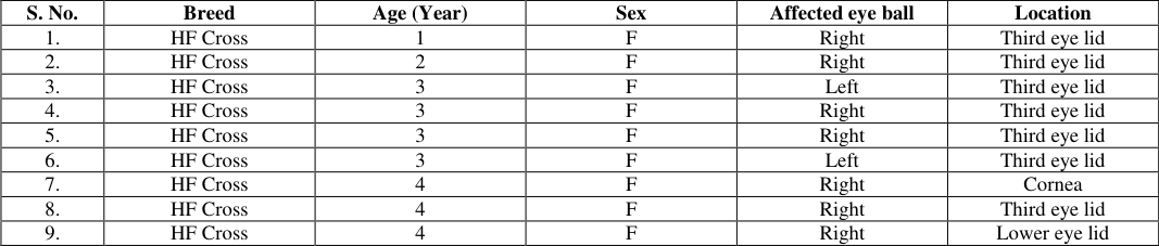 Table 1
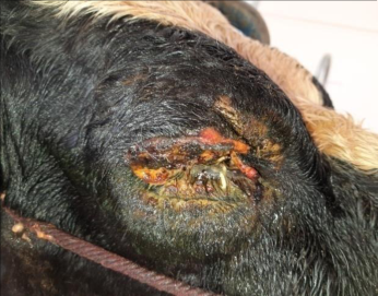 Figure 1
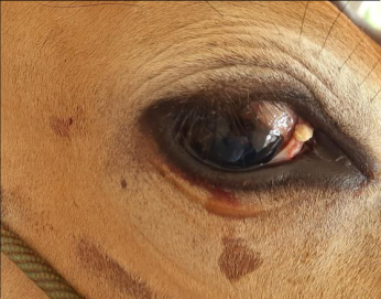 Figure 2
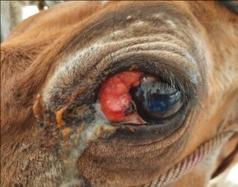 Figure 3
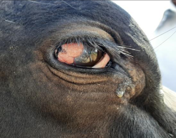 Figure 4
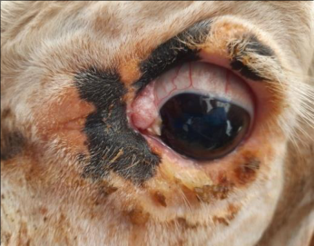 Figure 5
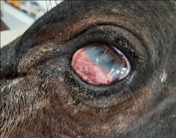 Figure 6
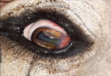 Figure 7
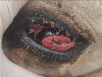 Figure 8
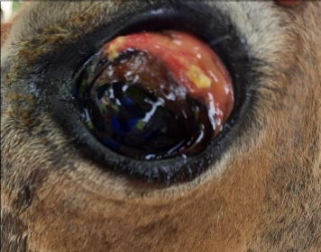 Figure 7
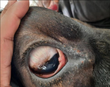 Figure 7
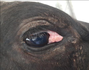 Figure 8
Introduction
Bovine ocular squamous cell carcinoma or cancer eye is the most commonly affecting tumours of the cattle eye ball, it is primarily an epithelial cell origin, the condition was reported worldwide and affects various ocular and peri-ocular tissues like surfaces of conjunctiva, sclerocorneal junction (limbus), nictitating membrane, cornea and the eyelid skin (Tsujita and Plummer, 2010) [1] . Etiology for the bovine ocular squamous cell carcinoma is unknown; however, heredity, nutrition level, age, UV light, less pigmented skin and some of the viruses contributes development of tumour (Reddy et al., 2017 and, Pazhanivel and Umarani, 2022) [2,12] . Various diagnostic methods are available to confirm and grade the conditions via cytology, histopathology and immunohistochemistry (Kumar et al., 2023) [5] . Regarding treatment option extirpation and enucleation remains simple, feasible and non-expensive method in various ocular pathological conditions affecting the bovine eye ball (Reddy et al., 2017) [2] .
Materials and Methods
Nineteen cattles presented to the large animal outpatient unit, Veterinary Clinical Complex, Veterinary College and Research institute, Salem from period June 2022 to February 2023 cattles were presented with the history of eye swellings/tumours were examined closely and the following parameter were recorded via breed, age, sex, affected eye ball, location of tumour in the eye ball and recurrence after treatment. Impression smears and tissue samples were collected for confirmatory diagnosis and grading of tumours before and after treatment respectively.
Results
Fifteen animals eyeballs were diagnosed as malignant stage of bovine ocular cell carcinoma eye balls were extirpated as per the standard protocol whereas four animal's eyeballs diagnosed as pre-malignant stage of bovine ocular cell carcinomas affected portion only removed with wide surgical margin. After the surgical excision eye balls and tumours were collected in 10% formalin and send it to histopathological examination for grading and confirming the tumour. Post operatively in both the cases antibiotics and analgesics were given for a week.
Breed affected: Out of nineteen animals twelve animals were Holstein Friesian cattle (63.16%) and remaining seven animals were jersey cross breed cow (36.84%) (Table 1, Figure 1 and Figure 2). Age: Out of nineteen animals nine animals are aged 1 to 3 calving (47.37%), seven animals aged 3-5 calving (36.84%) and three animals aged more than 5 calving 15.79%, Table 1.
| S. No. | Breed | Age (Year) | Sex | Affected eye ball | Location |
|---|---|---|---|---|---|
| 1. | HF Cross | 1 | F | Right | Third eye lid |
| 2. | HF Cross | 2 | F | Right | Third eye lid |
| 3. | HF Cross | 3 | F | Left | Third eye lid |
| 4. | HF Cross | 3 | F | Right | Third eye lid |
| 5. | HF Cross | 3 | F | Right | Third eye lid |
| 6. | HF Cross | 3 | F | Left | Third eye lid |
| 7. | HF Cross | 4 | F | Right | Cornea |
| 8. | HF Cross | 4 | F | Right | Third eye lid |
| 9. | HF Cross | 4 | F | Right | Lower eye lid |
Table text extracted from the original PDF; superscript letters are shown inline. The original table image is below.
View original table image
Location of the tumour and affected eye ball: Out of nineteen cattle's, thirteen (68.42%) cattles are affected by right eye and remaining six (31.85%) cattle are affected by left eye ball (Figure 3 and Figure 4). On closed ophthalmic examination out of nineteen cases twelve (63.16%) cases the lesions at third eye lids (Figure 5), cornea in one (5.26%) cases (Figure 6), in four cases at sclera-corneal junction (21.05%) (Figure 7) and in two cases lower eye lid (10.53%) (Table 1, Figure 8).
Gross pathology cytological examination:
Out of nineteen animals fifteen animals diagnosed as malignant (78.95%) Stage of bovine ocular squamous cell carcinomas and remaining four cases (21.05%) as premalignant stage of BOSCC by their gross pathology viaplaques, keratomas, papillomas and carcinomas (Image 1, 1a, 2, 2a, 3, 3a, 4 and 4a). The cytological examination of the fifteen animals diagnoses as premalignant stage of BOSCC revealed Clusters of monomorphic layers of cells with normochromic and normocytic nucleus whereas malignant stage of BOSCC appeared as Clusters of pleomorphic, polyhedral cells with large nuclei and some cells showed multinucleated cells. Neoplastic cells showing anisokaryosis, anisocytosis, intense deep blue cytoplasm with increased nucleus: cytoplasmic ratio and some neoplastic cells showing mitotic divisions also.
Histopathology:
The histopathological examination of the premalignant stage of BOSCC results revealed cluster of squamous neoplastic cells with some cells showing anaplastic changes and mitotic figures whereas malignant stage of BOSCC revealed Cords of neoplastic squamous cells observed as large, ovoid cells contained vesicular nuclei and central prominent nucleolus. Neoplastic areas showed numerous mitotic figures and keratin pearls or cell nest areas.
Tumour recurrence: Out of nineteen cases, six cases underwent surgical enucleation two cases had tumour recurrence after six months (Figure 1).
Discussion
The study results revealed incidence was higher in Holstein Friesian cattle (63.16%) followed by jersey crossbred cows (36.84%). This result was in accordance with the study results of Lakshmi et al. (2020) [7] , Pazhanivel and Umarani R (2022) [12] who recorded the highest incidence in Holstein Friesian crossbred cows followed by Jersey crossbred cows and nondescript cows. The age wise incidence revealed higher in animals aged between 1 to 3 calving (47.37%) followed by 3-5 calving (36.84%) and more than 5 calving (15.79%). This results was partially agreement with the study results of Lakshmi et al. (2020) [7] , Pazhanivel and Umarani R (2022) [12] , Gharagozlou et al. (2007) [10] . They observed higher incidence in age group of above 5 years followed by 2-5 years. In this study third eyelid (63.16%) was mostly affected followed by Limbus (21.05%), Lower eye lid (10.53%) and cornea (5.26%) this result was in conjunction with results of Gharagozlou et al. (2007) [10] . the author found that 70%of the animals had lesion at the nictitating membrane and palpebral conjunctiva. Cytological examination of this study result prompt with the observation of Hoffmann et al. (1978) [4] who recorded the following nuclear changes in the smears prepared from histologically confirmed cases of bovine ocular squamous cell carcinomas viz condensation of nuclear chromatin, prominent nucleoli, anisonucleosis, bizarre mitoses, multinucleation and increased nucleocytoplasmic ratio. Histologically the benign tumour revealed cluster of squamous neoplastic cells, some cells showing anaplastic changes and mitotic figures whereas malignant stage appeared cords of neoplastic squamous cells observed as large, ovoid cells contained vesicular nuclei and central prominent nucleolus. Neoplastic areas showed numerous mitotic figures and keratin pearls or cell nest areas. The same result was observed by Srivastav et al., (2022) [13] and Kumar et al. (2023) [5] . The author opined that presence of cell nest, large cells with eosinophilic cytoplasm and a large vesicular nucleus is suggestive of squamous cell carcinoma. Similar results of cell nest with high degree of keratinization with anaplasia, numerous mitotic figures, tumour islands with severe inflammation, neovascularization, hemorrhages in well differentiated carcinoma. Whereas absence of keratin pearls and abundance of fibrous tissue is characteristics of Poorly differentiated OSCC was observed by Kumar et al. (2023) [5] and Pazhanivel and Umarani (2022) [12] . In this study all the animals underwent surgical extirpation and enucleation except four cases diagnosed as premalignant stage of BOSCC. The later one was underwent tumour excision alone, hence itis a simple, feasible and least expensive method of treatment option for various ocular pathologies as stated by Pazhanivel and Umarani (2022) [12] . The prognosis and outcomes mainly depends on early detection, presence of metastasis and the timing of tumour removal (Ozcan-Martz et al., 2021) [11] . In the current study recurrence of tumours occurs in two cases. This could be due to late diagnosis and invasion of the tumours cells to the adjacent peri-ocular tissue.
Conclusion
Based on the above study results we conclude that incidence of bovine ocular squamous cell carcinoma was highest in Holstein Friesian cattle (63.16%) followed by Jersey cross bred cows (36.84%) and animals aged between 1 to 3 calving (47.37%) followed by 3-5 calving (36.84%) and more than 5 calving (15.79%). mostly right eye ball was affected (68.42%) than that of the left eye ball (31.85%). The tissue mostly affected was third eyelid (63.16%), this could be due to prolonged exposure of third eye lid of right side than the left side to ultra violet radiation. Furthers studies is require to correlate this finding.
Acknowledgement
AcknowledgmentThe author thanks the Dean, Veterinary College and Research Institute, TANUVAS for the facilities provided.
References
10 in the original paper- Tsujita H, Plummer CE. Bovine ocular squamous cell carcinoma. Vet Clin Food Anim. 2010;26(3):511-529.
- Reddy NVK, Reddy KJ, Mallesh P, Raju G. Surgical management of recurrent squamous cell carcinoma in cattle: A case report. Pharm Innov J. 2017;6(8):161-162.
- Fornazari GA, Kravetz J, Kiupel M, Sledge D, De Barros Filho IR, Montiani-Ferreira F et al. Ocular squamous cell carcinoma in Holstein cows from the South of Brazil. Vet World. 2017;10(12):1413.
- Hoffmann D, Spradbrow PB, Wilson BE. An evaluation of exfoliative cytology in the diagnosis of bovine ocular squamous cell carcinoma. J Comp Pathol. 1978;88(4):497-504.
- Kumar V, Jolhe DK, Ghosh RC, Sonkusale PM, Sharma S. Pathology and diagnosis of ocular squamous cell carcinoma in bovines. Indian J Vet Sci Biotechnol. 2023;19(4):43-49.
- Reddy NVK, Reddy KJ, Mallesh P, Raju G. Surgical management of recurrent squamous cell carcinoma in cattle: A case report. Pharm Innov J. 2017;6(8):161-162.
- Lakshmi MP, Veena P, Kumar RS, RaniPrameela D. Clinical, pathological and immunohistochemical studies on bovine eye cancer. Pharm Innov J. 2020;9:353-355.
- Mirza U, Singh S, Farooq UB. Transpalpebral exenteration of eye for recurrent squamous cell carcinoma in a Nili-Ravi buffalo: A case report. Buffalo Bull. 2023;42(2):185-192.
- Gharagozlou MJ, Hekmati P, Ashrafihelan J. A clinical and histopathological study of ocular neoplasms in dairy cattle. Vet Arhiv. 2007;77(5):409.
- Martz ÖA, Martz P, Ebert F, Bittner L, Wippermann W, Kaiser M, et al. Case reports of squamous cell carcinoma in cows. Vet Ital. 2021;57(2):161-168.
Cited by 2
From OpenAlex, OpenCitations and reference lists read by Scinovex
- Bovine Ocular Squamous Cell Carcinoma—A Descriptive Epidemiological Survey in the Azores, Portugal ↗
Beatriz Bilhastre, Helena Vala, Ana Clara Ribeiro, Sara Faria, Ana Oliveira, Sandra Branco et al. · Veterinary Sciences · 2026
Bovine ocular squamous cell carcinoma (BOSCC) is the most common ocular tumour in cattle, with a multifactorial aetiology involving ultraviolet (UV) radiation, genetic factors, pigmentation, and management practices. A detailed epidemiological characterisation of BOSCC in the Azores, Portugal, is provided, with particular emphasis on its spatial distribution and potential risk determinants. Data were obtained through an epidemiological questionnaire completed by…
❝ 1PDF - Bovine Ocular Squamous Cell Carcinoma ↗
Beatriz Bilhastre, Helena Vala, Carlos Pinto, Sandra Branco · Springer eBooks · 2026
❝ 0