
Research article · 2024 · Full text
International Journal of Biology SciencesA comprehensive review on the molecular basis of microbial pathogenesis of viral and bacterial infections
Volume 6(2) · Pages 157–161 · DOI: 10.33545/26649926.2024.v6.i2c.247 ↗
Research areasBacteriophages and microbial interactions
Abstract
Author-writtenResearch on microbial infections determines both the immediate and long-term interactions between specific microorganism and their hosts. Microbial pathogenesis is a complex phenomenon involving the interaction between microbial virulence factors and host cellular responses. These virulence factors include invasins, capsules, siderophores, enzymes, endotoxins, and exotoxins. Though the main problem in the current scenario is the coinfection. Beyond initiating infection, these factors also help the pathogenic microbes survive and thrive within the host's environment. Understanding the molecular mechanisms of microbial infections is essential for developing effective treatments, preventive measures, and vaccines. This review delves into how microbial pathogens invade host cells, evade the immune system, and establish infections. It also covers mechanisms of immune evasion, such as antigenic variation, mimicry, and the manipulation of host signaling pathways, utilizing advanced molecular biology techniques like CRISPR-Cas9 gene editing, transcriptomics, and proteomics. Additionally, it focuses on the key molecular interactions between viral and bacterial pathogen induced infections like respiratory co-infection and secondary bacterial pneumonia in patients with covid-19.
Summary
Source-checkedPlain-language reading of the full paper, prepared with AI and checked against the text. Each point links to the authors' own words.
Why it matters
- Microbial pathogenesis occurs through a complex interaction between the host's cellular responses and the virulence factors of the microbe. View evidence ↗
- Key virulence factors that allow pathogens to survive and thrive in a host include enzymes, capsules, invasins, siderophores, and both endotoxins and exotoxins. View evidence ↗
- Studying the molecular mechanisms of how microbes infect hosts is critical for the creation of vaccines and effective medical treatments. View evidence ↗
The question
- The review aims to examine the processes by which microbial pathogens establish infections, invade cells, and bypass the host's immune system. View evidence ↗
- The authors specifically focus on the molecular interactions involved in respiratory co-infections and secondary bacterial pneumonia in COVID-19 patients. View evidence ↗
What they did
- The review discusses the use of proteomics, transcriptomics, and CRISPR-Cas9 gene editing to understand immune evasion mechanisms. View evidence ↗
- The authors' commentary is derived from available clinical data and previous experiences with SARS-CoV and influenza. View evidence ↗
What they found
- Limited data on SARS-CoV-2 indicates that the incidence of viral co-infection varies between 0% and 19% across different case series. View evidence ↗
- In the previous SARS pandemic, MRSA was responsible for 47% of secondary bacterial pneumonia cases. View evidence ↗
- Bacterial pneumonia is typically marked by neutrophilic leukocytosis, whereas COVID-19 patients generally show lymphopenia and a normal white blood cell count. View evidence ↗
- In cases of isolated SARS-CoV-2 infection, procalcitonin levels are consistently reported as low or normal. View evidence ↗
- The use of tocilizumab for rheumatologic diseases is associated with a consistent increase in the risk of serious bacterial infections. View evidence ↗
- Tocilizumab may prevent the rise of white blood cell counts and C-reactive protein, which are typical responses to secondary bacterial infections. View evidence ↗
What it means
- The authors conclude that COVID-19 management should be based on the overall clinical presentation and illness trajectory, drawing from data on SARS-CoV and influenza. View evidence ↗
- The authors recommend starting broad-spectrum antifungal treatment, such as voriconazole, immediately upon the detection of invasive pulmonary aspergillosis. View evidence ↗
- The authors note that there is currently no clear pattern or established set of recommendations for managing viral co-infections or secondary bacterial pneumonia in COVID-19. View evidence ↗
Visual summary
1 visualsIllustrations explain the paper; they do not show study data.
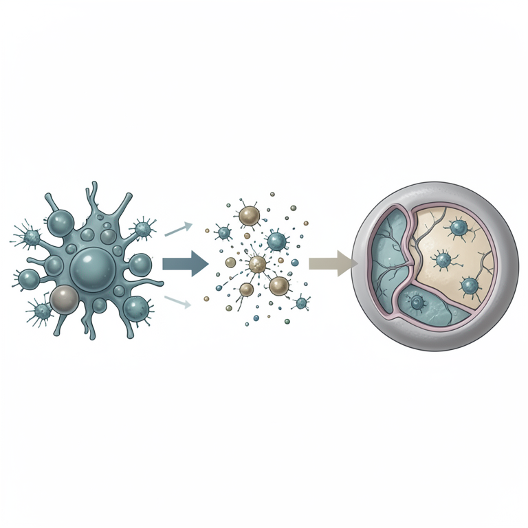
Microbial Pathogenesis Interaction
AI-generated conceptual illustration (a generic mammalian host cell and a mixture of bacteria and viruses). It explains the idea and does not show study data.
View source evidence ↗Introduction
The pathogenesis of Coronavirus Disease 2019 (COVID19), which is caused by the coronavirus type-2 (SARS-CoV-2) virus, is still not fully known despite the disease's global expansion. A thorough description has been provided of respiratory infections brought on by multiple viruses (viral co-infection) or by bacteria and viruses together (combined viral and bacterial pneumonia). Secondary bacterial pneumonia may develop during the healing phase of a viral respiratory infection, or it may follow the original phase [1] . The overall incidence of viral co-infection has varied greatly from 0% to 19% in different case series, according to the limited data on SARS-CoV-2. The rates of combined viral and bacterial pneumonia also seem modest [3] [8-10] . Furthermore, little is known about the organisms and risk factors that cause the condition. The most well-studied cases of combined viral and bacterial pneumonia, secondary bacterial pneumonia caused by Staphylococcus aureus and other prevalent community-acquired pneumonia pathogens, and considerable morbidity and mortality are associated with seasonal and pandemic influenza [2,3] . During the previous pandemic of Severe Acute Respiratory Syndrome (SARS), Methicillin-Resistant S. aureus (MRSA) caused 47% of cases of secondary bacterial pneumonia, although there was serious concern about cross-transmission. This pneumonia occurred as ventilator-associated pneumonia in 25% of patients at a single center [7] . The following commentary is based on available clinical data and experience with similar viruses, such as influenza and SARS-CoV, because there isn't a clear pattern or set of guidelines for viral co-infection, combined viral and bacterial pneumonia, or secondary bacterial pneumonia in the context of SARS-CoV-2. Based on our current understanding, a comprehensive approach to COVID-19 would ideally encompass both the entire presentation and the illness trajectory.
All patients with co-infection with the influenza A or B viruses should receive treatment with oseltamivir or a different medication, regardless of the severity of their illness [11] . If there is a clear exposure or risk factor, empirical treatment for influenza virus co-infection may be explored while test results are awaited. Therapy alternatives are restricted and only work in certain situations, including immunosuppression or hypogammaglobulinemia, if viral coinfection with another respiratory virus, such as respiratory syncytial virus, is found [12,13] . It is highly advised to visit an infectious disease specialist to weigh the advantages of this course of treatment against the possibility of aggravating COVID-19-related organ failure and the possible side effects of the prescribed medicine or medications.
Bacterial pneumonia
A strong index of suspicion is needed to diagnose secondary bacterial pneumonia with COVID-19 or combination viral and bacterial pneumonia. Even while viral and bacterial symptomatology significantly overlap [2][3][4][5][6][7][8][9][10][14][15][16][17][18][19][20][21][22][23][24][25][26][27][28][29] , some aspects of bacterial infection may still be distinguishable. Bacterial pneumonia is characterized by neutrophilicleukocytosis, but COVID-19 patients usually exhibit lymphopenia along with a normal white blood cell count [5,8,14,15] .
Mode of Infection
In order to distinguish between the two possible causes of community-acquired pneumonia, procalcitonin is neither sensitive nor specific [11] . But procalcitonin levels in isolated SARS-CoV-2 infection have been regularly reported to be normal (low) in various COVID-19 case series. This has led to its widespread, albeit unvalidated, usage to "rule out" combination viral and bacterial pneumonia, but the precise cutoff is still unknown. This comment emphasizes how important it is to take into account every factor within the framework of the clinical setting. Antibiotics can be safely delayed in patients with mild to moderate respiratory failure consistent with COVID-19 presentation and without clear evidence of bacterial infection. This is because there is little chance of mixed viral and bacterial pneumonia in these individuals. In this instance, the advancement of COVID-19 is more likely to be the cause of the steadily deteriorating respiratory failure during the first week of presentation than the emergence of a newly superimposed secondary bacterial pneumonia. This covers patients who begin receiving supplemental oxygen support in a non-invasive manner before eventually needing invasive mechanical ventilation. Even if respiratory discomfort is getting worse, medicines shouldn't be started if there isn't any sign of bacterial pneumonia. Unless otherwise demonstrated, nosocomial acquisition of secondary bacterial infection that is, hospitalacquired pneumonia, infection at an extrapulmonary site, or both is likely if a patient experiences new or sharply worsening respiratory failure, sepsis, or both following an initial period of consistent improvement (considered to be days). Although COVID-19 can induce acute respiratory decompensation on its own, there is little information on the potential involvement of secondary bacterial pneumonia in this condition. Consequently, until this secondary infection is ruled out, using empiric antibiotics based on guidelines may be prudent. One or more of the following symptoms could indicate secondary bacterial pneumonia: new or recrudescent fever; fresh onset or change in sputum characteristics; fresh neutrophilia or leukocytosis (or both); fresh pertinent imaging results; and fresh or rising oxygen demand. It's crucial to manage these patients appropriately and take into account any additional potential causes of hospital-acquired infections, such as urinary tract or indwelling central venous catheters. Empirical treatment for all potential causes at the outset is crucial for a critically ill patient who has been admitted and is experiencing acute respiratory failure. Because procalcitonin levels in patients with multiorgan failure can be mistakenly increased, this is very crucial [30,31] , therefore it may be difficult to distinguish between concealed bacterial infection consolidation and bilateral infiltrates of acute respiratory distress syndrome using imaging investigations.
Possible mode of management
The American Thoracic Society (ATS) and Infectious Diseases Society of America (IDSA) guidelines, host risk factors, and previous microbiologic data should all be taken into consideration when developing an empirical treatment plan for community-acquired pneumonia [19] . For every patient, respiratory samples (tracheal aspirate is preferred over sputum in those on mechanical ventilation) and blood cultures should be submitted, ideally prior to the initiation of medications. All patients who come with severe communityacquired pneumonia should have a test for the urine antigen of streptococcus pneumoniae. Depending on the clinical setting and epidemiology, samples for Legionella pneumophila urine antigen and Mycoplasma pneumoniaeIgM and IgG antibodies may be provided. If there are no symptoms of bacterial pneumonia, a positive respiratory culture may indicate colonization, particularly in those who have already had pneumonia caused by the same organism in the past or have changed airway anatomy. This distinction can be made with the aid of laboratory markers, radiologic characteristics, and quantitative and semiquantitative culture techniques [19] . While the presentation of secondary bacterial pneumonia in a patient receiving invasive mechanical ventilation resembles that of hospital-acquired pneumonia, guidelines dictate the aggressive use of empiric broad-spectrum antibiotics that cover MRSA, Pseudomonas aeruginosa, and potentially other multidrug-resistant organisms [19] . The adverse effects of institutional antibiograms and antibiotics must also be taken into account. In addition to frequently exhibiting lower-grade fevers and higher secretions, patients with ventilator-associated tracheobronchitis may also have lowgrade fevers and prove challenging to wean off of ventilator assistance. Only a careful case-based review is necessary because the evidence supporting antibiotic therapy for this clinical entity is weak. For community-acquired pneumonia [32] , antibiotic therapy should be administered for 5 to 7 days; in the case of hospital-acquired pneumonia and ventilator-associated pneumonia [19] , it should be administered for 7 days, provided no problems arise. In particular, if side effects are observed, think about reducing the period if patients show symptoms of clinical stability. Based on the procalcitonin level trend over the course of 24 to 48 hours, determining the procalcitonin level at presentation will aid in the deescalation of antibiotics [33] . It is permissible to stop all antibiotics if, after 48 hours of testing, a microbiological source cannot be found and the procalcitonin level is less than 0.5 μg/L or drops by 80% or more from peak concentration [19] . Because interleukin 6 (IL-6) inhibitors, such tocilizumab, reduce frequent indications of sepsis, using them for COVID-19-related cytokine activation syndrome poses a special problem. When tocilizumab is used for rheumatologic illnesses, there is a consistently observed increased risk of serious bacterial infections [34,35,36,37] . After using tocilizumab, C-reactive protein and other acute-phase reactants, such as white blood cell count, may not rise in response to a secondary bacterial infection [35,38,39] . It's unclear exactly how long this impact lasts after taking one or two doses. Although procalcitonin may be less influenced by IL-6 inhibitors, there is insufficient data to distinguish between bacterial and viral pneumonia in this setting, and more research is needed to make this determination [40][41][42][43][44] .
Conclusion
Combining SARS-CoV-2 with a bacterial pathogen (combined viral and bacterial pneumonia) or SARS-CoV-2 with another virus (viral co-infection) can cause respiratory infections in COVID-19. Either during the healing phase or after the initial viral respiratory illness, secondary bacterial pneumonia can develop. Regarding viral co-infection, combined viral and bacterial pneumonia, or subsequent bacterial pneumonia in COVID-19, there isn't a clear pattern or set of recommendations. The management strategy for COVID-19 should, ideally, take into account the overall presentation and the progression of illness based on available clinical data and experience with viruses related to influenza and SARS-CoV. Last but not least, invasive pulmonary aspergillosis is linked to high rates of morbidity and mortality and has been reported in critically ill patients with seasonal and pandemic influenza. Patients with acute respiratory distress syndrome linked with COVID-19 have also been documented to have invasive pulmonary aspergillosis. High-risk patients include those with immunocompromised states, prior or concurrent influenza viral co-infection, clinical worsening despite proper antibiotic treatment, and positive fungal indicators such as galactomannan on culture. These patients should be monitored closely for this consequence. Treatment with a wide antifungal, such as voriconazole, should be started as soon as invasive pulmonary aspergillosis is detected, after consulting with colleagues in infectious diseases.
Acknowledgement
AcknowledgmentsThe authors are highly thankful to the Department of Medical Laboratory Technology, Swami Vivekananda University, Barrackpore. The authors also declare that no funds or grants were received during the preparation of this article.
References
44 in the original paper- Van der Sluijs KF, van der Poll T, Lutter R, Juffermans NP, Schultz MJ. Bench-to-bedside review: bacterial pneumonia with influenza pathogenesis and clinical implications. Crit Care. 2010;14(2):219. DOI:10.1186/cc8893.
- Pongpirul WA, Mott JA, Woodring JV, et al. Clinical characteristics of patients hospitalized with coronavirus disease, Thailand. Emerg Infect Dis. 2020;26(7):15801585. DOI:10.3201/eid2607.200598.
- Arentz M, Yim E, Klaff L, et al. Characteristics and outcomes of 21 critically ill patients with COVID-19 in Washington State. JAMA. 2020;323(16):1612-1614. DOI:10.1001/jama.2020.4326.
- Wu C, Chen X, Cai Y, et al. Risk factors associated with acute respiratory distress syndrome and death in patients with coronavirus disease 2019 pneumonia in Wuhan, China. JAMA Intern Med. 2020;180(7):1-11. DOI:10.1001/jamainternmed.2020.0994.
- Ding Q, Lu P, Fan Y, Xia Y, Liu M. The clinical characteristics of pneumonia patients coinfected with 2019 novel coronavirus and influenza virus in Wuhan, China. J Med Virol. 2020 Mar 20. DOI:10.1002/jmv.25781.
- Khodamoradi Z, Moghadami M, Lotfi M. Co-infection of coronavirus disease 2019 and influenza A: a report from Iran. Arch Iran Med. 2020;23(4):239-243. DOI:10.34172/aim.2020.04.
- Touzard-Romo F, Tapé C, Lonks JR. Co-infection with SARS-CoV-2 and human metapneumovirus. R I Med J (2013). 2020;103(2):75-76. PMID: 32192233.
- Chen N, Zhou M, Dong X, et al. Epidemiological and clinical characteristics of 99 cases of 2019 novel coronavirus pneumonia in Wuhan, China: a descriptive study. Lancet. 2020;395(10223):507-513. DOI:10.1016/S0140-6736(20)30211-7.
- Wang L, He W, Yu X, et al. Coronavirus disease 2019 in elderly patients: characteristics and prognostic factors based on 4-week follow-up. J Infect. 2020 Mar 30. DOI:10.1016/j.jinf.2020.03.019.
- Zhou F, Yu T, Du R, et al. Clinical course and risk factors for mortality of adult inpatients with COVID-19 in Wuhan, China: a retrospective cohort study. Lancet. 2020;395(10229):1054-1062. DOI:10.1016/S01406736(20)30566-3.
- Uyeki TM, Bernstein HH, Bradley JS, et al. Clinical practice guidelines by the Infectious Diseases Society of America: 2018 update on diagnosis, treatment, chemoprophylaxis, and institutional outbreak management of seasonal influenza. Clin Infect Dis. 2019;68(6):e1-e47. DOI:10.1093/cid/ciy866.
- Beaird OE, Freifeld A, Ison MG, et al. Current practices for treatment of respiratory syncytial virus and other non-influenza respiratory viruses in high-risk patient populations: a survey of institutions in the Midwestern Respiratory Virus Collaborative. Transpl Infect Dis. 2016;18(2):210-215. DOI:10.1111/tid.12510.
- Beigel JH, Nam HH, Adams PL, et al. Advances in respiratory virus therapeutics a meeting report from the 6th Isirv Antiviral Group conference. Antiviral Res. 2019;167:45-67. DOI:10.1016/j.antiviral.2019.04.006.
- Ruan Q, Yang K, Wang W, Jiang L, Song J. Clinical predictors of mortality due to COVID-19 based on an International Journal of Biology Sciences https://www.biologyjournal.net analysis of data of 150 patients from Wuhan, China. Intensive Care Med. 2020 Mar 3. DOI:10.1007/s00134020-05991-x.
- Yang X, Yu Y, Xu J, et al. Clinical course and outcomes of critically ill patients with SARS-CoV-2 pneumonia in Wuhan, China: a single-centered, retrospective, observational study. Lancet Respir Med.; 2020. DOI:10.1016/S2213-2600(20)30079-5.
- Huang C, Wang Y, Li X, et al. Clinical features of patients infected with 2019 novel coronavirus in Wuhan, China. Lancet. 2020;395(10223):497-506. DOI:10.1016/S0140-6736(20)30183-5.
- Lippi G, Plebani M. Procalcitonin in patients with severe coronavirus disease 2019 (COVID-19): a metaanalysis. Clin Chim Acta. 2020;505:190-191. DOI:10.1016/j.cca.2020.03.004.
- Rule JA, Hynan LS, Attar N, et al. Procalcitonin identifies cell injury, not bacterial infection, in acute liver failure. PLoS One. 2015;10(9):e0138566. DOI:10.1371/journal.pone.0138566.
- Metlay JP, Waterer GW, Long AC, et al. Diagnosis and treatment of adults with community-acquired pneumonia. An official clinical practice guideline of the American Thoracic Society and Infectious Diseases Society of America. Am J Respir Crit Care Med. 2019;200(7):e45-e67. DOI:10.1164/rccm.2019081581ST.
- Xu X, Han M, Li T, et al. Effective treatment of severe COVID-19 patients with tocilizumab. Available from: http://www.chinaxiv.org/abs/202003.00026. Accessed April 24, 2020.
- Bloomfield M, Parackova Z, Cabelova T, et al. AntiIL6 autoantibodies in an infant with CRP-less septic shock. Front Immunol. 2019;10:2629. DOI:10.3389/fimmu.2019.02629.
- Gaensbauer JT, Press CA, Hollister JR, Asturias EJ. Procalcitonin: a marker of infection not subverted by treatment with interleukin-6 receptor inhibition. Pediatr Infect Dis J. 2013;32(9):1040. DOI:10.1097/INF.0b013e318295a3d0.
- Sato H, Tanabe N, Murasawa A, et al. Procalcitonin is a specific marker for detecting bacterial infection in patients with rheumatoid arthritis. J Rheumatol. 2012;39(8):1517-1523. DOI:10.3899/jrheum.111601.
- Luo P, Liu Y, Qiu L, Liu X, Liu D, Li J et al. Tocilizumab treatment in COVID-19: a single center experience. J Med Virol. 2020 Apr 6. DOI:10.1002/jmv.25801.
- Michot JM, Albiges L, Chaput N, et al. Tocilizumab, an anti-IL6 receptor antibody, to treat COVID-19-related respiratory failure: a case report. Ann Oncol. 2020;31(7):961-964. DOI:10.1016/j.annonc.2020.03.300.
- Wong HYF, Lam HYS, Fong AH, et al. Frequency and distribution of chest radiographic findings in COVID19 positive patients. Radiology. 2020;296(2):E72-E78. DOI:10.1148/radiol.2020201160.
- Kanne JP, Little BP, Chung JH, Elicker BM, Ketai LH. Essentials for radiologists on COVID-19: an updateradiology scientific expert panel. Radiology. 2020;296(2):E113-E114. DOI:10.1148/radiol.2020200527.
- Pan F, Ye T, Sun P, et al. Time course of lung changes at chest CT during recovery from coronavirus disease 2019. Radiology. 2020;295(3):715-721. doi:10.1148/radiol.2020200370.
- Giamarellos-Bourboulis EJ, Mega A, Grecka P, et al. Procalcitonin: A marker to clearly differentiate systemic inflammatory response syndrome and sepsis in the critically ill patient? Intensive Care Med. 2002;28(9):1351-1356. DOI:10.1007/s00134-002-1398z.
- Wee LE, Ko KKK, Ho WQ, Kwek GTC, Tan TT, Wijaya L et al. Community-acquired viral respiratory infections amongst hospitalized inpatients during a COVID-19 outbreak in Singapore: co-infection and clinical outcomes. J Clin Virol. 2020 Jul;128:104436. DOI:10.1016/j.jcv.2020.104436.
- Hui L, Zhang X, An X, et al. Higher serum procalcitonin and IL-6 levels predict worse diagnosis for acute respiratory distress syndrome patients with multiple organ dysfunction. Int J Clin Exp Pathol. 2017;10(7):7401-7407. PMID: 31966582.
- Kalil AC, Metersky ML, Klompas M, et al. Management of adults with hospital-acquired and ventilator-associated pneumonia: 2016 clinical practice guidelines by the Infectious Diseases Society of America and the American Thoracic Society. Clin Infect Dis. 2016;63(5):e61-e111. DOI:10.1093/cid/ciw353.
- Lam SW, Bauer SR, Fowler R, Duggal A. Systematic review and meta-analysis of procalcitonin-guidance versus usual care for antimicrobial management in critically ill patients: focus on subgroups based on antibiotic initiation, cessation, or mixed strategies. Crit Care Med. 2018;46(5):684-690. DOI:10.1097/CCM.0000000000002953.
- Campbell L, Chen C, Bhagat SS, Parker RA, Östör AJ. Risk of adverse events including serious infections in rheumatoid arthritis patients treated with tocilizumab: a systematic literature review and meta-analysis of randomized controlled trials. Rheumatology (Oxford). 2011;50(3):552-562. DOI:10.1093/rheumatology/keq343.
- Chiu YM, Chen DY. Infection risk in patients undergoing treatment for inflammatory arthritis: nonbiologics versus biologics. Expert Rev Clin Immunol. 2020;16(2):207-228. DOI:10.1080/1744666X.2019.1705785.
- Yamamoto K, Goto H, Hirao K, et al. Longterm safety of tocilizumab: results from 3 years of follow-up postmarketing surveillance of 5,573 patients with rheumatoid arthritis in Japan. J Rheumatol. 2015;42(8):1368-1375. DOI:10.3899/jrheum.141210.
- Calderón-Goercke M, Loricera J, Aldasoro V, et al. Tocilizumab in giant cell arteritis. Observational, openlabel multicenter study of 134 patients in clinical practice. Semin Arthritis Rheum. 2019;49(1):126-135. DOI:10.1016/j.semarthrit.2019.01.003.
- Nguyen MT, Pødenphant J, Ravn P. Three cases of severely disseminated Staphylococcus aureus infection in patients treated with tocilizumab. BMJ Case Rep. 2013 Jan 2;2013:bcr2012007413. DOI:10.1136/bcr2012-007413.
- Bari SF, Khan A, Lawson T. C reactive protein may not be reliable as a marker of severe bacterial infection in patients receiving tocilizumab. BMJ Case Rep. 2013 Oct 31. DOI:10.1136/bcr-2013-010423.
- Vanderbeke L, Spriet I, Breynaert C, Rijnders BJ, Verweij PE, Wauters J et al. Invasive pulmonary aspergillosis complicating severe influenza: epidemiology, diagnosis and treatment. Curr Opin Infect Dis. 2018;31(6):471-480. DOI:10.1097/QCO.0000000000000504.
- Schauwvlieghe AFAD, Rijnders BJA, Philips N, et al. Invasive aspergillosis in patients admitted to the intensive care unit with severe influenza: a retrospective cohort study. Lancet Respir Med. 2018;6(10):782-792. DOI:10.1016/S2213-2600(18)30274-1.
- Yu Z, Zhang Q, Gu B, et al. Clinical features of fatal pandemic influenza A/H1N1 infection complicated by invasive pulmonary fungal infection. Mycopathologia. 2020;185(2):319-329. DOI:10.1007/s11046-019-00421z.
- Hwang DM, Chamberlain DW, Poutanen SM, Low DE, Asa SL, Butany J et al. Pulmonary pathology of severe acute respiratory syndrome in Toronto. Mod Pathol. 2005;18(1):1-10. DOI:10.1038/modpathol.3800247.
- Koehler P, Cornely OA, Bottiger BW, et al. COVID-19 associated pulmonary aspergillosis. Mycoses. 2020;63(6):528-534. DOI:10.1111/myc.13096.