
Research article · 2024 · Full text
International Journal of Biology SciencesExploring the pathogenesis of fungal infection in the gastrointestinal tract: Emerging insights and diagnostic challenges
Volume 6(2) · Pages 162–166 · DOI: 10.33545/26649926.2024.v6.i2c.248 ↗
Research areasHelicobacter pylori-related gastroenterology studiesEosinophilic EsophagitisMicroscopic Colitis
Abstract
Author-writtenIn nature, fungi are abundant and can be found in extremely close contact to food, soil, mammals, and insects. These are among the most prevalent infections that can affect any organ in the body, including the gastrointestinal system in individuals with weakened immune systems, and they can have a serious negative impact on morbidity and death. The gastrointestinal system is regularly colonized and infected by various yeast and other fungus due to its constant exposure to fungi. In addition, it is the source of numerous disseminated fungal infections. The number of individuals with impaired immune systems has increased along with the occurrence of invasive fungal infections, especially in the gastrointestinal tract. Since the immunocompromised patients may exhibit unusual clinical characteristics, therefore diagnosis of fungal infections in these patients can be especially challenging. This review will focus on several significant emerging fungal infections on the GI tract in immunocompromised patients, even though Aspergillus and Candida species account for the majority of fungi diagnosed in this population of patients with compromised immune systems.
Summary
Source-checkedPlain-language reading of the full paper, prepared with AI and checked against the text. Each point links to the authors' own words.
Why it matters
- The gastrointestinal (GI) tract is frequently colonized and infected by fungi because of constant exposure, and it often serves as a starting point for infections that spread throughout the body. View evidence ↗
- There has been a rise in invasive fungal infections in the GI tract, coinciding with an increase in the number of immunocompromised individuals. View evidence ↗
- Diagnosing fungal infections in immunocompromised patients is difficult because these patients often present with atypical clinical features. View evidence ↗
The question
- The review aims to examine significant emerging fungal infections of the GI tract in immunocompromised patients, moving beyond the most common Aspergillus and Candida species. View evidence ↗
What they did
- Culture remains the gold standard for diagnosing and identifying fungal species, although it can be time-consuming. View evidence ↗
- Polymerase chain reaction (PCR) and sequencing of the 18S rRNA are increasingly used for the direct identification of fungi from culture material. View evidence ↗
- The galactomannan assay is a specific serological tool used to identify invasive aspergillosis, as it is typically negative for other invasive fungal diseases. View evidence ↗
- The beta D glucan assay is a broad serological test that is positive for most disseminated fungal diseases, including Pneumocystis, but negative for mucormycosis. View evidence ↗
What they found
- The general death rate for mucormycosis is approximately 40%, with higher mortality observed in bone marrow transplant recipients and patients with hematologic malignancies. View evidence ↗
- Gastrointestinal zygomycosis is rare compared to pulmonary disease and has a high mortality rate, reported to be between 40% and 50%. View evidence ↗
- In cases of disseminated Histoplasma capsulatum infection, more than 80% of patients experience involvement of the gastrointestinal tract. View evidence ↗
- Penicilliopsis marneffei typically measures 2-5 microns, though it can occasionally reach 20 microns in length and is characterized by a central septum. View evidence ↗
- Aspergillus can be distinguished from Mucor by its uniformly widened septate hyphae that branch at acute angles. View evidence ↗
- Pneumocystis jirovecii infection has been linked to the use of infliximab, a medication used for Crohn's disease and rheumatoid arthritis. View evidence ↗
What it means
- The authors conclude that medical professionals can better diagnose and treat GI fungal infections by understanding the specific epidemiology, risk factors, and pathological findings of different fungi. View evidence ↗
- Despite the benefits of appropriate antifungal therapy, aspergillosis and zygomycosis continue to have high fatality rates. View evidence ↗
- The authors state that there is still significant room for improvement in the effectiveness of serology-based testing. View evidence ↗
Visual summary
1 visualsIllustrations explain the paper; they do not show study data.
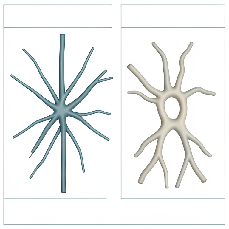
Morphological Comparison of Fungal Hyphae
AI-generated conceptual illustration (Aspergillus and Mucor fungal hyphae). It explains the idea and does not show study data.
View source evidence ↗Introduction
Immunocompromised patients frequently experience fungal infections as a consequence, and endemic mycoses are among the leading infectious causes of morbidity and mortality in the and environmental factors including the patient's residence and the kinds and amounts of exposure [5,6] . Additionally, the host is the only source of the inflammatory response to the organism; the differential diagnosis for each given mycosis is created by the patient's surroundings, history of exposure, and particular immune system deficiencies. This analysis will concentrate on a number of significant new fungal infections that affect immunocompromised people.
Infection by filamentous fungi
Fungi in the order Mucorales are the source of the potentially fatal infection known as mucormycosis [7] . As previously mentioned, Aspergillus species are known to be the most prevalent filamentous fungus in patients with impaired immune systems. Mucormycosis is becoming more common, especially in people with diabetes, hematologic cancers, and bone marrow transplant recipients. The Zygomycetes order was recently eliminated and replaced with the order Mucorales in the subphylum Mucormycotina [8] . Diabetes and other metabolic acidosis-causing conditions, deferoxamine medication, organ or bone marrow transplant, neutropenia, deterioration of the skin and soft tissues, intravenous drug usage, neonatal preterm, and malnourishment are all linked to mucormycosis. Remarkably, there doesn't seem to be a correlation between this infection and HIV/AIDS. It seems that the incidence is rising, particularly among cancer patients. In general, the death rate is about 40%, and it is significantly greater for patients who have hematologic malignancies and those who have had a bone marrow transplant [9,10] . The most common gross manifestation of gastrointestinal zygomycosis is ulceration, which is often large and has rolled, irregular borders that may resemble like malignancies. These fungi can also superinfect previously ulcerated tissues, and disseminated disease can result from a primary gastrointestinal infection. While pulmonary disease is common, gastrointestinal zygomycosis is relatively uncommon and often fatal, with mortality reportedly approaching 40-50% [11] . Studies have shown that the histological similarities between the lesions induced by Mucor and related species and aspergillosis are considerable [12] . Aspergillus and Mucor are both angiogenic bacteria that cause necrosis, infarction, and thrombosis. Although the degree of inflammation varies, neutrophils are typically seen in large quantities. Spores are not produced by Mucor. Seldom observed are segmented aberrant forms with interior components and varying size, round to oval appearances. [13] . Differential diagnosis of Mucormycosis mostly involves various fungal illnesses. As previously mentioned, mucormycosis and aspergillosis can resemble one another quite a bit in terms of morphology and clinical presentation. Aspergillus, on the other hand, has uniformly widened septate hyphae that branch at acute angles, unlike Mucor. Fusarium is a newly discovered filamentous fungal infection that is also linked to neutropenia. It is morphologically identical to Aspergillus and closely resembles aspergillosis in both clinical and radiographic aspects [14] .
Accessory diagnostic measure
Since it greatly influences the usage of antifungal medications, accurate speciation of fungal infections is essential. For the diagnosis and speciation of fungal diseases, culture is still the gold standard; nevertheless, the growth and speciation of cultures might take a long period. More importantly, culture material is not always available because to the therapeutic situation. There is also growing availability for the direct identification of fungi using the 18S rRNA sequence using polymerase chain reaction and sequencing, which may be done straight from culture material. In the event that no culture material is available, either the patient should undergo another biopsy or molecular assays should be tried. Two serological laboratory tests are useful for differentiating between the different filamentous fungus that can be seen on a slide, in addition to molecular testing. During growth, a component of the Aspergillus cell wall called galactomannan is released. Because the galactomannan assay is positive for invasive aspergillosis but negative for other invasive fungal diseases, it is a useful serologic test for identifying invasive aspergillosis. However, Penicillium marneffei will cross-react with the galactomannan assay. Additionally useful is the beta D glucan assay, which is negative for mucormycosis but positive for almost all disseminated fungal diseases, including Pneumocystis infection [15] .
Differential diagnostic measure of yeast
When one thinks of gastrointestinal infections involving yeast forms, one typically thinks of species of Candida, and the most common cause of fungal infection-related death in the HIV community is still Cryptococcus [16] . But in the population of immunocompromised patients, numerous other important but less prevalent yeasts are becoming recognized as serious infections.
The dimorphic fungus Penicillium marneffei is native to several regions of East Asia [17] . One of the most prevalent opportunistic illnesses among Asian AIDS patients is Penicilliopsis. Rarely have cases of this virus been reported in immunocompetent individuals, and patients with it who are HIV-negative frequently also suffer from other immunocompromising illnesses such autoimmune diseases, hematologic malignancies, malnourishment, or other incapacitating infections. The lungs and liver are the organs most frequently affected by P. marneffei, then the GI tract. Within a few weeks, dissemination can happen and can be lethal very quickly, particularly in people with impaired immune systems [18,19] . After infecting the mononuclear phagocyte system, the organism grows inside histiocytes, which expand to hold more and more organisms. There are three possible inflammatory responses: granulomatous, suppurative, and mixed. The spherical to ovoid creatures pack and spread out the macrophages that are engaged. Necrosis becomes more prevalent as the lesions enlarge, and the macrophages may lyse and release free organisms. The fungus is usually small (2-5 microns), resembling Histoplasma; however, occasionally, they can be up to 20 microns long, elongated, or curved, with a central septum and rounded ends. Furthermore, P. marneffei split by fission at their central septum, meaning they do not bud [20] .
Differential diagnosis of some common fungi
Histoplasmosis: Since both organisms can be intracellular and have comparable sizes, Histoplasma capsulatum is the frequent organism in the morphological differential diagnosis. Although Histoplasma capsulatum is occasionally endemic in certain regions, it is also known to occur in other nonendemic places. Over 80% of individuals with disseminated infection experience GI involvement. Diffuse lymphohistiocytic infiltrates and nodules, typically affecting the mucosa and submucosa with ulceration, are among the histologic findings. Numerous organisms may be observed in immunocompromised patients with almost no tissue reaction. Histoplasma, on the other hand, can be distinguished from buds by their pointed pole [21,22] .
Cryptococcosis:
In the population of immunocompromised patients, Cryptococcus neoformans is a yeast that is frequently found and is an uncommon but significant source of gastrointestinal infection [23] . The majority of patients with GI cryptococcosis also have meningeal and pulmonary diseases that are related with their multisystem organ involvement and hematogenously disseminated disease. The type and degree of the inflammatory response vary depending on the host's immunological status; it can be suppurative, necrotizing, and frequently granulomatous, or it might be almost nonexistent, as in the case of anergic hosts. Compared to P. marneffei, Cryptococcus is bigger and usually exhibits a wide range of sizes. Round to oval in shape, Cryptococcus frequently has a clearing "soap-bubble" surrounding it, which on H&E sections represents the capsule's weak staining. In addition, it lacks P. marneffei's transverse septum and frequently budding. However, mucicarmine staining will frequently yield a negative result for capsule-deficient Cryptococcus neoformans and Cryptococcus gattii, a new variety of Cryptococcus [24,25] .
Pneumocystosis
Another significant entity in this patient cohort is Pneumocystis jirovecii. Even though P. jiroveci's life cycle more closely resembles that of a protozoan, there is strong molecular evidence that P. jirovecii is more similar to fungi. In addition to patients with AIDS, Pneumocystis infection has infrequently been recorded in the context of organ transplant, hematologic malignancy, other immunodeficiency conditions, and steroid therapy. Extrapulmonary, or GI, involvement is not uncommon in the immunocompromised population. Additionally, there have been reports of a correlation between pneumocystis infection and infliximab therapy, an immunosuppressive medication used to treat rheumatoid arthritis and Crohn's disease [26,27] . Mucosal arteries or the lamina propria may exhibit microscopic, granular, foamy eosinophilic casts resembling those observed in lung Pneumocystis infection. Similar to the lung, a wide range of inflammatory reactions could manifest, such as necrosis, granulomatous inflammation, and significant macrophage infiltrates. Many have distinctive interior structures that are paired or single comma-shaped [27] . Candida glabrata, which has microscopic budding yeast forms comparable in size to Histoplasma and P. marneffei but without hyphae or pseudohyphae, is one of the other organisms in the differential diagnosis. In addition, C. glabrata have more buds than Histoplasma, are frequently extracellular, and do not have the "halo" that Histoplasma has in tissue slices. Unlike P. marneffei, C. glabrata does not normally pack and spread out macrophages and does not have a transverse septum. On Gram stain, yeast forms of the Candida species will also stain Gram positive0 [12] .
Subjective diagnostic measure
Similar to filamentous fungi, precise speciation of these invasive yeasts is essential, as it greatly impacts therapy; cultures are very useful when material is available, but tissue sections may have diagnostic features that enable therapy to be instituted without waiting for the results of mycological cultures; molecular assays are becoming more and more accessible for many of these yeasts, and they can frequently be carried out straight from the paraffin block; immunohistochemistry and in-situ hybridization assays are available for some of these entities, but they are not easily obtained. Serologic testing may also be useful in this type of fungal infections, as previously mentioned [15] . Due to their distinct tissue appearances, P. marneffei and Aspergillus will crossreact in the galactomannan assay used to diagnose Aspergillus infections. This could be useful. Because the beta D glucan assay is positive for pneumocystis infection, it is also beneficial [28] .
Advance serological diagnostic measure
Making diagnostic decisions is made easier by serological testing, which offers a speedier method of identifying the fungi causing the disease. These tests are performed to show the presence of an antigen or antibody in serum or other bodily fluids related to a possible fungal infection. As a potential prognostic marker, serology-based tests have the benefit of being non-invasive and providing fast findings, in contrast to culture procedures [29] . Samples such as blood, urine, sputum, etc. can be used for these tests. Even in cases when the fungus species is nonculturable, the culture test is negative, or the patient is difficult to obtain a sample from because of an underlying medical condition, a serology test may yield a positive result [30,31] . Immunocompromised or immunosuppressive patients are a key factor in restriction for antibody-based testing as they are unable to elicit enough amounts of antibodies and may produce false negative results. Fungal antigen detection in these cases, however, provides the answer. Secreted during fungal growth, polysaccharides or proteins (fungal antigens) can end up in a variety of physiological fluids, which makes them excellent for detecting as potential disease markers in both immunocompetent and immunocompromised individuals. However, there is still significant space for improvement in serology-based testing, as has been addressed under each subheading [32] .
Advance molecular based diagnostic measure
Recently, the science of mycology has witnessed significant advancements in molecular techniques to support the diagnosis and identification of fungi. Compared to culture tests, molecular methods offer a detection approach with fewer variances and a high-performance output, providing data faster. The fact that there are numerous commercial assays for fungal identification demonstrates how welldeveloped, standardized, and validated fungal PCR tests have become. The current section will go over the latest advancements in molecular-based methods that make them more accurate in the identification of fungal pathogens, step-by-step. From talking about the new PCR assays and related advances to using newer approaches like DNA metabarcoding, evolution of new sequencing, and bioinformatic tools.
Conclusion
The literature on fungus-related infections of the gastrointestinal tract is the primary topic of the present study. It offers fundamental knowledge about causes, symptoms, and treatment options and is simple to utilize in therapeutic settings. When patients experience colonic symptoms, especially in cases when there is immunocompromised patient, physicians should be mindful of this fungal organism. While numerous fungal infections, such as aspergillosis, candidiasis, cryptococcosis, histoplasmosis, paracoccidioidomycosis, penicilliosis, pneumocystosis, scedosporiosis, and zygomycosis, have been linked to colonic disease, understanding the variations in epidemiology, risk factors, clinical manifestations, and pathological findings will assist medical professionals in diagnosing and treating these infections. Even while appropriate antifungal medication substantially improves results, cases of aspergillosis and zygomycosis still have high fatality rates.
References
31 in the original paper- Malcolm TR, Chin-Hong PV. Endemic mycoses in immunocompromised hosts. Curr Infect Dis Rep. 2013;15(6):536-543.
- Fleming RV, Walsh TJ, Anaissie EJ. Emerging and less common fungal pathogens. Infect Dis Clin North Am. 2002;16(4):915-vii.
- Dave M, Purohit T, Razonable R, Loftus EV Jr. Opportunistic infections due to inflammatory bowel disease therapy. Inflamm Bowel Dis. 2014;20(1):196212.
- Bitar D, Van Cauteren D, Lanternier F, Dannaoui E, Che D, Dromer F, et al. Increasing incidence of zygomycosis (mucormycosis), France, 1997-2006. Emerg Infect Dis. 2009;15(9):1395-1401.
- Parize P, Rammaert B, Lortholary O. Emerging invasive fungal diseases in transplantation. Curr Infect Dis Rep. 2012;14(6):668-675.
- Schwesinger G, Junghans D, Schröder G, Bernhardt H, Knoke M. Candidosis and aspergillosis as autopsy findings from 1994 to 2003. Mycoses. 2005;48(3):176180.
- Dictar MO, Maiolo E, Alexander B, Jacob N, Verón MT. Mycoses in the transplanted patient. Med Mycol. 2000;38 Suppl 1:251-258.
- Hibbett DS, Binder M, Bischoff JF, Blackwell M, Cannon PF, Eriksson OE, et al. A higher-level phylogenetic classification of the Fungi. Mycol Res. 2007;111(Pt 5):509-547.
- Gonzalez CE, Rinaldi MG, Sugar AM. Zygomycosis. Infect Dis Clin North Am. 2002;16(4):895-vi.
- Spellberg B, Edwards Jr J, Ibrahim A. Novel perspectives on mucormycosis: pathophysiology, presentation, and management. Clin Microbiol Rev. 2005;18(3):556-569.
- Radhakrishnan N, Yadav SP, Oberoi J, Kulshreshta R, Bhalla S, Sachdeva A et al. Intestinal mucormycosis: a rare entity in pediatric oncology. Pediatr Hematol Oncol. 2013;30(3):178-183.
- Young RC, Bennett JE, Vogel CL, Carbone PP, DeVita VT. Aspergillosis. The spectrum of the disease in 98 patients. Medicine. 1970;49(2):147-173.
- Martino P, Gastaldi R, Raccah R, Girmenia C. Clinical patterns of Fusarium infections in immunocompromised patients. J Infect. 1994;28 Suppl 1:7-15.
- Hope WW, Walsh TJ, Denning DW. Laboratory diagnosis of invasive aspergillosis. Lancet Infect Dis. 2005;5(10):609-622.
- Walsh TJ, Groll A, Hiemenz J, Fleming R, Roilides E, Anaissie E et al. Infections due to emerging and uncommon medically important fungal pathogens. Clin Microbiol Infect. 2004;10 Suppl 1:48-66.
- Bulterys PL, Le T, Quang VM, Nelson KE, LloydSmith JO. Environmental predictors and incubation period of AIDS-associated Penicillium marneffei infection in Ho Chi Minh City, Vietnam. Clin Infect Dis. 2013;56(9):1273-1279.
- Borradori L, Schmit JC, Stetzkowski M, Dussoix P, Saurat JH, Filthuth I et al. Penicilliosis marneffei infection in AIDS. J Am Acad Dermatol. 1994;31(5 Pt 2):843-846.
- Wong KF. Marrow penicilliosis: a readily missed diagnosis. Am J Clin Pathol. 2010;134(2):214-218.
- Lamps LW, Lai KK, Milner DA Jr. Fungal infections of the gastrointestinal tract in the immunocompromised host: an update. Adv Anat Pathol. 2014;21(4):217-227.
- Lamps LW, Molina CP, West AB, Haggitt RC, Scott MA. The pathologic spectrum of gastrointestinal and hepatic histoplasmosis. Am J Clin Pathol. 2000;113(1):64-72.
- Cappell MS, Mandell W, Grimes MM, Neu HC. Gastrointestinal histoplasmosis. Dig Dis Sci. 1988;33(3):353-360.
- Washington K, Gottfried MR, Wilson ML. Gastrointestinal cryptococcosis. Mod Pathol. 1991;4(6):707-711.
- Bonacini M, Nussbaum J, Ahluwalia C. Gastrointestinal, hepatic, and pancreatic involvement International Journal of Biology Sciences https://www.biologyjournal.net with Cryptococcus neoformans in AIDS. J Clin Gastroenterol. 1990;12(3):295-297.
- Harris JR, Lockhart SR, Debess E, Marsden-Haug N, Goldoft M, Wohrle R, et al. Cryptococcus gattii in the United States: clinical aspects of infection with an emerging pathogen. Clin Infect Dis. 2011;53(12):11881195.
- Dieterich DT, Lew EA, Bacon DJ, Pearlman KI, Scholes JV. Gastrointestinal pneumocystosis in HIVinfected patients on aerosolized pentamidine: report of five cases and literature review. Am J Gastroenterol. 1992;87(12):1763-1770.
- Kaur N, Mahl TC. Pneumocystis jiroveci (carinii) pneumonia after infliximab therapy: a review of 84 cases. Dig Dis Sci. 2007;52(6):1481-1484.
- Van Cutsem J, Meulemans L, Van Gerven F, Stynen D. Detection of circulating galactomannan by Pastorex Aspergillus in experimental invasive aspergillosis. Mycoses. 1990;33(2):61-69.
- Kozel TR, Wickes B. Fungal diagnostics. Cold Spring Harb Perspect Med. 2014;4(4):a019299.
- Richardson M, Page I. Role of serological tests in the diagnosis of mold infections. Curr Fungal Infect Rep. 2018;12(3):127-136.
- Cota GF, de Sousa MR, Demarqui FN, Rabello A. The diagnostic accuracy of serologic and molecular methods for detecting visceral leishmaniasis in HIV infected patients: meta-analysis. PLoS Negl Trop Dis. 2012;6(5):e1665.
- Theel ES, Doern CD. β-D-glucan testing is important for diagnosis of invasive fungal infections. J Clin Microbiol. 2013;51(11):3478-3483.