
Research article · 2024 · Full text
International Journal of Biology SciencesCandida albicans infection: Virulence factors and pathogenesis
Volume 6(2) · Pages 215–220 · DOI: 10.33545/26649926.2024.v6.i2c.254 ↗
Research areasAntifungal resistance and susceptibilityMedical and Biological Ozone ResearchFungal Infections and Studies
Abstract
Author-writtenThere are over 100 species in the genus Candida, however only few of them are harmful to humans. A widespread commensal fungus called Candida albicans colonizes the skin of healthy people, the gastrointestinal and vaginal tracts, and the oropharynx, especially in immunocompromised people. C. albicans is a normal component of the microbiota's flora in 50% of the population. Candida can present with a wide range of clinical symptoms before developing into an opportunistic infection. These symptoms might include localized, superficial mucocutaneous problems or invasive, potentially fatal diseases involving several organ systems. The expression of virulence factors like adhesins and invasins, the secretion of hydrolytic enzymes, the transition from yeast (at 37°C) to filamentous hyphae (molds) (at 22°C), and the capacity to form biofilms are characteristics of the interaction between Candida albicans and host cells. These characteristics taken together lead to cell adhesion, invasion, and damage. Several virulence factors that contribute to disease are expressed by Candida albicans. These include morphogenesis (the reversible change between unicellular yeast cells and filamentous, growing forms), secreted aspartyl proteases, and phospholipases. Host recognition biomolecules include adhesins. Primary and secondary candidiasis is the two categories of oral candidiasis, which have different clinical presentations. The gastrointestinal tract serves as the body's primary source of Candida albicans, and immune system malfunction, mucosal barrier degradation, and dysbiosis of the home microbiota all contribute to the development of infections. Candida infections that cause invasive candidemia are linked to the prevalence of Candida albicans in the blood.
Summary
Source-checkedPlain-language reading of the full paper, prepared with AI and checked against the text. Each point links to the authors' own words.
Why it matters
- Candida albicans is a common fungus that lives as a commensal on the skin and in the gastrointestinal, vaginal, and oropharyngeal tracts of healthy individuals. View evidence ↗
- C. albicans is the most common cause of mucosal and systemic fungal infections globally, accounting for approximately 70% of such cases. View evidence ↗
- Species within the Candida genus are responsible for up to 400,000 systemic fungal infections. View evidence ↗
The question
- The objective of this review is to synthesize current data on the virulence factors of C. albicans and explore how these factors relate to the symptoms of infection. View evidence ↗
What they did
- The review examines key interaction characteristics including the use of adhesins, invasins, hydrolytic enzymes, biofilm formation, and temperature-dependent transitions between yeast and hyphal forms. View evidence ↗
What they found
- C. albicans exhibits high environmental flexibility, capable of growing in temperatures from 20°C to 42°C and across a pH range of 4.5 to 8.5. View evidence ↗
- The ability to shift from yeast to hyphal form is critical for pathogenicity, as strains that cannot form filaments are avirulent. View evidence ↗
- Filamentation is tissue-specific; in mouse models of systemic candidiasis, filaments form in the kidney but not in the liver or spleen. View evidence ↗
- The proteins Als3 and Ssa1 act as adhesins and invasins that bind to E-cadherin and N-cadherin on host cells to trigger induced endocytosis. View evidence ↗
- The peptide toxin candidalysin promotes pathogenicity by creating pore-like structures that degrade host cell membranes. View evidence ↗
- C. albicans uses six Superoxide Dismutases (SODs) to neutralize reactive oxygen species, protecting the fungus from oxidative death caused by the host immune system. View evidence ↗
- The molecular chaperone Hsp90 is essential for the fungus to maintain pathogenicity and develop resistance to drugs. View evidence ↗
- C. albicans can evade the immune system by masking its cell wall components (PAMPs) in response to metabolic signals. View evidence ↗
What it means
- The authors conclude that understanding the interaction between virulence factors and the host immune response is key to developing better diagnostics and treatments. View evidence ↗
- The authors state that further research is needed to fully uncover the molecular mechanisms of pathogenesis and identify new antifungal drug targets. View evidence ↗
Visual summary
1 visualsIllustrations explain the paper; they do not show study data.
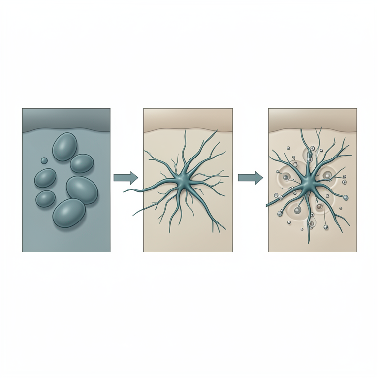
Pathogenesis Cycle of C. albicans
AI-generated conceptual illustration (Candida albicans fungi and human epithelial cells). It explains the idea and does not show study data.
View source evidence ↗Introduction
Fungal infections are a well-known indicator of compromised immunity. Numerous microorganisms with a wide range of traits make up microscopic fungus. A few numbers of them, particularly in immunocompromised patients, can infect humans and cause disease [1,40] . Due to their high associated mortality and low cost, opportunistic invasive fungal infections have drawn the attention of public health systems worldwide [2,3] . Fungal infections can impact the skin, nails, respiratory system, and internal organs. They are caused by a variety of fungi, including Cryptococcus, Candida, Aspergillus, and Dermatophytes. These infections are a serious health concern since they can range from superficial, moderate illnesses to severe systemic disorders. In order to effectively manage these infections, prompt identification and proper antifungal treatment are essential, particularly for those with weakened immune systems. The most prevalent fungi that infect people are species of Candida. These species are real opportunistic pathogens; they enter the circulation and deep tissues by taking advantage of the host's compromised state. There are over 100 species in the genus Candida, however only few of them are harmful to humans. A serious infection in immunocompromised hosts, candidiasis is caused by Candida albicans and infrequently by other species. Numerous pathogens, including Candida albicans, can cause fungal infections. They can damage the skin and mucosal surfaces and can infect the entire body. Up to 400,000 different types of systemic fungal infections are caused by species of Candida [4] . Approximately 70% of fungal infections worldwide are caused by Candida albicans, which is the most frequent causal agent of systemic and mucosal infections worldwide [5] . For the previous few decades, it has been the main cause of invasive infections that can be fatal.
The death rate is about 40% even with treatment, particularly in hospital settings [6,7] . There are various morphological forms of Candida albicans, including blastospores, pseudohyphae, and hyphae. By budding, bacterospores divide asexually [8] . On the surface of the blastospore, new cell material forms during that process. The new bud, which starts the growth phase, develops from a tiny, carefully chosen blastospore and is typically found away from the location of a birth scar. Cells divide when the growth phase is over, with the daughter cell forming a partition to keep itself apart from the parent cell [9] . Pseudohyphae are made up of chains of elongated yeast cells, while hyphae are made up of branched chains of tubular cells that do not narrow at the septation sites [10] . Higher than 37°C temperatures, an alkali pH, serum, and high CO 2 concentrations all promote filamentation [11] . In the same way, when N-acetylglucosamine (GlcNAc) is present, it is also improved by a deficiency of carbon and nitrogen [12] . Numerous transcription factors are among the complex regulatory network of signal pathways that are activated during this blastospore-to-hypha transition [13] . The primary distinction in composition between yeast and hyphae is that the hypha wall contains somewhat more chitin than yeast [14] . Chitin, protein, and glucan comprise the cell wall. Its function is to shield the cell from environmental stressors such osmotic shifts, dehydration, and temperature swings as well as from the host's immune system [15,16] . Sterols are found in the cell membrane of fungus and are unique to them. It is produced on lipid bodies and the endoplasmic reticulum [17] . There is a phospholipid bilayer in the cell membrane that contains proteins that function as receptors as well as those that are involved in signal transduction and transport [18] . Candida albicans employs amino acids as a supply of nitrogen and glucose as a source of carbon in its metabolism [19] . This study's main goals are to compile the most recent information on Candida albicans virulence factors and investigate the connection between these factors and infection symptoms.
Characteristics of
Characteristics of
Various morphogenesis
Although Candida albicans is a single type of fungus, it can take on a variety of shapes or morphologies, such as:
1. Form of yeast: an oval-shaped single-celled organism that procreates by budding.
Pseudohyphal form: A multicellular filamentous form
with a yeast cell-like chain resemblance.
Hyphal form:
A real filamentous form with lengthy filaments that branch out. 4. Chlamydospore form: A resting cell shape with robust walls that can withstand abrasive environments.
Apart from these physical variations, Candida albicans has the ability to transition between distinct phenotypic states, such as:
1. White form: The morphology of a smooth, white colony.
Opaque form:
The morphology of a rough, grayishwhite colony. 3. The GUT: (Gastrointestinal tract) form is a unique variation designed to endure in the GI system. 4. Because of its ability to adapt to varied habitats and host niches, Candida albicans can exist in a variety of forms and states, which increases its virulence and capacity to cause disease.
Pathogenesis Virulence factors of Candida albicans
A number of virulence factors that Candida albicans carries enhance its capacity to spread infection and disease: Because of these virulence characteristics, Candida albicans is an effective opportunistic pathogen that may colonize, invade, and survive in host tissues. Because of its virulence characteristics, Candida actively contributes to the pathophysiology of infection onset and progression. An infection begins when colonization occurs, which is caused by one set of virulence factors, and spreads with the aid of another group [20] .
Flexibility in transitioning between several morphogenic stages
The variety of cellular development states that Candida albicans exhibits aids in its establishment and persistence in many mammalian tissues. Different filamentous or yeastlike morphologies, colony characteristics, and unique genetic profiles are linked to these cell phenotypic variances [21] . The capacity of Candida albicans to transform its shape between the yeast and hyphal forms is particularly significant in its pathogenic life cycle, as it allows the fungus to penetrate mucosal barriers and cause invasive illness. Yeast, commonly regarded as the commensal form, has the ability to colonize superficial commensal habitats [22,23] . On the other hand, the hyphal form is generally considered to be the invasive form of the fungus, which permits Candida albicans to pierce host barriers and enter deeply seated tissues [24] . The morphological state of Candida albicans is influenced by many environmental stimuli, such as pH, host temperature, food availability, and quorum sensing mechanisms [25][26][27][28] . It is noteworthy that several anatomical regions in the mouse gut contain distinct hyphal morphotypes and C. albicans yeast [29] . The fact that nonfilamentous C. albicans strains are avirulent illustrates the significance of the morphogenic shift from yeast to hyphae [30] . The tissue's capacity to manage infection is correlated with C. albicans' in vivo filament production during systemic candidiasis in mice. As a result, during systemic candidiasis, C. albicans forms filaments in the mouse kidney, but neither the liver nor the spleen exhibit filamentation. This tissue-specific propensity to filament is associated with the liver's and spleen's ability to control the infection, in contrast to the kidney, which is unable to control fungal proliferation and gradually loses function [31] .
Adhesion, invasion, and host cell damage
The variety of surfaces that Candida albicans can colonize, from host mucosal tissues to medical devices and tools, emphasizes the significance of adhesion and invasion elements for the pathogen's ability to succeed. The yeast cells of Candida albicans attach themselves to the epithelium and/or endothelium in tissue, causing hyphal extension and subsequent active host cell penetration. Members of the Als and Hwp1 families' adhesin and invasin mediate the process [32][33][34] . Fungal ligands interact with epithelial cells during mucosal infection, causing induced endocytosis and active penetration by Candida albicans. Active penetration during systemic infection can open blood arteries, allowing fungal cells to travel to distant body locations. After then, colonization and the spread of illness are started by endothelium penetration [35] . The adhesins and invasins Als3 and Ssa1 bind host cell N-cadherin on endothelial cells and E-cadherin on oral epithelial cells, thereby mediating induced endocytosis [36] . Developmentally down-regulated protein 9 (NEDD9) signaling and platelet-derived growth factor BB (PDGF BB) are activated in oral epithelial cells through induced endocytosis [37] . Hyphales that are formed after Candida albicans adheres to epithelial cells are able to aggressively penetrate the plasma membrane of these cells [38, 39] . The expression of hypha-associated proteins, which are known to cause harm and activate the immune system, coincides with the development of hyphae [64] . It has recently been demonstrated that Candidalysin, a recently identified peptide toxin generated from hyphae, is a key factor in Candida albicans pathogenicity. The Ece1p protein is encoded by ECE1, which is expressed by hyphal filaments. Kex2p and Kex1p metabolize Ece1p to produce mature candidalysin, which is subsequently secreted. High quantities of candidalysin cause membrane degradation by interacting with cell membranes to produce pore-like structures [41, 42] . Rapid necrotic as opposed to apoptotic cell death is the outcome of the oxidative stress and subsequent calcium influx that are caused in the host cells [43] . Albumin, which functions as an anti-toxin through hydrophobic interactions, neutralizes the toxin, perhaps providing an immunological defense against chronic candidalysin damage [44] .
Stress resistance
Differential stress resistance responses must be induced by Candida albicans in response to the numerous stresses that it faces from host immune cells and varied cues from its surroundings. For instance, C. albicans has six Superoxide Dismutases (SODs) that help detoxify Reactive Oxygen Species (ROS) by converting O 2 -into molecular oxygen and hydrogen peroxide, allowing the fungus to evade oxidative death by immune cells [56] . As a result, C. albicans strains lacking Sod had lower pathogenicity [57] . Attenuating oxidative stress is aided by additional fungal antioxidant proteins and DNA damage repair genes [58,59] . Additional stressors include treatment with antifungal drugs, nitrosative stress, temperature and osmolarity variations, and changes in pH [60,61,62] . Heat shock proteins (Hsps), especially Hsp90, regulate a number of the fungal stress responses.
Stabilizing various signal transducers is the function of Hsp90, an ATP-dependent molecular chaperone that is widely distributed and conserved. Drug resistance and fungal pathogenicity are made possible by C. albicans Hsp90. The stress-activated protein kinase Hog1/Sty1, the mitogen-activated kinase Mck1, and/or the protein phosphatase calcineurin are all modulated to produce this effect [63] .
Immune evasion by masking cell wall components
The identification of fungal Pathogen-Associated Molecular Patterns (PAMPs) by host epithelial and immune cell Pattern Recognition Receptors (PRRs) is the initial stage in the formation of an immunological response to Candida albicans. Chitin, β-glucan, and mannoproteins make up the cell wall of Candida albicans.
References
48 in the original paper- Brown GD, Denning DW, Gow NA, Levitz SM, Netea MG, White TC. Hidden killers: Human fungal infections. Sci Transl Med. 2012;4(165):165rv13.
- Drgona L, Khachatryan A, Stephens J, Charbonneau C, Kantecki M, Haider S, et al. Clinical and economic burden of invasive fungal diseases in Europe: focus on pre-emptive and empirical treatment of Aspergillus and Candida species. Eur J Clin Microbiol Infect Dis. 2014;33(1):7-21.
- Menzin J, Meyers JL, Friedman M, Korn JR, Perfect JR, Langston AA, et al. The economic costs to United States hospitals of invasive fungal infections in transplant patients. Am J Infect Control. 2011;39(4):e15-e20.
- Mukaremera L, Lee KK, Mora-Montes HM, Gow NAR. Candida albicans yeast, pseudohyphal, and hyphal morphogenesis differentially affects immune recognition. Front Immunol. 2017;8:629.
- Morad HOJ, Wild AM, Wiehr S, Davies G, Maurer A, Pichler BJ, et al. Pre-clinical imaging of invasive candidiasis using ImmunoPET/MR. Front Microbiol. 2018;9:1996.
- Chen H, Zhou X, Ren B, Cheng L. The regulation of hyphae growth in Candida albicans. Virulence. 2020;11(1):337-348.
- Basmaciyan L, Bon F, Paradis T, Lapaquette P, Dalle F. Candida albicans interactions with the host: Crossing the intestinal epithelial barrier. Tissue Barriers. 2019;7(2):1612661.
- Molero G, Díez-Orejas R, Navarro-García F, Monteoliva L, Pla J, Gil C, et al. Candida albicans: genetics, dimorphism and pathogenicity. Int Microbiol. 1998;1(2):95-106.
- Kornitzer D. Regulation of Candida albicans hyphal morphogenesis by endogenous signals. J Fungi (Basel). 2019;5(1):21.
- Basso V, d'Enfert C, Znaidi S, Bachellier-Bassi S. From genes to networks: the regulatory circuitry controlling Candida albicans morphogenesis. Curr Top Microbiol Immunol. 2019;422:61-99.
- Kornitzer D. Regulation of Candida albicans hyphal morphogenesis by endogenous signals. J Fungi (Basel). 2019;5(1):21.
- Garcia-Rubio R, de Oliveira HC, Rivera J, TrevijanoContador N. The fungal cell wall: Candida, Cryptococcus, and Aspergillus species. Front Microbiol. 2020;10:2993.
- Höfs S, Mogavero S, Hube B. Interaction of Candida albicans with host cells: virulence factors, host defense, escape strategies, and the microbiota. J Microbiol (Seoul). 2016;54(3):149-169.
- Ciurea CN, Kosovski IB, Mare AD, Toma F, PinteaSimon IA, et al. Candida and Candidiasis opportunism versus pathogenicity: a review of the virulence traits. Microorganisms. 2020;8(6):857.
- Jordá T, Puig S. Regulation of ergosterol biosynthesis in Saccharomyces cerevisiae. Genes. 2020;11(7):795.
- Cho I, Jackson MR, Swift J. Roles of cross-membrane transport and signaling in the maintenance of cellular homeostasis. Cell Mol Bioeng. 2016;9:234-246.
- Viana R, Dias O, Lagoa D, Galocha M, Rocha I, Teixeira MC et al. Genome-scale metabolic model of the human pathogen Candida albicans: A promising platform for drug target prediction. J Fungi (Basel). 2020;6(3):171.
- Deorukhkar SC. Virulence traits contributing to pathogenicity of Candida species. J Microbiol Exp. 2017;5. DOI: 10.15406/jmen.2017.05.00140.
- Noble SM, Gianetti BA, Witchley JN. Candida albicans cell-type switching and functional plasticity in the mammalian host. Nat Rev Microbiol. 2017;15(2):96-108.
- Böhm L, Torsin S, Tint SH, Eckstein MT, Ludwig T, Pérez JC et al. The yeast form of the fungus Candida albicans promotes persistence in the gut of gnotobiotic mice. PLoS Pathog. 2017;13(10):e1006699.
- Pande K, Chen C, Noble SM. Passage through the mammalian gut triggers a phenotypic switch that promotes Candida albicans commensalism. Nat Genet. 2013;45(9):1088-1091.
- Desai JV. Candida albicans hyphae: From growth initiation to invasion. J Fungi (Basel). 2018;4(1):10.
- Lu Y, Su C, Liu H. Candida albicans hyphal initiation and elongation. Trends Microbiol. 2014;22(12):707714.
- Vylkova S, Carman AJ, Danhof HA, Collette JR, Zhou H, Lorenz MC et al. The fungal pathogen Candida albicans autoinduces hyphal morphogenesis by raising extracellular pH. mBio. 2011;2(3):e00055-e11.
- Flanagan PR, Liu NN, Fitzpatrick DJ, Hokamp K, Köhler JR, Moran GP. The Candida albicans TORactivating GTPases Gtr1 and Rhb1 coregulate starvation responses and biofilm formation. mSphere. 2017;2(6):e00477-17.
- Lindsay AK, Deveau A, Piispanen AE, Hogan DA. Farnesol and cyclic AMP signaling effects on the hypha-to-yeast transition in Candida albicans. Eukaryot Cell. 2012;11(10):1219-1225.
- Witchley JN, Penumetcha P, Abon NV, Woolford CA, Mitchell AP, Noble SM et al. Candida albicans morphogenesis programs control the balance between gut commensalism and invasive infection. Cell Host Microbe. 2019;25(3):432-443.e6.
- Lo HJ, Köhler JR, DiDomenico B, Loebenberg D, Cacciapuoti A, Fink GR et al. Nonfilamentous C. albicans mutants are avirulent. Cell. 1997;90(5):939949.
- Lionakis MS, Lim JK, Lee CC, Murphy PM. Organspecific innate immune responses in a mouse model of invasive candidiasis. J Innate Immun. 2011;3(2):180199.
- Liu Y, Filler SG. Candida albicans Als3, a multifunctional adhesin and invasin. Eukaryot Cell. 2011;10(2):168-173.
- Nobile CJ, Nett JE, Andes DR, Mitchell AP. Function of Candida albicans adhesin Hwp1 in biofilm formation. Eukaryot Cell. 2006;5(10):1604-1610.
- Swidergall M, Filler SG. Oropharyngeal candidiasis: fungal invasion and epithelial cell responses. PLoS Pathog. 2017;13(1):e1006056.
- Filler SG, Swerdloff JN, Hobbs C, Luckett PM. Penetration and damage of endothelial cells by Candida albicans. Infect Immun. 1995;63(3):976-983.
- Phan QT, Myers CL, Fu Y, Sheppard DC, Yeaman MR, Welch WH, et al. Als3 is a Candida albicans invasin that binds to cadherins and induces endocytosis by host cells. PLoS Biol. 2007;5(3):e64.
- Liu Y, Shetty AC, Schwartz JA, Bradford LL, Xu W, Phan QT, et al. New signaling pathways govern the host response to C. albicans infection in various niches. Genome Res. 2015;25(5):679-689.
- McCall AD, Pathirana RU, Prabhakar A, Cullen PJ, Edgerton M. Candida albicans biofilm development is governed by cooperative attachment and adhesion maintenance proteins. NPJ Biofilms Microbiomes. 2019;5(1):21.
- Wächtler B, Wilson D, Haedicke K, Dalle F, Hube B. From attachment to damage: defined genes of Candida albicans mediate adhesion, invasion and damage during interaction with oral epithelial cells. PLoS One. 2011;6(2):e17046.
- Pal M, Hofmeister M, Gutama KP, Paula CR, Leite DP Jr. Growing role of Candida albicans as an important cause of nosocomial infection. J Adv Microbiol Res. 2022;3(1):47-52.
- Moyes DL, Wilson D, Richardson JP, Mogavero S, Tang SX, Wernecke J, et al. Candidalysin is a fungal peptide toxin critical for mucosal infection. Nature. 2016;532(7597):64-68.
- Naglik JR, Gaffen SL, Hube B. Candidalysin: discovery and function in Candida albicans infections. Curr Opin Microbiol. 2019;52:100-109.
- Blagojevic M, Camilli G, Maxson M, Hube B, Moyes DL, Richardson JP, et al. Candidalysin triggers epithelial cellular stresses that induce necrotic death. Cell Microbiol. 2021;23(10):e13371.
- Austermeier S, Pekmezović M, Porschitz P, Lee S, Kichik N, Moyes DL, et al. Albumin neutralizes hydrophobic toxins and modulates Candida albicans pathogenicity. mBio. 2021;12(3):e0053121.
- Frohner IE, Bourgeois C, Yatsyk K, Majer O, Kuchler K. Candida albicans cell surface superoxide dismutases degrade host-derived reactive oxygen species to escape innate immune surveillance. Mol Microbiol. 2009;71(1):240-252.
- Hwang CS, Rhie GE, Oh JH, Huh WK, Yim HS, Kang SO et al. Copper-and zinc-containing superoxide dismutase (Cu/ZnSOD) is required for the protection of Candida albicans against oxidative stresses and the expression of its full virulence. Microbiology (Reading, England). 2002;148(Pt 11):3705-3713.
- Loll-Krippleber R, d'Enfert C, Feri A, Diogo D, Perin A, Marcet-Houben M, et al. A study of the DNA damage checkpoint in Candida albicans: uncoupling of the functions of Rad53 in DNA repair, cell cycle regulation and genotoxic stress-induced polarized growth. Mol Microbiol. 2014;91(3):452-471.
- Smith DA, Nicholls S, Morgan BA, Brown AJ, Quinn J. A conserved stress-activated protein kinase regulates a core stress response in the human pathogen Candida albicans. Mol Biol Cell. 2004;15(9):4179-4190.
- Danhof HA, Vylkova S, Vesely EM, Ford AE, Gonzalez-Garay M, Lorenz MC et al. Robust extracellular pH modulation by Candida albicans during growth in carboxylic acids. mBio. 2016;7(6):e01646-16.
- Enjalbert B, Smith DA, Cornell MJ, Alam I, Nicholls S, Brown AJ, et al. Role of the Hog1 stress-activated protein kinase in the global transcriptional response to stress in the fungal pathogen Candida albicans. Mol Biol Cell. 2006;17(2):1018-1032.
Cited by 3
From OpenAlex, OpenCitations and reference lists read by Scinovex
- Fungal Phagocytosis in the Absence of Oxidative Stress Response Proteins ↗
Minerva Nájera-Martínez, Armando Vega López and Israel Lara Vega · Biochemistry · 2026
Reactive oxygen species (ROS) are crucial in cell metabolism and homeostasis. ROS can damage biomolecules and organelles. To date, the paradigm is that ROS are integral components of cellular signaling and various cellular functions. The widely used concept of “oxidative stress” has been refined into “oxidative distress,” wherein ROS reach damaging levels, and “oxidative eustress,” wherein ROS do not cause…
❝ 1PDF - Internal Transcribed Spacer (ITS) and Hyphal Wall Protein 1 (HWP1): From Species Identification to Infection Diagnosis of Candida ↗
Moaamal Khalaf Hadi, Ali Abdul Hussein S. AL‐Janabi, Hameedah Hadi Abdulwahid · Microbiology and Biotechnology Letters · 2026
The Candida genus has several pathogenic species that cause candidiasis in the human body.Identification and differentiation of these species requires precise methods.Such methods can also be used for diagnosing candidiasis by detecting the causative agents.Genetic variation in the internal transcribed spacer (ITS) region and hyphal wall protein 1 (HWP1), cell wall components of Candida, can be used for the diagnosis…
❝ 0PDF - The relationship between candida spp. isolates and immune response in patients with respiratory tract infections ↗
Shaymaa Amer Jalal, Batol Imran Dheeb · AIP conference proceedings · 2026
❝ 0