
Research article · 2024 · Full text
International Journal of Veterinary Sciences and Animal HusbandryMolecular study of Hydatid Cyst isolated from human and domestic animal in Babylon province, Iraq
Volume 9(6) · Pages 199–203 · DOI: 10.22271/veterinary.2024.v9.i6d.1864 ↗
Research areasParasitic infections in humans and animalsParasitic Infections and DiagnosticsCongenital Anomalies and Fetal Surgery
Abstract
Author-writtenThis study included collecting 50 cyst samples of human from different areas of in Babylon province in Iraq. The human hydatid cysts collected from patients infected by hydatidosis after surgical operation from hospitals and labratory of babylon province. Through a period from September 2023to June2024. Traditional methodes and polymerase chain reaction testing have been used to detected of genotyping of hydatid cyst in human. According to the geographical areas in human infections were highes in Al-mahaweel district which were (30%) and the lowest infection rate was in Al-hashimia district which was (4%), There was highly significant differences between the regions when analysis statistically at the probility level (p
Summary
Source-checkedPlain-language reading of the full paper, prepared with AI and checked against the text. Each point links to the authors' own words.
Why it matters
- Hydatidosis is a zoonotic disease caused by the larval stage of Echinococcus granulosus, which creates significant medical, veterinary, and economic burdens for humans and domestic animals. View evidence ↗
- Iraq is recognized as one of the countries severely impacted by this endemic condition. View evidence ↗
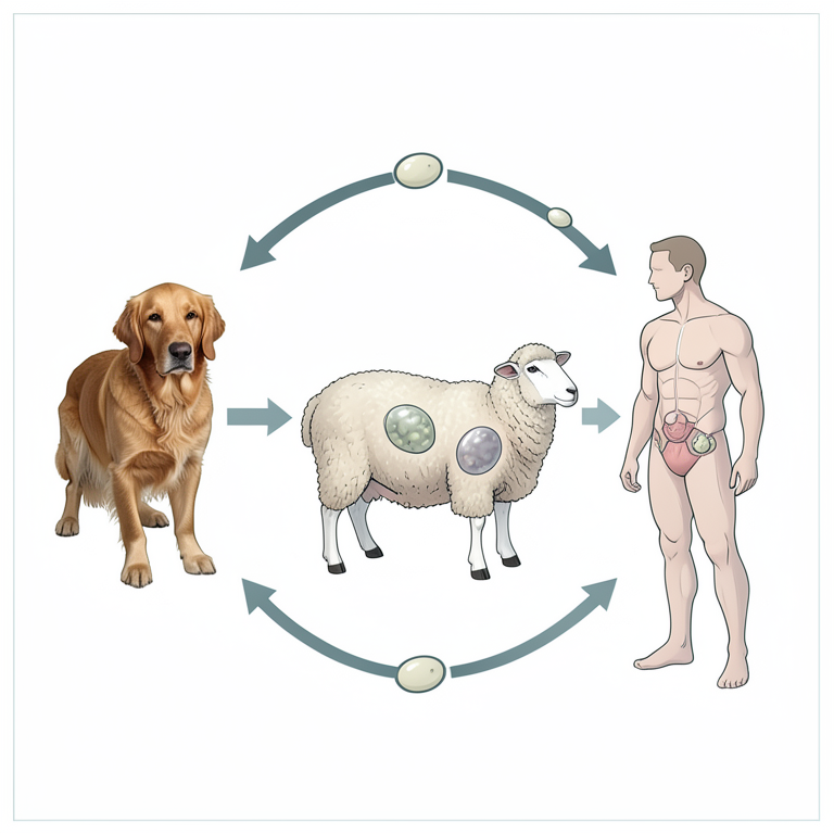
- Human infection occurs when eggs excreted in the feces of infected dogs contaminate food, water, or the environment and are subsequently ingested. View evidence ↗
The question
- The study aimed to determine the prevalence of hydatid cyst infections in humans in the Babylon province using molecular analysis and PCR assays. View evidence ↗
What they did
- The researchers collected 50 human cyst samples from various regions of Babylon province, Iraq, between September 2023 and June 2024. View evidence ↗
- Genotyping was performed using traditional methods and PCR targeting the mitochondrial cytochrome c oxidase subunit 1 (COX1) gene. View evidence ↗
- Of the samples sent for sequencing, six provided good sequences, and three human samples were subjected to bioinformatics analysis. View evidence ↗
What they found
- Geographically, the highest prevalence of hydatid cysts was found in the Al-mahaweel district (30%), while the lowest was in the Al-hashimia district (4%). View evidence ↗
- The highest infection rate occurred in the 30-40 year age group (44%), significantly higher than in those over 40 years (20%). View evidence ↗
- Females had a higher infection rate (62%) compared to males (38%), although this difference was not statistically significant. View evidence ↗
- Rapid PCR genotyping identified eight genotypes, with G6 being the dominant strain at 32%. View evidence ↗
- Bioinformatics analysis of three human samples confirmed the presence of Echinococcus granulosus. View evidence ↗
- The 50 isolated cysts were found in the liver, lung, and thoracic areas, appearing white, soft, spherical or ovoid, and ranging in size from 5cm to 12cm. View evidence ↗
What it means
- The authors concluded that hydatidosis is a widespread zoonotic disease in Iraq with different strains affecting humans and animals. View evidence ↗
- The authors suggest that females are more susceptible to infection due to direct contact with pathogens while handling and cooking meat. View evidence ↗
Visual summary
3 visualsIllustrations explain the paper; charts are replotted from the paper's own table values.
Transmission Cycle of E. granulosus
AI-generated conceptual illustration (Dogs, sheep, and humans). It explains the idea and does not show study data.
View source evidence ↗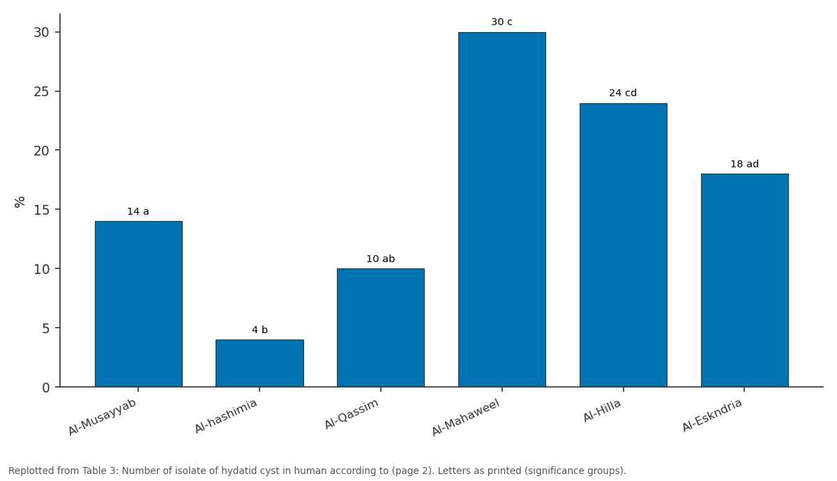
%: Al-Musayyab vs Al-hashimia vs Al-Qassim vs Al-Mahaweel vs Al-Hilla
Replotted from Table 3 (page 2) of the original paper. Every bar is the paper's own table value.
View source evidence ↗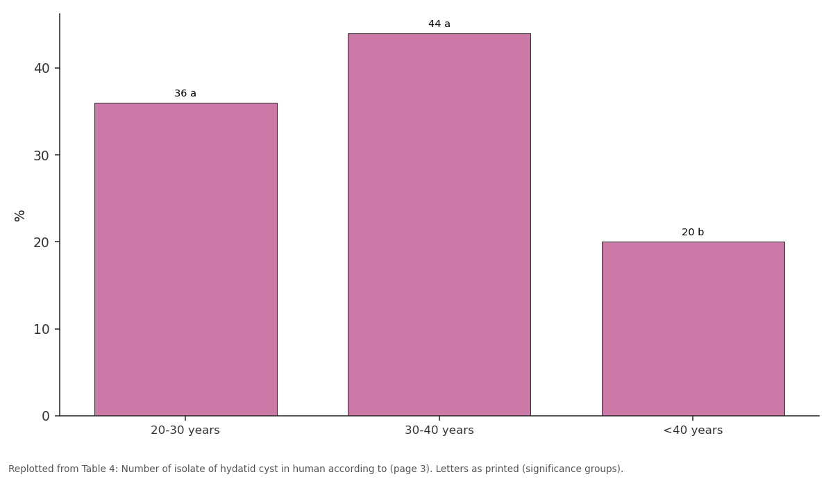
%: 20-30 years vs 30-40 years vs <40 years
Replotted from Table 4 (page 3) of the original paper. Every bar is the paper's own table value.
View source evidence ↗Figures & tables
From the original paper Table 1
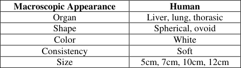 Table 2
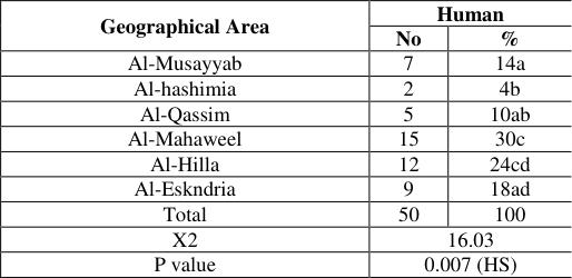 Table 3
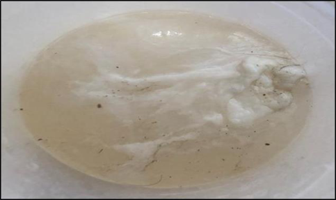 Figure 1
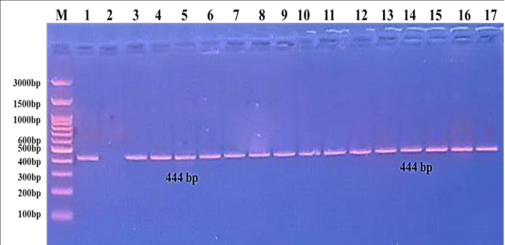 Figure 2
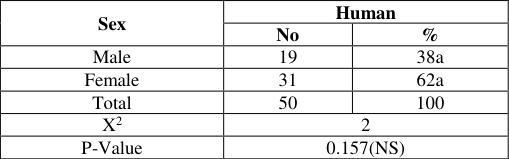 Table 5
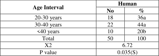 Table 4
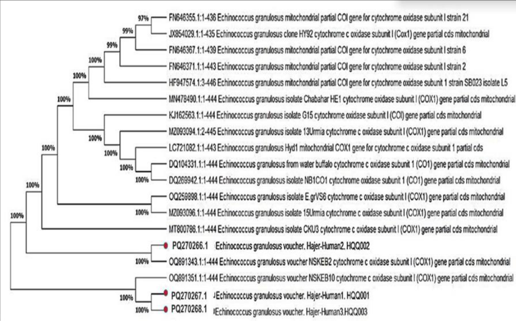 Figure 3
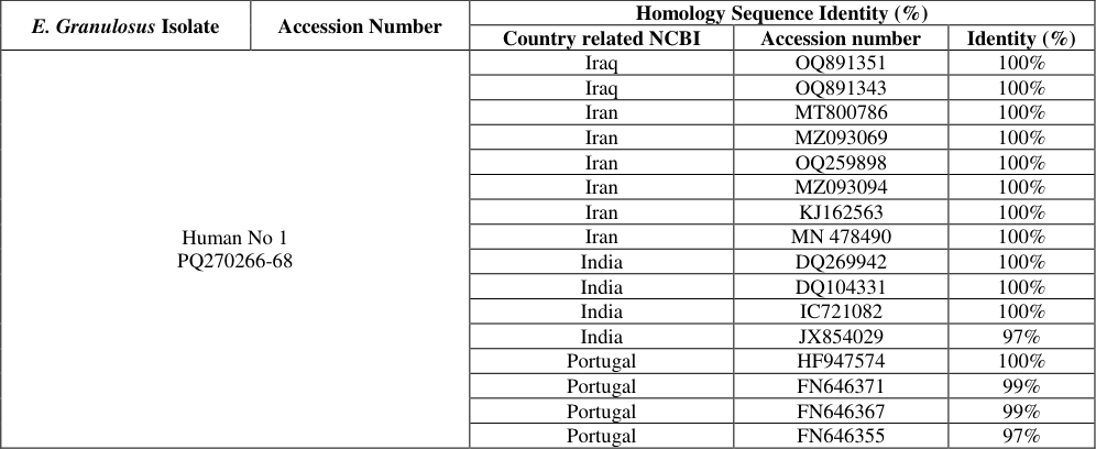 Table 6
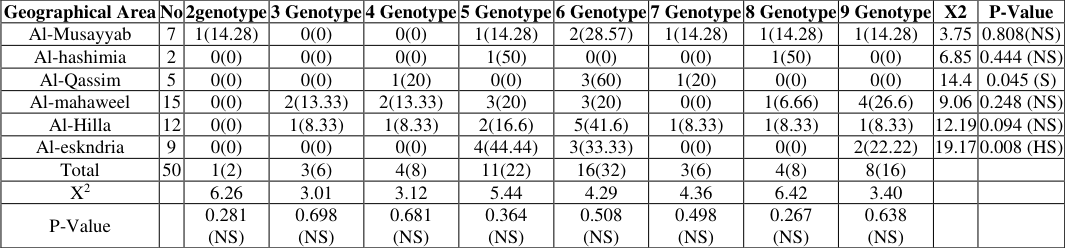 Table 7
Introduction
Echinococcus granulosus larval stage causes hydatidosis, a zoonotic disease that significantly impacts both human and domestic animal health, leading to various medical, veterinary, and economic challenges. Iraq is identified as one of the countries severely affected by this endemic condition (ALhadidi, Al-Hamairy et al. 2022) [6] . Hydatid disease is prevalent globally, largely due to the high population of stray dogs infected with adult worms. This disease is referred to by several names, including Hydatidosis, Echinococcosis, and Cystic Echinococcosis. Carnivorous animals serve as the definitive hosts for the parasite, with the canine family such as dogs, hyenas, wolves, leopards, and other carnivorous species playing a key role. Herbivorous animals, including sheep, cows, camels, buffaloes, and horses, act as intermediate hosts. The disease primarily affects organs such as the liver, lungs, spleen, brain, and other muscles in both humans and intermediate hosts, sparing areas like hair and nails (Al-Nuaimi and Al-Hassani 2023) [4] . Humans can become infected through contact with infected dogs that carry adult E. granulosus in their intestines, leading to the excretion of eggs in feces. Consequently, humans and other intermediate hosts may ingest eggs that contaminate food, water, or the environment. While the disease is often asymptomatic, it can present as a complicated cyst, with the most common complications being compression or rupture of pericystic structures. Additionally, hypersensitivity reactions may occur due to circulating immune complexes or activation of the alternative complement pathway, potentially resulting in glomerulonephritis or an anaphylactoid reaction. Furthermore, the cysts can become superinfected with bacterial or fungal pathogens (Baraak 2014) [8] . According to the WHO (2021), the incidence of human hydatidosis in endemic areas can exceed significant thresholds.
The incidence rates of human hydatidosis in endemic areas can exceed 50 cases per 100,000 individuals annually, with prevalence figures ranging from 5% to 10% in certain regions of Peru, Argentina, China, Central Asia, and East Africa. Globally, the prevalence of echinococcosis varies between 9% and 66%. The cysts associated with this condition grow slowly, filled with clear fluid and numerous protoscolices. On average, they increase in size by about 1 cm each year and can attain diameters of up to 20 cm. These cysts primarily develop in the liver (70%) and lungs (20%), while the remaining 10% can manifest in various other locations, including the brain, skeletal muscles, kidneys, bones, heart wall, and eye socket.
Materials and Methods:
The research involved the collection of samples from 50 individuals in the Babylon province. These samples underwent molecular analysis using PCR assays to determine the prevalence of hydatid cyst infections in humans.
Sample Collection:
Samples were gathered from 50 individuals of varying ages and both sexes across different regions of the Babylon province, including Al-Musayyab, Al-Qassim, Al-Mahaweli, and Al-Hilla, during the period from September 2023 to June 2024.
Primers Utilized in This Study:
The primers and their sequences, along with the product size, pertain to the mitochondrial subunit 1 of the cytochrome c oxidase 1 (cox1) gene (refer to Table 1). Elyasi H, Golmohammadi R, (2020) [9] . R TAAAGAAAGAACATAATGAAAATG
| No | (COX1) Primer sequence 5´ to 3´ | Amplicon size | Annealing TM | Ref |
|---|---|---|---|---|
| F | Ttttttgggcatcctgaggtttat | 444 Bp | 54 | Elyasi H, Golmohammadi R, (2020) [9]. |
| R | TAAAGAAAGAACATAATGAAAATG |
Table text extracted from the original PDF; superscript letters are shown inline. The original table image is below.
View original table image
Results and Discussion
Results and Discussion
Macroscopic appearance in human
The present study identified from 50 patients the isolated parasitic cysts 50 from different organs (Liver, lung, thorasic), while The characteristics macroscopic appearance of hydatid cyst sizes Large 12cm, Medium 10cm,Small 5cm and shape cyst Spherical, ovoid, color cyst White, Consistency cyst Soft as shown in Table 2. These results in agreement with results of (Issa et al. 2022; Mustafa, et al., 2019) [16,19] . The main reasons that control to size or diameter of hydatid cysts attributed to host species, physiological and histological nature of infected tissue, site of cystic development in organ (superficial or deep), host immune response that restricted cystic growth, early detection of the cyst and may be strains variation that infects inappropriate host.
| Macroscopic Appearance | Human |
|---|---|
| Organ | Liver, lung, thorasic |
| Shape | Spherical, ovoid |
| Color | White |
| Consistency | Soft |
| Size | 5cm, 7cm, 10cm, 12cm |
Table text extracted from the original PDF; superscript letters are shown inline. The original table image is below.
View original table image
Number of isolate of hydatid cyst 3.3.1 Number of isolate of hydatid cyst in human according to the geographical area
The prevalence of infections in Hydatid Cysts this study was highest in Al-mahaweel districts which were (30c%). while the lower prevalence of Hydatid Cysts Al-hashimia district which was (4b%). The statistical analysis reveals that there was significant (p>0.05) difference in the prevalence of parasite among geographical areas in (Table 3-2). The current study agreement with study in Punjab (32.45%) Muqaddas, Hira, et al. (2019) [19] . It was nearby the percentage that recorded in Karbala, Iraq (2.54%), India (10%) and Iran (3.84%) by Al-Gharrawi, (2008) [2] consecutively. The curreent study disagreement with rate infection Hydatid cyst was (54.2%) in Duhok, the present results contradict other studies in Iran as they reported higher infection rates, with 61% and 89.77%, respectively. These results are disagreement with Salem et al., 2011) in Mauritania, (Al-Bosely 2013) [1] in Zakho (Kurdistan, Iraq), (Piccoli et al., 2013) in Romania and (Khalf et al., 2014) [14] in Baghdad (Iraq) as they reported rates of 76%, 90%, 92% and 47%, respectively (Table 3).
| Geographical Area | Human | — |
|---|---|---|
| No | % | |
| Al-Musayyab | 7 | 14a |
| Al-hashimia | 2 | 4b |
| Al-Qassim | 5 | 10ab |
| Al-Mahaweel | 15 | 30c |
| Al-Hilla | 12 | 24cd |
| Al-Eskndria | 9 | 18ad |
| Total | 50 | 100 |
| X2 | 16.03 | |
| P value | 0.007 (HS) |
Table text extracted from the original PDF; superscript letters are shown inline. The original table image is below.
View original table image
Number of isolate of hydatid cyst in human according to the age
In the present study the incidence of hydatid cysts was different according to age of the host. In human more prevalence rate (44a%) was recorded in (30-40 years) while little prevalence rate (20b%) was in ( > 40) years people with a Significant difference at p<0.05 as shown in (Table 3-3). These results in agreement with result of (Al-Marsomy 2021) [3] found to be the infection rate of hydatid cysts increases in the age group (31-40) years, while the lowest were at the age range (41-50) and (51-60) year. These result is disagreement with study that about (36.5%) of patients were at age 40 years (Fadhil and Baiee 2018) [10] . This finding disagreement with a previous study the highest rate of infection was found among the age class of 21-30 years (Issa et al. 2022) [16] , (Table 4).
| Age Interval | Human | — |
|---|---|---|
| No | % | |
| 20-30 years | 18 | 36a |
| 30-40 years | 22 | 44a |
| <40 years | 10 | 20b |
| Total | 50 | 100 |
| X2 | 6.72 | |
| P value | 0.035(S) |
Table text extracted from the original PDF; superscript letters are shown inline. The original table image is below.
View original table image
Number of isolate of hydatid cyst in human according to the sex
The result of infection with hydatidosis according to sex revealed that females were exposed to infection more than males in human with No Significant difference at P<0.05 (table 3-4). The results of the current agreement with study Al-Marsomy WA, (2021) [3] showed that the percentage of female infections (70%) was higher that of males (30%) and (Ismail et al., 2023) [15] . The findings also revealed that females are infected at a higher rate than males. The rate of infection in females (61%) more than males (39%).Compared to males Mohamed et al., 2021 [17] . may be due to females responsible for cooking and preparing food, which makes them in direct contact with the pathogens. These result is disagreement with studies (Gulsun, S et al., 2010, Bakal et al., 2015) [12,7] there were higher rate males than females Table 5.
| Sex | Human | — |
|---|---|---|
| No | % | |
| Male | 19 | 38a |
| Female | 31 | 62a |
| Total | 50 | 100 |
| X2 | 2 | |
| P-Value | 0.157(NS) |
Table text extracted from the original PDF; superscript letters are shown inline. The original table image is below.
View original table image
Sequencing Phylogenetic tree analysis in human
According to the gene sequence analysis only 3 samples gave good sequence that which subjected to bioinformatics analysis, and their result revealed that of analyzed samples were E. granulosus, that which recorded in gene bank under accession numbers of PQ270266, PQ270267, PQ270268 (Figure 3), (Table 6). In the present study, a total of 50 samples of human were liver and lung thorasicas were examined by rapid PCR detection, Hydatid Cyst, showed only eigher genotypes G2 and G9. The human strain G6 was the dominant 32% shown in Table 7.
| E. Granulosus Isolate | Accession Number | Homology Sequence Identity (%) | — | — |
|---|---|---|---|---|
| Country related NCBI | Accession number | Identity (%) | ||
| Human No 1 PQ270266-68 | Iraq | OQ891351 | 100% | |
| Iraq | OQ891343 | 100% | ||
| Iran | MT800786 | 100% | ||
| Iran | MZ093069 | 100% | ||
| Iran | OQ259898 | 100% | ||
| Iran | MZ093094 | 100% | ||
| Iran | KJ162563 | 100% | ||
| Iran | MN 478490 | 100% | ||
| India | DQ269942 | 100% | ||
| India | DQ104331 | 100% | ||
| India | IC721082 | 100% | ||
| India | JX854029 | 97% | ||
| Portugal | HF947574 | 100% | ||
| Portugal | FN646371 | 99% | ||
| Portugal | FN646367 | 99% | ||
| Portugal | FN646355 | 97% |
Table text extracted from the original PDF; superscript letters are shown inline. The original table image is below.
View original table image
These study current agreement with (Nikmanesh et al., 2014) [20] the isolates with G6 genotype was obtained from lung samples. Genotyping of Echinococcus granulosus Isolates from Human Clinical Samples in Iran, Tehran study disagreement with study (Gholami et al., 2012) [12] in Golestan province in Iran, of E. granulosus showed presence of G1 genotype in all 30 human samples. These study current disagreement with (Hedayati, et al., 2019) [13] study in Mazandaran Province, North of Iran founded G1 and G2-G3 genotypes, respectively the Human Cystic Echinococcosis.
The differences results between this study and others could be due to differences in animal management, nutrition, natural resistance, treatment programs, and local geo-climatic and international factors.
Conclusion
The study was concluded that the disease is zoontic and widespread in Iraq and the results showed that the different strain in human and animal. Females in humans and animals are more susceptible to infection, and the reason is due to the direct contact of women with the handling and cooking of meat. Infections in rural areas are more than in urban areas due to the spread of dogs, the main carrier of the disease
Figures and tables from the paper
References
18 in the original paper- Al-Gharrawi AK. Epidemiological study of hydatid disease in patients underwent surgical treatment in AlHussain General Hospital/Karbala. Karbala Sci Uni J. 2008;6(3):87-93.
- Al-Marsomy WA. Epidemiology of hydatid disease in Iraq: A study of hydatidosis patients in Baghdad Province. Indian Journal of Forensic Medicine & Toxicology. 2021;15(2):3525-3530.
- Al-Nuaimi NZK, Al-Hassani SJM. Molecular investigation of Echinococcus granulosus in infected patients in Babylon province. Pakistan Heart Journal. 2023;56(2):20-28.
- Al-Saeed ATM, Al-Mufty KSA. Human hydatidosis in Duhok-Kurdistan Region-North of Iraq. Med J Babylon. 2016;13:125-133.
- Alhadidi RM, Al-Hamairy AK, Altameme HJM. Gene sequencing of hydatid cysts isolated from human and sheep in Central Euphrates Provinces, Iraq. Journal of University of Babylon for Pure and Applied Sciences. 2022:67-75.
- Bakal U, Simsek S, Kazez A. Surgical and molecular evaluation of pediatric hydatid cyst cases in eastern Turkey. Korean J Parasitol. 2015;53(6):785.
- Elyasi H, Golmohammadi R. Genetic identification of Echinococcus granulosus isolates from domestic animals in Sabzevar, Northeast Iran. Iranian Journal of Parasitology. 2020;15(4):621-623. https://doi.org/10.18502/ijpa.v15i4.4883.
- Fadhil AA, Baiee HA. Epidemiological and clinical features of hydatid cyst disease in Babylon Province during the years 2010-2015. of University of Babylon for Pure and Applied Sciences. 2018;26(2):185199.
- Gholami Sh, Sosari M, Fakhar M, Sharif M, Daryani A, Hashemi M, et al. Molecular characterization of Echinococcus granulosus from hydatid cysts isolated from human and animals in Golestan Province, North of Iran. Iranian J Parasitol. 2012;7(4):8-16.
- Gulsun S, Cakabay B, Kandemır MN, Aslan S, Atalay BASA, Sogutcu N, et al. Retrospective analysis of echinococcosis in an endemic region of Turkey: a review of 193 cases. Iranian Journal of Parasitology. 2010;5(3):20.
- Hedayati Z, Daryani A, Sarvi S, Gholami S, Sharif M, Pirestani M, et al. Molecular genotyping of human cystic echinococcosis in Mazandaran Province, North of Iran. Iranian Journal of Parasitology. 2019;14(1):151.
- Khalf MS, AlTaie LH, AlFaham MA. The incidence of hydatid cyst in humans in Baghdad governorate. IOSR J Pharm Biol Sci. 2014;9:11-14.
- Ismail MA, Mohammed SJ, Hassan NH. The epidemiology of hydatid cysts liver diagnosis in Najaf governorate. AIP Conference Proceedings. 2023;2776(1):AIP Publishing.
- Issa AR, Arif SH, Mohammed AA, Santolamazza F, Santoro A, Mero WMS, et al. Insights into human cystic echinococcosis in the Kurdistan Region, Iraq: characteristics and molecular identification of cysts. Pathogens. 2022;11(4):408.
- Mohamed RJ, Alsaedi ANN, Taher JH. Epidemiology of hydatid disease in Najaf province/Iraq. Journal of Physics: Conference Series. 2021;1879(2):1.
- Muqaddas H, Arshad M, Ahmed H, Mehmood N, Khan A, Simsek S. Retrospective study of cystic echinococcosis (CE) based on hospital records from five major metropolitan cities of Pakistan. Acta Parasitol. 2019;64:866-872.
- Mustafa JY, Al-Azizz SA, Lazim AR. Molecular study of hydatid cysts in human, sheep and donkeys at Basra city, southern Iraq. Biochem Cell Arch. 2019;19(2).
- Nikmanesh B, Mirhendi H, Ghalavand Z, Alebouyeh M, Sharbatkhori M, Beigom EKIA, et al. Genotyping of Echinococcus granulosus isolates from human clinical samples based on sequencing of mitochondrial genes in Iran, Tehran. Iranian J Parasitol. 2014;9(1):20.