
Research article · 2024 · Full text
International Journal of Veterinary Sciences and Animal HusbandryHistomorphological study of esophagus in adult guinea pigs (Cavia porcellus)
Volume 9(6) · Pages 141–144 · DOI: 10.22271/veterinary.2024.v9.i6c.1850 ↗
Research areasCongenital heart defects researchDiet and metabolism studiesTissue Engineering and Regenerative Medicine
Abstract
Author-writtenThe research elucidated the histological and morphological characteristics of the esophagus during adulthood. A selection of fifteen healthy guinea pigs was obtained from the public market for this study. The animals were euthanized using ketamine at a dosage of 30 ml/kg body weight via intramuscular injection. The esophagus was examined topographically in situ. Morphologically, the esophagus is a muscular tube divided into three segments: cervical, thoracic, and abdominal. It extends from the oropharynx to the cardiac region of the stomach, where it connects at the distal end. Histological analysis revealed that the esophagus comprises four layers: mucosa, submucosa, muscularis, and adventitia or serosa, and it lacks glandular structures. This study enhances the understanding of both the gross and microscopic morphometrics of the esophagus in guinea pigs.
Summary
Source-checkedPlain-language reading of the full paper, prepared with AI and checked against the text. Each point links to the authors' own words.
Why it matters
- The esophagus acts as a narrow muscular tube that connects the oropharynx to the cardiac region of the stomach. View evidence ↗
- The keratinization of the esophageal epithelium varies by species and diet, which is important for the process of converting food into energy. View evidence ↗
- Studying the histomorphological features of the adult guinea pig's esophagus helps highlight the animal's importance in biomedical research. View evidence ↗
The question
- The study aims to examine and describe the histomorphological characteristics of the esophagus in adult guinea pigs. View evidence ↗
What they did
- The researchers used fifteen healthy adult guinea pigs obtained from a public market. View evidence ↗
- Animals were euthanized via intramuscular injection of ketamine at a dose of 30 ml/kg body weight. View evidence ↗
- Tissue samples were fixed in 10% neutral buffered formalin for two days, with the solution changed daily. View evidence ↗
- Histological analysis was performed using Hematoxylin Eosin, Masson's trichrome, PAS, and Alcian blue stains. View evidence ↗
What they found
- The esophagus of the adult guinea pig measured 12.53 cm in length and weighed 0.36 grams. View evidence ↗
- The esophagus is morphologically divided into three distinct segments: the cervical, thoracic, and abdominal regions. View evidence ↗
- The abdominal segment of the esophagus measured 3.20±0.06 cm in length and weighed 0.07±0.003 gm. View evidence ↗
- The esophagus consists of four histological layers, mucosa, submucosa, muscularis, and adventitia/serosa, and does not contain glandular structures. View evidence ↗
- The entire length of the esophagus is lined with keratinized stratified squamous epithelium. View evidence ↗
- The muscular layer (tunica muscularis) transitions from striated muscle in the cranial region to mixed fibers in the intermediate region, and finally to smooth fibers in the caudal region. View evidence ↗
- The muscularis consists of three layers: a thin outer longitudinal layer, a thick middle circular layer, and an inner longitudinal layer covering less than half the circumference. View evidence ↗
What it means
- The authors conclude that this study improves the understanding of the gross and microscopic morphometrics of the guinea pig esophagus. View evidence ↗
Visual summary
2 visualsIllustrations explain the paper; they do not show study data.
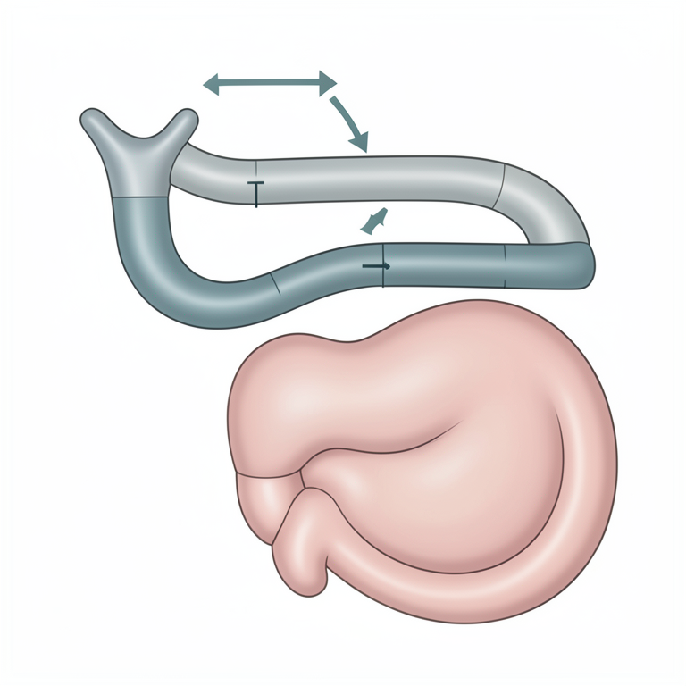
Gross Anatomy of the Guinea Pig Esophagus
AI-generated conceptual illustration (adult guinea pig (Cavia porcellus) esophagus). It explains the idea and does not show study data.
View source evidence ↗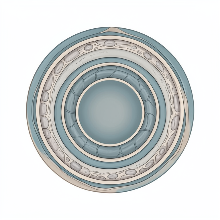
Histological Layers of the Esophagus
AI-generated conceptual illustration (adult guinea pig (Cavia porcellus) esophagus). It explains the idea and does not show study data.
View source evidence ↗Figures & tables
From the original paper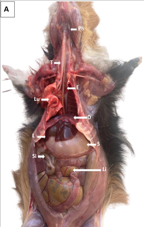 Figure 1
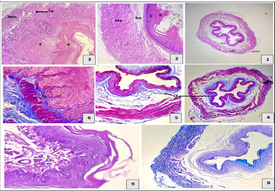 Figure 2
Introduction
The esophagus is a narrow, muscular conduit and serves to connect with cardiac region of the stomach in a linear fashion. It traverses from oropharynx to stomach cardiac sphincter. Consequently, it is categorized into three distinct macroscopic sections: cervical, thoracic, and abdominal. The esophagus leads to the fundus of the stomach through the cardiac section, which is lined histologically by a thick, stratified squamous epithelium. The keratinization of this epithelium is influenced by food and species diversity, playing a vital role in the conversion of food into the energy necessary for bodily functions. This study aims to elucidate occurring in these organs at this stage and to underscore the significance of the adult guinea pig as biomedical research by examining the histomorphological features of its esophagus.
Materials and Methods
Numbers of good health animals measuring 15.030±0.17 cm in length and weighing 66.87±0.50 g from the public market [3] . These animals were anesthesia by injection about 30 mg/kg bw ketamine. As soon as the esophagus was isolated from the abdominal cavity, they were carefully dissected, and measured for length, and weight. The study involved histological and histochemical analyses. The pieces were fixed for two days using 10% neutral buffered formalin, which was changed daily. The samples underwent histological and histochemical staining after successfully passing through routine histological sectioning with a thickness of 5 micrometers [4,5] . Hematoxylin Eosin and Masson's trichrome stains were used on the specimens to determine micromorphometric parameters and get a general histological characteristic. The esophageal were stained and identified using PAS and Alcian blue [6] .
Results and Discussion
Results and Discussion
Esophagus
Esophagus is a skeletal muscle tube that connects with cardiac orifice of stomach. It measures 12.53 cm in length and 0.36 grams in weight. Its color varies from white to gray. Approximately 12.53 by 0.16 cm and 0. 00g in weight, it varies in color from white to pink. The esophagus is divided into the neck can be divided into three part cervical, thoracic and abdominal (Fig. 1a,b). These results agree with [7] . The thoracic esophagus extends from the thoracic inlet dorsally. (Fig 1-a, b). who reported that the esophagus was situated ventral to ventral cervical muscles of the cats and dorsally to the trachea. On the posterior surface of the left lobe of the liver's esophageal groove, the short abdominal segment of the esophagus is located. measuring 3.20±0.06 cm in length and 0.07±0.003 gm in weight. At the base of the esophagus, it extended from the esophageal hiatus of the diaphragm to the gastroesophageal at the stomach cardiac orifice. It is surrounded by a thicker region of muscle fibers making a circular muscle known as the esophageal sphincter (Fig 1-B). These results agree with [8,9,12,13] . The histological description, it's has the mucosa, submucosa, muscularis, and adventitia or serosa formed the four tunics of the esophagus. This observation is similar to that made by [15] , which noted that the rabbit esophageal lumen appears star-shaped from the cross-section. This mucosa consists of the epithelium, lamina propria, and muscularis mucosae. Throughout the length of the esophagus, the epithelial lining is characterized by keratinized stratified squamous epithelium. (Fig 2,3). This epithelium is interspersed with a well-developed lamina propria, which consists of relatively dense connective tissue, scattered lymphocytes, and vascular structures (Fig 236). This observation is consistent with the findings reported in reference [10] . These findings contrast with those reported by [16] , which indicated that the lamina muscularis in grey mongooses was thin and comprised scattered, interrupted bundles of skeletal muscle in the cervical area. Submucosa have no esophageal glands and characteristic via loose connective tissue abundant in collagen fibers, a small amount of elastic fibers, and the presence of blood vessels and lymphocytes (Fig 2-A; B 1, 2, 3). This observation, which noted the presence of some elastic and collagen fibers but no distinct submucosal glands, aligned with the findings of [10] . In the cranial region, the tunica muscularis was composed of striated muscle fibers that transitioned to mixed fibers in the intermediate region and smooth fibers in the caudal region. It consisted of three layers arranged from outermost to innermost: a thinner outer layer of longitudinally oriented muscle fibers, a thicker middle layer with circularly arranged muscle fibers, and an internal layer of longitudinal muscle fibers that covered less than half of the esophageal circumference, consistent with [16] . The adventitia was loose connective tissue that was highly vascularized, it covered by simple squamous epithelium which called serosa (Fig 2-1), similar to the observations of [9,16] . (1) Lumen (L), keratin (K), mucosa (M), submucosa (Sm), muscularis (Me), and adventia (Ad). X4 H&E stain.
(2) Keratin (K), stratified squamous epithelium (E), submucosa (Sm), and muscularis (Me). X10 H&E stain.
References
19 in the original paper- Erman L, Mingli W, Liping W. A histomorphological study on the digestive system of adult hedgehog. J Forestry Res. 1998;9: 111-4.
- Kadhim KK. Histomorphology and histochemical study of esophagus and stomach in grey mongoose (Herpestes edwardsii) in Iraq. Indian J Nat Sci. 2019;9(52):1645875.
- Vdoviaková K, Petrovová E, Maloveská M, Krešáková L, Teleky J, Elias MZJ, Petrášová D. Surgical anatomy of the gastrointestinal tract and its vasculature in the laboratory rat. Gastroenterol Res Pract. 2016;2016: 1-9.
- Amin AA, Atiyah AA. Evaluation of general anesthesia induced by propofol, ketamine protocol in rabbits premedicated with diazepam. Iraqi J Vet Med. 2014;38(2):100-7.
- Bonhomme V, Staquet C, Montupil J, Defresne A, Kirsch M, Martial C, Gosseries O. General anesthesia: a probe to explore consciousness. Front Syst Neurosci. 2019;13: 36.
- Luna LG. Manual of histologic staining methods of armed forces institute of pathology. 3rd ed. New York, U.S.A.: McGraw-Hill; 1968. p. 123.
- Al-Harith HH, Suhaib AH. Histological study of small intestine during twenty-day age in Iraqi pigeon (Columba livia). Plant Arch. 2020;20(2):5323-7.
- AL-Taai SAH, Nsaif RH, Almayahi MS. Histomorphological study of esophagus in squirrel (Sciurus anomalus). Biochem Cell Arch. 2021;21(1):1391-4.
- Al-Shreefy MN, Al-Taai SAH. Histomorphological and histochemical study of esophagus and stomach in neonate guinea pigs (Cavia porcellus). J Anim Health Prod. 2024;12(2):205-11. Available from: http://dx.doi.org/10.17582/journal.jahp/2024/12.2.205.21
- Al-Shreefy MN, Al-Taai SAH. Histomorphological and histochemical study of esophagus and stomach in adult guinea pigs (Cavia porcellus). Wasit J Pure Sci. 2024;3(2):306-14. Available from: https://doi.org/10.31185/wjps.412
- AL-Taai SAH. Pharynx -The incredible rendezvous sites of gas. IntechOpen. 2022. Available from: https://www.intechopen.com/chapters/80497
- P, Ranjan R. Gross morphology and histo-architecture of rabbit esophagus. Department of Veterinary Anatomy and Histology, West Bengal University of Animal and Fishery Sciences. 2016;93(05):40-4.
- Abd Elkader NA, Emam IA, Farghali HA, Salem NY. Oesophageal foreign bodies in cats: Clinical and anatomic findings. PLoS One. 2020;15(6).
- Al-Saffar FJ, Al-Samawy ERM. Histomorphological and histochemical study of stomach of domestic pigeon (Columba livia domestica). Iraqi J Vet Med. 2016;40(1):89-96.
- Salih AN, Hamza LO. Histological and histochemical study of stomach in neonatal cats. Ann For Res. 2022;65(1):6441-52.
- Davies RR, Davies JAR. Rabbit gastrointestinal physiology. Vet Clin Exot Anim Pract. 2003;6(1):13953.
- Cai X, Bao D, Ye G, Chu B, Tang Z, Hua R, Hua L. The complex and well-developed morphological and histological structures of the gastrointestinal tract of the plateau zokor improve its digestive adaptability to highfiber foods. Animals. 2022;12(18):2447.
- Igbokwe CO, Obinna J. Oesophagal and gastric morphology of the African rope squirrel (Funisciurus anerythrus). J Appl Life Sci Int. 2016;4(2):1-9.
- Calamar CD, Patruica S, Dumitrescu G, Bura M, Dunea IB, Nicula M. Morphohistological study of the digestive tract and the annex glands of Chinchilla laniger. Anim Sci Biotech. 2014;47(1):269-74.