
Research article · 2024 · Full text
International Journal of Veterinary Sciences and Animal HusbandryDevelopment and evaluation of an indirect ELISA for detecting sialodacryoadenitis virus antibodies in laboratory rats
Volume 9(5) · Pages 808–810 · DOI: 10.22271/veterinary.2024.v9.i5l.1849 ↗
Research areasAnimal Virus Infections StudiesVirus-based gene therapy researchBacteriophages and microbial interactions
Abstract
Author-writtenSialodacryoadenitis virus (SDAV), a coronavirus that commonly infects laboratory rats, causes respiratory illness and inflammation of the salivary and lacrimal glands. This study focuses on the development and evaluation of an indirect enzyme-linked immunosorbent assay (ELISA) for detecting antibodies against SDAV. The ELISA demonstrated promising results for rapid and reliable diagnosis of SDAV infection. The assay was applied to a panel of positive and negative serum against SDAV and has demonstrated a 100% positive and negative predictive value. The clear distinction between negative and positive samples suggests high sensitivity and specificity of the assay. This indirect ELISA shows potential as a valuable tool for SDAV diagnosis in laboratory settings, offering rapid and reliable results that could significantly enhance disease monitoring and control strategies in animal facilities. In conclusion, the developed indirect ELISA for SDAV antibody detection demonstrates promising performance and could contribute to improved animal welfare and research quality in laboratory settings.
Summary
Source-checkedPlain-language reading of the full paper, prepared with AI and checked against the text. Each point links to the authors' own words.
Why it matters
- Sialodacryoadenitis virus (SDAV) is a coronavirus that commonly infects laboratory rats, leading to inflammation of the lacrimal and salivary glands as well as respiratory illness. View evidence ↗
- Managing SDAV in laboratory rats is essential to ensure research animal health and avoid confounding variables in scientific experiments. View evidence ↗
- Enzyme-linked immunosorbent assay (ELISA) is a preferred diagnostic tool because it allows for simultaneous processing of many samples with high specificity and sensitivity. View evidence ↗
The question
- The objective of this study was to develop and evaluate an indirect ELISA capable of detecting antibodies against the sialodacryoadenitis virus (SDAV). View evidence ↗
What they did
- The SDAV antigen was prepared by propagating the virus on L2P cells and purifying the antigen from the infected cell supernatant using a 15-45% sucrose density gradient centrifuged at 20,000 rpm. View evidence ↗
- The study used a serum panel consisting of 20 negative samples and 10 known positive samples. View evidence ↗
- ELISA plates were coated with 200ng of SDAV antigen per well in a pH 9.6 carbonate bicarbonate buffer, incubated overnight at 4°C, and then blocked with 0.1% Bovine serum albumin. View evidence ↗
- Serum samples were added at a 1:200 dilution, detected using HRP-conjugated goat anti-rat antibodies (1:1000) with TMB substrate, and the optical density (OD) was measured at 450nm. View evidence ↗
What they found
- Checkerboard titration determined that the optimal coating concentration was 0.20 μg/well and the optimal serum dilution was 1:100. View evidence ↗
- Negative serum samples showed optical density (OD) values ranging from 0.123 to 0.289, with a mean OD of 0.209. View evidence ↗
- Using the formula ODNeg + 3*SD, the study established a diagnostic cut-off value of 0.348. View evidence ↗
- All positive samples were identified as strongly positive, with a mean OD of 2.906 and a range between 1.868 and 3.267. View evidence ↗
- The indirect ELISA demonstrated 100% positive and negative predictive values when applied to the serum panel. View evidence ↗
What it means
- The authors conclude that the developed indirect ELISA is a promising tool for the rapid and reliable diagnosis of SDAV, which can improve disease monitoring and control in animal facilities. View evidence ↗
Visual summary
2 visualsIllustrations explain the paper; charts are replotted from the paper's own table values.
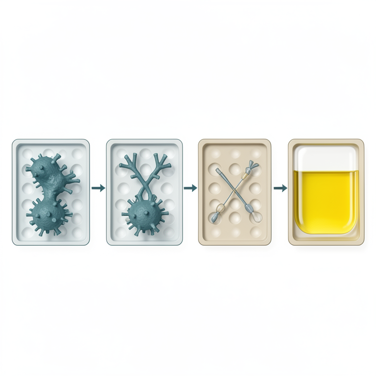
Indirect ELISA Workflow
AI-generated conceptual illustration (laboratory rats and a microtiter plate). It explains the idea and does not show study data.
View source evidence ↗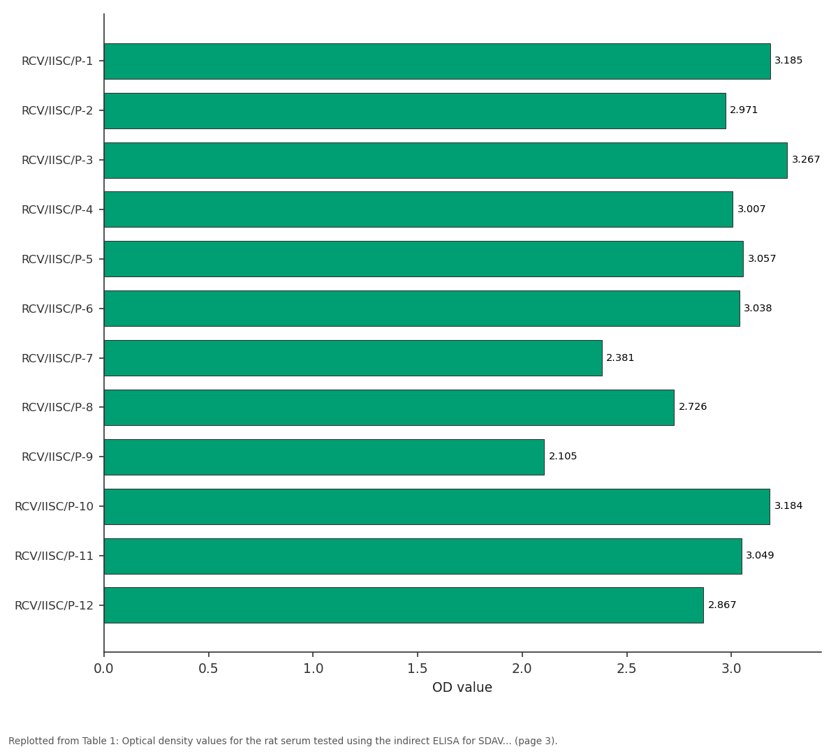
OD value: RCV/IISC/P-1 vs RCV/IISC/P-2 vs RCV/IISC/P-3 vs RCV/IISC/P-4 vs RCV/IISC/P-5
Replotted from Table 1 (page 3) of the original paper. Every bar is the paper's own table value.
View source evidence ↗Figures & tables
From the original paper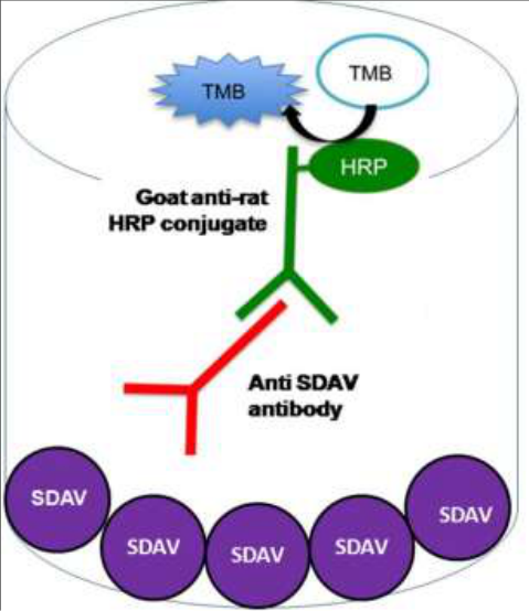 Figure 1
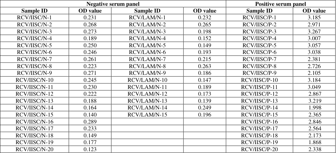 Table 1
Introduction
Sialodacryoadenitis virus (SDAV) is a coronavirus that commonly infects laboratory rats, causing respiratory illness and inflammation of the salivary and lacrimal glands ( (Bartak et al., 2021; Yoo et al., 2000) [1,10] . SDAV infection leads to a range of clinical manifestations, including lesions in the nasal cavity, respiratory epithelium, and olfactory epithelium (Bihun & Percy, 1995) [3] . The virus can replicate in various cell types, including Clara cells, ciliated cells in the bronchial airway, and alveolar type I and II cells in the lung parenchyma (Funk et al., 2009) [5] . Interestingly, SDAV can also infect mice, causing interstitial pneumonia and antibody responses, despite the animals remaining asymptomatic (Bhatt et al., 1977) [2] . This cross-species infection potential highlights the importance of understanding SDAV's characteristics and zoonotic potential, especially given the ongoing pandemic and the role of the Coronaviridae family in human zoonoses (Bartak et al., 2021) [1] . The study of SDAV has been challenging due to the lack of a widely available continuous cell line for virus propagation. However, recent advancements have shown that SDAV can be propagated in LBC and L-2 cell lines, facilitating further research and comparison with other murine coronaviruses (D. H. Percy et al., 1991) [6] . Understanding SDAV infection in laboratory rats is crucial for maintaining the health of research animals and preventing potential confounding effects in experimental studies (Kemp et al., 1991; Yu et al., 2011) [6,12] . Enzyme-linked immunosorbent assay (ELISA) has revolutionized the detection of various pathogens, including viruses, bacteria, and parasites (Ellens, 1981; Richardson & Warnock, 1983) [4,8] . This versatile technique can be adapted for the detection of sialodacryoadenitis virus (SDAV) infection, a coronavirus that affects rodents. ELISA offers several advantages over traditional diagnostic methods, including high sensitivity, specificity, and the ability to process multiple samples simultaneously (Suzuki et al., 1982; Young et al., 2021) [9,11] . In this study we have developed an indirect ELISA for the detection of antibodies against SDAV. This assay would enable rapid and reliable diagnosis of SDAV infection, facilitating better management and control of the disease in laboratory animal facilities.
Materials and Methods
Materials and Methods
Cells
Rat lung epithelial cell line (L2P) cells were cultured at 37 ℃ in high-glucose Dulbecco's modified Eagle's medium (Gibco) supplemented with 10% fetal bovine serum (FBS) and penicillin/streptomycin.
Virus culture and antigen preparation
The SDAV culture was a kind gift from Dr. Nagendra Hegde, National Institute of Biotechnology, Hyderabad. SDAV was propogated on L2P cells. Experiments were performed at a Biosafety Level-2 according to the institutional guidelines. For antigen preparation, the infected cell supernatant was centrifuged at 20000 rpm on a 15-45% sucrose density gradient and the pure antigen interface ring was collected and stored at -80 ℃ until use
Serum
Rat serum samples used in this study were collected from Laboratory animal medicine, Tamil Nadu veterinary and animal sciences university, Chennai and Central animal facility, Indian institute of science, Bengaluru. The serum panel comprised of 10 known positive serum and 20 negative serum. The serum samples were inactivated for 30 min in a heat block at 56 ℃ and stored at -20 ℃ until use.
ELISA
ELISA plates (Nunc, USA) were coated with SDAV antigen (200ng per well) in carbonate bicarbonate buffer (pH 9.6) and incubated overnight at 4 ℃, and blocked with 0.1% Bovine serum albumin. After washing three times with PBS containing 0.05% Tween20, serum samples were added at a dilution of 1:200. The antibodies against SDAV present in the serum was detected using goat anti-rat conjugated to HRP (1:1000; Sigma), with TMB as the substrate followed by addition of. The optical density was measured at 450nm (Fig 1).
Results and Discussion
The format of the ELISA is shown in Fig 1 . Following a checkerboard titration the best coating concentration of to be 0.20 μg/well. The best serum dilution concentration was arrived at 1: 100, and the determination results of 20 negative sera are shown in Table1. The optical density values of the negative sample ranged between 0.123 -0.289. The mean optical density value of the negative samples was 0.209. The cut off value was calculated by the formula ODNeg + 3*SD and was arrived as 0.348. The assay was then used on twenty number of serum samples positive for RCV (Table 1). The assay was able to detect all the positive samples as strongly positive. The mean optical density value of the positive samples was 2.906 (Range 1.868 -3.267). This study successfully developed an indirect ELISA for detecting antibodies against Sialodacryoadenitis virus (SDAV) in laboratory rats. The assay demonstrates promising results for rapid and reliable diagnosis of SDAV infection, which could significantly improve disease management and control in laboratory animal facilities. When applied to 20 serum samples positive for Rat Coronavirus (RCV), the assay successfully identified all samples as strongly positive. The OD values for these samples were substantially higher than the defined cut-off, with a mean of 2.906 (Range 1.868 -3.267). This clear distinction between negative and positive samples suggests high sensitivity and specificity of the assay. These results indicate that our indirect ELISA method is effective in differentiating between SDAV-positive and SDAV-negative samples. The wide gap between the cut-off value (0.348) and the lowest positive sample OD (1.868) further supports the robustness of the assay. In conclusion, this indirect ELISA shows promise as a valuable tool for SDAV diagnosis in laboratory settings. Its ability to provide rapid and reliable results could significantly enhance disease monitoring and control strategies in animal facilities, ultimately improving animal welfare and research quality.
| Negative serum panel | — | — | — | Positive serum panel | — |
|---|---|---|---|---|---|
| Sample ID | OD value | Sample ID | OD value | Sample ID | OD value |
| RCV/IISC/N-1 | 0.231 | RCV/LAM/N-1 | 0.232 | RCV/IISC/P-1 | 3.185 |
| RCV/IISC/N-2 | 0.268 | RCV/LAM/N-2 | 0.265 | RCV/IISC/P-2 | 2.971 |
| RCV/IISC/N-3 | 0.273 | RCV/LAM/N-3 | 0.198 | RCV/IISC/P-3 | 3.267 |
| RCV/IISC/N-4 | 0.189 | RCV/LAM/N-4 | 0.152 | RCV/IISC/P-4 | 3.007 |
| RCV/IISC/N-5 | 0.250 | RCV/LAM/N-5 | 0.149 | RCV/IISC/P-5 | 3.057 |
| RCV/IISC/N-6 | 0.246 | RCV/LAM/N-6 | 0.193 | RCV/IISC/P-6 | 3.038 |
| RCV/IISC/N-7 | 0.261 | RCV/LAM/N-7 | 0.215 | RCV/IISC/P-7 | 2.381 |
| RCV/IISC/N-8 | 0.223 | RCV/LAM/N-8 | 0.263 | RCV/IISC/P-8 | 2.726 |
| RCV/IISC/N-9 | 0.271 | RCV/LAM/N-9 | 0.186 | RCV/IISC/P-9 | 2.105 |
| RCV/IISC/N-10 | 0.245 | RCV/LAM/N-10 | 0.147 | RCV/IISC/P-10 | 3.184 |
| RCV/IISC/N-11 | 0.230 | RCV/LAM/N-11 | 0.189 | RCV/IISC/P-11 | 3.049 |
| RCV/IISC/N-12 | 0.222 | RCV/LAM/N-12 | 0.173 | RCV/IISC/P-12 | 2.867 |
| RCV/IISC/N-13 | 0.188 | RCV/LAM/N-13 | 0.139 | RCV/IISC/P-13 | 3.219 |
| RCV/IISC/N-14 | 0.164 | RCV/LAM/N-14 | 0.249 | RCV/IISC/P-14 | 1.998 |
| RCV/IISC/N-15 | 0.140 | RCV/LAM/N-15 | 0.196 | RCV/IISC/P-15 | 2.365 |
| RCV/IISC/N-16 | 0.289 | RCV/IISC/P-16 | 2.846 | ||
| RCV/IISC/N-17 | 0.233 | RCV/IISC/P-17 | 2.564 | ||
| RCV/IISC/N-18 | 0.149 | RCV/IISC/P-18 | 2.173 | ||
| RCV/IISC/N-19 | 0.177 | RCV/IISC/P-19 | 1.868 | ||
| RCV/IISC/N-20 | 0.123 | RCV/IISC/P-20 | 2.338 |
Table text extracted from the original PDF; superscript letters are shown inline. The original table image is below.
View original table image
References
11 in the original paper- Bartak M, Bańbura MW, Słońska A, Cymerys J. SDAV, the Rat Coronavirus-How Much Do We Know about It in the Light of Potential Zoonoses. Viruses. 2021;13(10):1995. DOI:10.3390/v13101995
- Bhatt PN, Jonas AM, Jacoby RO. Respiratory infection in mice with sialodacryoadenitis virus, a coronavirus of rats. Infect Immun. 1977;18(3):823-827. DOI:10.1128/iai.18.3.823-827.1977
- Bihun CGD, Percy DH. Morphologic Changes in the Nasal Cavity Associated with Sialodacryoadenitis Virus Infection in the Wistar Rat. Vet Pathol. 1995;32(1):1-10. DOI:10.1177/030098589503200101
- Funk CJ, Leete J, Manzer R, Miura TA, Holmes KV, Travanty EA, et al. Rat respiratory coronavirus infection: replication in airway and alveolar epithelial cells and the innate immune response. J Gen Virol. 2009;90(12):29562964. DOI:10.1099/vir.0.014282-0
- Kemp DR, Hayes MA, Percy DH. Lack of effects of viral sialoadenitis and depletion of epidermal growth factor on initiation of hepatic carcinogenesis in the rat. Toxicol Pathol. 1991;19(2):156-163. DOI:10.1177/019262339101900210
- Percy D, Bond S, Macinnes J. Replication of sialodacryoadenitis virus in mouse L-2 cells. Arch Virol. 1989;104(3-4):323-333. DOI:10.1007/bf01315553
- Richardson MD, Warnock DW. Enzyme-linked immunosorbent assay and its application to the serological diagnosis of fungal infection. Med Mycol. 1983;21(1):1-14. DOI:10.1080/00362178385380031
- Suzuki T, Ueda S, Samejima T. Enzyme-linked immunosorbent assay for diagnosis of equine infectious anemia. Vet Microbiol. 1982;7(4):307-315. DOI:10.1016/0378-1135(82)90010-4
- Yoo D, Christie N, Cooper M, Pei Y. Primary structure of the sialodacryoadenitis virus genome: sequence of the structural-protein region and its application for differential diagnosis. Clin Diagn Lab Immunol. 2000;7(4):568-573. DOI:10.1128/cdli.7.4.568-573.2000
- Young VP, Spencer JV, Faure L, Mariano MC. Detection of Cytomegalovirus Interleukin 10 (cmvIL-10) by Enzyme-Linked Immunosorbent Assay (ELISA). Methods Mol Biol. 2021;2244:291-299. DOI:10.1007/978-1-0716-1111-1_15
- Yu VM, Brenner MJ, Mackinnon SE, Hunter DA. Effect of sialodacryoadenitis virus infection on axonal regeneration. Microsurgery. 2011;31(6):458-464. DOI:10.1002/micr.20914.