
Research article · 2024 · Full text
International Journal of Veterinary Sciences and Animal HusbandryEstablishment of a polymerase chain reaction assay for monitoring mycoplasma infection in laboratory mice colony
Volume 9(6) · Pages 135–137 · DOI: 10.22271/veterinary.2024.v9.i6c.1846 ↗
Research areasMicrobial infections and disease researchAquaculture disease management and microbiotaHerpesvirus Infections and Treatments
Abstract
Author-writtenThis study presents the development of an in-house polymerase chain reaction (PCR) assay designed for detecting Mycoplasma infections in laboratory mouse colonies. The 16S rRNA gene, conserved among four Mycoplasma species commonly found in mice, was targeted to ensure broad detection. The assay demonstrated high sensitivity, with a detection limit of 0.1 pg, and excellent specificity, as no amplification was observed with genomic DNA from other organisms, including Mus musculus and various bacterial pathogens. Screening 30 lung tissue samples from laboratory mice confirmed the absence of Mycoplasma infection, indicating the assay’s potential utility in routine health monitoring of animal colonies. This PCR method offers an efficient, specific, and sensitive alternative for pathogen surveillance, supporting improved animal welfare and research reliability.
Summary
Source-checkedPlain-language reading of the full paper, prepared with AI and checked against the text. Each point links to the authors' own words.
Why it matters
- Health monitoring in laboratory mouse facilities is essential to ensure animal welfare and the reliability of research outcomes. View evidence ↗
- Mycoplasma infections are a major challenge in biomedical research because they can compromise the reproducibility and reliability of experiments. View evidence ↗
- Traditional serological monitoring can be unreliable, potentially producing false positives from past infections or false negatives in immune-compromised mice. View evidence ↗
The question
- The researchers aimed to develop an in-house polymerase chain reaction (PCR) assay to monitor Mycoplasma infections in laboratory mouse colonies. View evidence ↗
What they did
- The assay targeted the 16S rRNA gene, which is conserved across four common mouse Mycoplasma species: M. pulmonis, M. arthritidis, M. neurolyticum, and M. collis. View evidence ↗
- Lung tissue samples for validation were sourced from the Laboratory animal medicine unit of Tamil Nadu Veterinary and Animal Sciences University. View evidence ↗
What they found
- Positive PCR amplification of the targeted Mycoplasma DNA resulted in a product of 147 base pairs (bp). View evidence ↗
- The assay demonstrated high sensitivity, with a detection limit of 0.1 pg of template DNA. View evidence ↗
- The assay was highly specific, showing no amplification when tested against genomic DNA from Mus musculus (Balb/c) or various bacterial pathogens including S. aureus, S. pyogenes, K. pneumonia, E. coli, and C. jejuni. View evidence ↗
What it means
- The authors conclude that the in-house PCR assay is a promising tool for routine screening in animal facilities due to its high sensitivity and specificity. View evidence ↗
- The authors note that the screening results are based on a limited sample size and may not represent the entire colony or facility. View evidence ↗
- Further validation and optimization are required to ensure the assay's effectiveness across diverse research settings. View evidence ↗
Figures & tables
From the original paper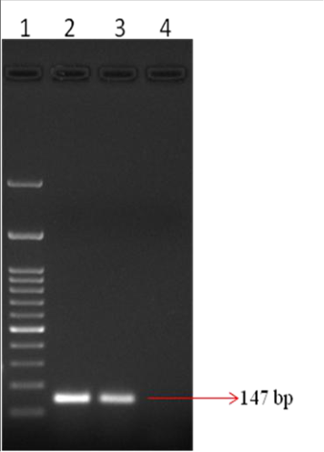 Figure 1
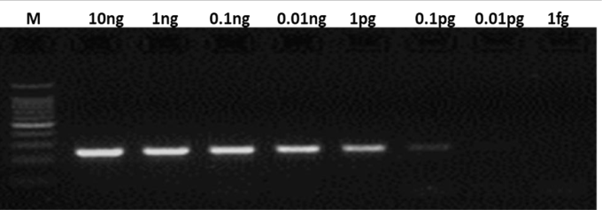 Figure 2
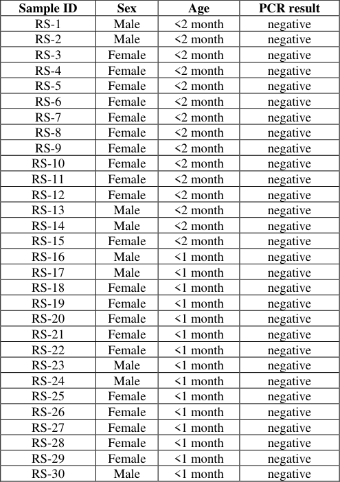 Table 1
Introduction
Health monitoring is crucial for maintaining animal welfare and ensuring reliable research results in laboratory mouse facilities. Polymerase chain reaction (PCR) has emerged as a powerful tool for detecting and identifying various pathogens in mouse colonies (Kim et al., 2022) [5] . PCR allows for the amplification of specific DNA sequences, enabling the detection of even low numbers of organisms present in pathological lesions or body fluids (Lebech, 1994) [6] . PCR-based assays have revolutionized laboratory diagnostics over the past two decades, offering advantages such as high sensitivity, specificity, and the ability to detect multiple targets simultaneously (Dale & Dragon, 1994; Yu et al., 2019) [2,10] . In the context of mouse health monitoring, PCR has proven particularly valuable for screening prevalent infectious pathogens like mouse parvoviruses (Wang et al., 2013) [9] . The technique can be adapted to detect both DNA and RNA pathogens through variations such as reverse transcription PCR (Cárdenas & Alby, 2016; Jalali et al., 2017) [1,4] . The application of PCR in mouse health monitoring programs has led to improved efficiency and reliability in pathogen detection. For instance, the exhaust air dust (EAD) PCR method has been shown to be a reliable complementary approach to traditional soiled bedding sentinel (SBS) monitoring, especially for pathogens that may not be efficiently transmitted in individually ventilated cage (IVC) systems (Kim et al., 2022) [5] . Furthermore, PCR-based methods can reduce the number of animals used in health monitoring, aligning with the 3R principles of animal research (Kim et al., 2022) [5] . Mycoplasma infection in laboratory mice poses a significant challenge to biomedical research, affecting the reliability and reproducibility of experimental results. These small, wall-less bacteria can colonize various tissues, with Mycoplasma pulmonis being a common respiratory pathogen (Schoeb, 2007) [7] . The susceptibility to mycoplasma infection can be influenced by factors such as estrogen levels, as demonstrated in studies with Mycoplasma hominis in BALB/c mice (Shkarupeta et al., 2004) [8] . Cross-reactivity between different mycoplasma species in serological tests can complicate diagnosis, as observed with M. arthritidis and M. pulmonis (Davidson et al., 1983) [3] . The impact of mycoplasma infection extends beyond the immediate health of the mice, potentially altering immune responses and experimental outcomes. In this study we have developed an in-house PCR assay to detect the presence of mycoplasma infection in laboratory mouse and validated using a panel of samples collected from a laboratory mouse colony.
Materials and Methods
Materials and Methods
Samples and Cultures
All mice lung tissue samples were obtained from the Laboratory animal medicine unit of Tamil Nadu Veterinary and Animal Sciences University. These were collected during routine necropsy examinations. The pure cultures of mycoplasma were a kind gift from Dr. S.G. Ramachandra, Indian Institute of Science. DNA from Staphylococcus aureus, Streptococcuss pyogenes, Klebsiella pneumonia, Escherichia coli and Camphylobacter jejuni were a kind gift from Prof. Shrikrishna Isloor, Bangalore Veterinary College, India
DNA extraction and Polymerase chain reaction
DNA was extracted from tissue samples, environmental swabs and mycoplasma cultures by DNA isolation reagent (TAKARA, India) following manufacturers instructions. The final DNA was reconstituted in nuclease free water and stored at -20C until use. PCR was performed in 25 μl volumes using GoTaq DNA Mastermix (Promega). The final reaction mix consisted of 1× mastermix, 1.5 pmoles each of forward (5'-ACACCATGGGAGCTGGTAAT-3') and reverse primers (5'-CCCACGTTCTCGTAGGGATA -3'). PCR thermal cycling condition was 95 °C for 2 min followed by 40 cycles of95 °C for 30 s, 55 °C for 30 s, and 72 °C for 30 s. Products were separated by electrophoresis in 2%TAE agarose gel.
Determination of sensitivity and specificity
For determining the specificity, the PCR reaction mix was spiked with a predetermined quantity of DNA from bacteria species. PCR amplification was done as detailed above. For determination of specificity, dilutions of Mycoplasma genomic DNA were subjected to PCR reaction as above.
Results and Discussion
Conventional methods such as gross pathology, parasitology and bacteriology continue to be used in monitoring the health of laboratory rodents. However these methods are time consuming and often needs separate laboratories and separate technical personnel to conduct the test. The other widely used method, serology looks for antibodies developed against the pathogen. This method is also suffers from the downside of giving false positives owing to the past infections and false negatives in immune-compromised animals. Polymerase chain reaction (PCR), has been widely used as a sensitive and specific method to detect infection on laboratory animals. In this study we developed an in house PCR assay to monitor the Mycoplasma infection in laboratory mice. The primers were designed on the 16S rRNA gene and is conserved in four species of Mycoplasma (M. pulmonis, M. arthritidis, M. neurolyticum, and M. collis) that commonly affect mice. The performance of the assay was evaluated by using the Mycoplasma genomic DNA as template in PCR reaction. The positive PCR amplification produced 147 bp amplification (fig. 1). The limit of detection of the assay was assessed by using serial dilution of the Mycoplasma genomic DNA in the PCR reaction. The limit of detection of the assay was found to be 0.1 pg (fig 2). -1). Of the thirty samples tested none showed amplification specific for Mycoplasma, indicating that the colony is free of mycoplasma infection The developed in-house PCR assay for monitoring Mycoplasma infection in laboratory mice demonstrates promising results in terms of sensitivity and specificity. The assay's design, targeting the conserved 16S rRNA gene across four common Mycoplasma species affecting mice, provides a broad detection range for potential infections. The limit of detection of 0.1 pg indicates high sensitivity, which is crucial for early detection of Mycoplasma infections in laboratory mice. The specificity of the assay is a notable strength, as demonstrated by the lack of amplification when testing against genomic DNA from various other organisms, including the host species (Mus musculus) and common bacterial pathogens. This high specificity reduces the likelihood of false-positive results, which is essential for accurate monitoring of Mycoplasma infections in mouse colonies.
The application of the optimized PCR assay to screen lung tissue samples from 30 mice yielded negative results for all samples. This outcome suggests that the tested colony is currently free of Mycoplasma infection. However, it is important to note that this result is based on a limited sample size and may not be representative of the entire colony or facility. Implementing regular screening of the mouse colony over time would help establish the assay's reliability for ongoing Mycoplasma surveillance.
In conclusion, the developed in-house PCR assay shows potential as a valuable tool for monitoring Mycoplasma infections in laboratory mice. Its high sensitivity and specificity make it a promising candidate for routine screening in animal facilities. However, further validation and optimization may be necessary to ensure its reliability and effectiveness in diverse research settings.
Conflict of Interest
Conflict of Interest
Financial Support
Financial Support
Figures and tables from the paper
| Sample ID | Sex | Age | PCR result |
|---|---|---|---|
| RS-1 | Male | <2 month | negative |
| RS-2 | Male | <2 month | negative |
| RS-3 | Female | <2 month | negative |
| RS-4 | Female | <2 month | negative |
| RS-5 | Female | <2 month | negative |
| RS-6 | Female | <2 month | negative |
| RS-7 | Female | <2 month | negative |
| RS-8 | Female | <2 month | negative |
| RS-9 | Female | <2 month | negative |
| RS-10 | Female | <2 month | negative |
| RS-11 | Female | <2 month | negative |
| RS-12 | Female | <2 month | negative |
| RS-13 | Male | <2 month | negative |
| RS-14 | Male | <2 month | negative |
| RS-15 | Female | <2 month | negative |
| RS-16 | Male | <1 month | negative |
| RS-17 | Male | <1 month | negative |
| RS-18 | Female | <1 month | negative |
| RS-19 | Female | <1 month | negative |
| RS-20 | Female | <1 month | negative |
| RS-21 | Female | <1 month | negative |
| RS-22 | Female | <1 month | negative |
| RS-23 | Male | <1 month | negative |
| RS-24 | Male | <1 month | negative |
| RS-25 | Female | <1 month | negative |
| RS-26 | Female | <1 month | negative |
| RS-27 | Female | <1 month | negative |
| RS-28 | Female | <1 month | negative |
| RS-29 | Female | <1 month | negative |
| RS-30 | Male | <1 month | negative |
Table text extracted from the original PDF; superscript letters are shown inline. The original table image is below.
View original table image
References
6 in the original paper- Cárdenas AM, Alby K. Nucleic acid amplification by polymerase chain reaction. In: American Society for Microbiology, editor. 2016. p. 129-36. Available from: https://doi.org/10.1128/9781555819156.ch11
- Dale B, Dragon EA. Polymerase chain reaction in infectious disease diagnosis. Lab Med. 1994;25(10):63741. Available from: https://doi.org/10.1093/labmed/25.10.637
- Davidson MK, Brown MB, Lindsey JR, Cassell GH, Boorman GA. Natural Mycoplasma arthritidis infection in mice. Curr Microbiol. 1983;8(4):205-8. Available from: https://doi.org/10.1007/bf01579547
- Kim H, Park J-H, Goo J-S, Jang Y-C, Jeon C-M. Evaluation of exhaust air dust polymerase chain reaction as a supplement method for soiled bedding sentinel monitoring in specific pathogen free mouse facility using two different individually ventilated cage racks. Lab Anim. 2022;57(1):40-49. Available from: https://doi.org/10.1177/00236772221123184
- Shkarupeta MM, Govorun VM, Lazarev VN. Experimental Mycoplasma hominis infection of the genital tract in BALB/c mice. Bull Exp Biol Med. 2004;137(1):53-5. Available from: https://doi.org/10.1023/b:bebm.0000024386.33789.0f
- Wang KW, Wang MH, Wan CH, Hsieh SC, Chou JY, Chang CY, et al. Multiplex polymerase chain reaction assay for the detection of minute virus of mice and mouse parvovirus infections in laboratory mice. Lab Anim. 2013;47(2):116-21. Available from: https://doi.org/10.1177/0023677213478298