
Research article · 2024 · Full text
International Journal of Veterinary Sciences and Animal HusbandryComparative histomorphometric study of the trachea between immature and mature domestic gazelle: Gazella Subgutturosa
Volume 9(5) · Pages 705–710 · DOI: 10.22271/veterinary.2024.v9.i5k.1793 ↗
Research areasAnimal Diversity and Health StudiesComparative Animal Anatomy Studiesmelanin and skin pigmentation
Abstract
Author-writtenThe study involved five immature and five mature gazelles in order to define the characteristics of the trachea, the mucosa, submucosa, hyaline cartilage, adventitia, and muscle fibers make up the complex tissue of the trachea. The cervical portion of the trachea is home to many simple alveolar mucous glands, the thoracic part of trachea has goblet cells that form
Summary
Source-checkedPlain-language reading of the full paper, prepared with AI and checked against the text. Each point links to the authors' own words.
Why it matters
- In ruminant animals, the trachea is a tube-like structure that extends from the cricoid cartilage to the point where it divides into the bronchi. View evidence ↗
- The primary function of the trachea is to provide the mechanical compliance necessary to handle changes in pressure during swallowing, coughing, and breathing. View evidence ↗
- There is a lack of available data regarding the microscopic structural organization of the trachea in Iraqi gazelles and how this structure influences the tissue's mechanical response. View evidence ↗
The question
- The study aims to determine the microstructure of tracheal elements and understand how this structure affects the mechanical response of the tissue. View evidence ↗
What they did
- The study used ten healthy indigenous Gazella subgutturosa gazelles from Sawa Station in Al-Muthanna City between January and May 2023, consisting of five one-year-old and five two-to-three-year-old animals. View evidence ↗
- Histological analysis employed Periodic acid/Schiff (PAS), Mallory, and Masson's trichrome stains to identify smooth muscles, collagen fibers, mucoproteins, glycoproteins, carbohydrates, and the basement membrane. View evidence ↗
- The thickness of the mucosa, submucosa, and adventitia was measured using an ocular micrometer across five sections per age group, with means and standard errors calculated from thirty slides per section. View evidence ↗
What they found
- The trachea of mature gazelles is composed entirely of ciliated cells, while the trachea of immature gazelles consists entirely of non-ciliated columnar epithelium. View evidence ↗
What it means
- The authors conclude that the thickness of histological layers and the type and quantity of goblet cells in the trachea vary according to the age of the gazelle. View evidence ↗
Visual summary
2 visualsIllustrations explain the paper; charts are replotted from the paper's own table values.
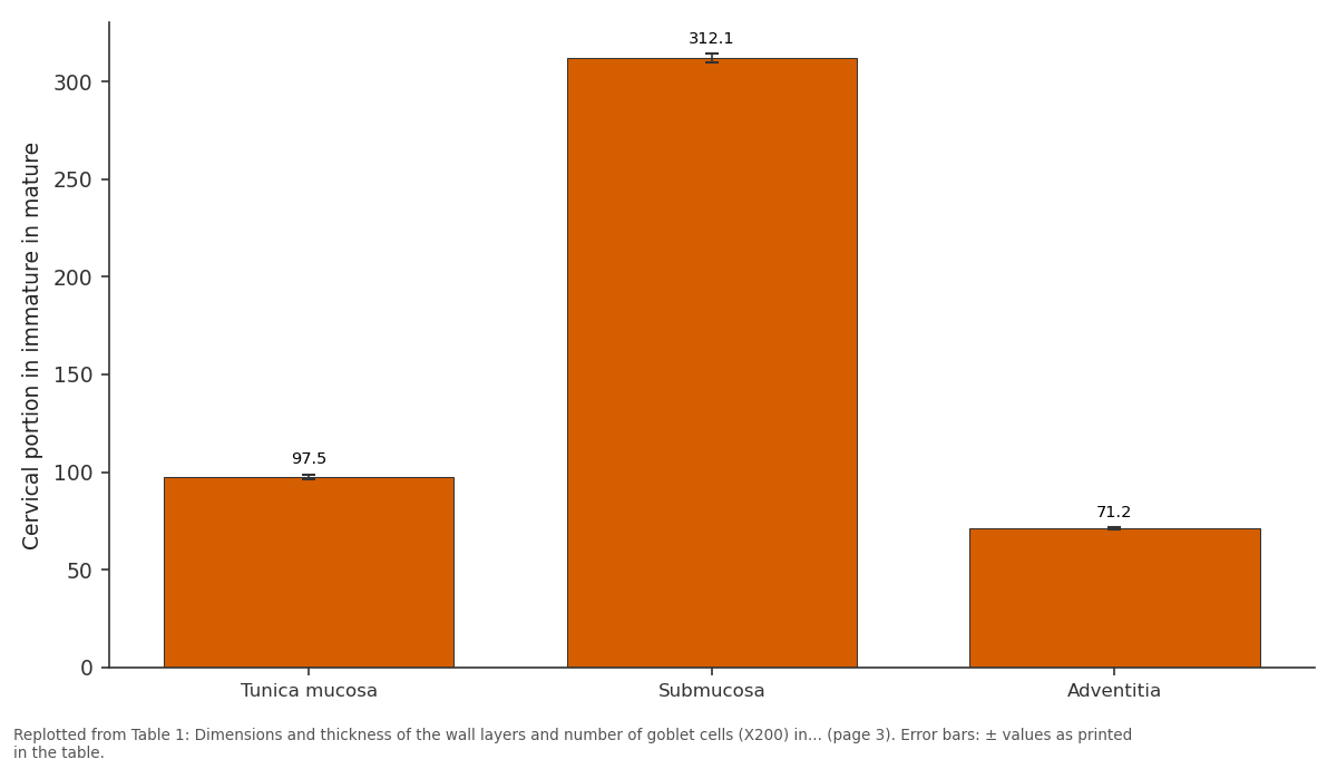
Cervical portion in immature in mature: Tunica mucosa vs Submucosa vs Adventitia
Replotted from Table 1 (page 3) of the original paper. Every bar is the paper's own table value.
View source evidence ↗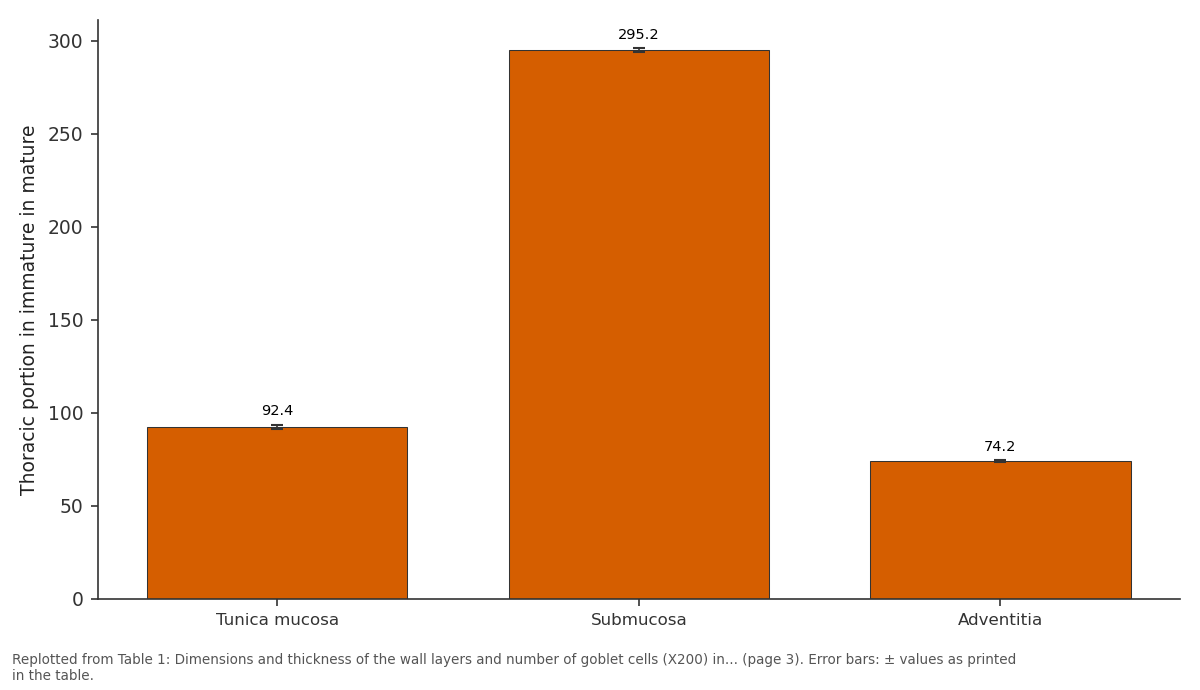
Thoracic portion in immature in mature: Tunica mucosa vs Submucosa vs Adventitia
Replotted from Table 1 (page 3) of the original paper. Every bar is the paper's own table value.
View source evidence ↗Figures & tables
From the original paper Table 1
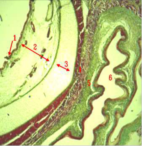 Figure 1
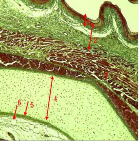 Figure 2
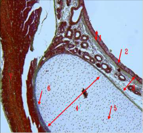 Figure 3
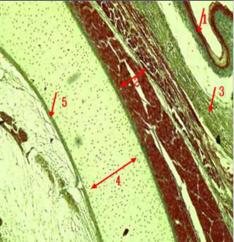 Figure 4
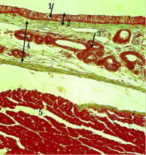 Figure 5
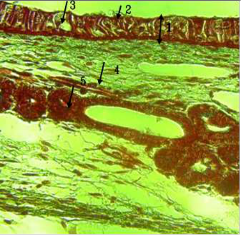 Figure 6
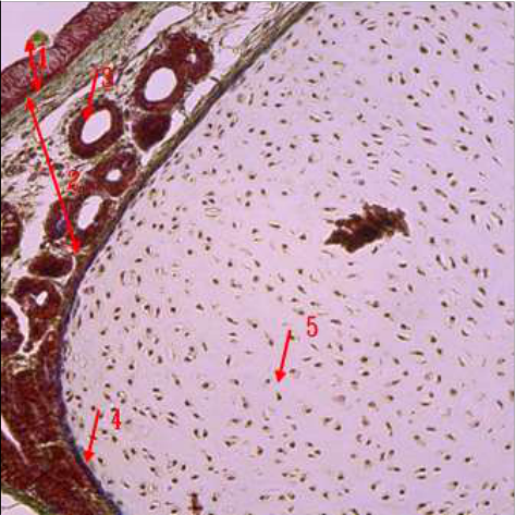 Figure 7
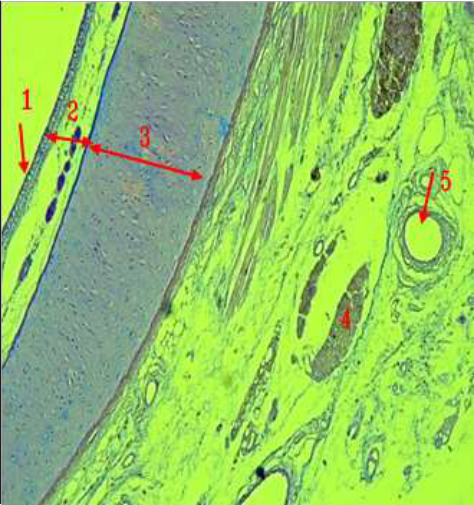 Figure 8
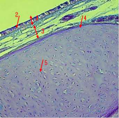 Figure 9
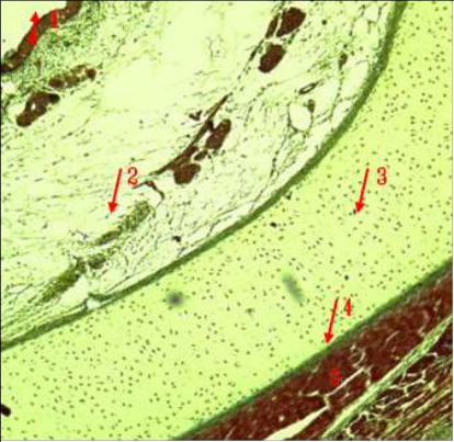 Figure 10
Introduction
The gazelles are graceful, herbivorous, ruminant, tiny, and herbivorous antelopes that are known as quick animals. They are primarily found in Africa's hot, deserts, arid savannas and grasslands. Their main habitats are the hot, arid savannas, deserts, and grasslands of Africa, the Indian subcontinent, and the regions of southwest and central Asia [1] . In addition to allowing gases to condense and exchange, the respiratory system also controls body temperature, creates and excretes a number of chemicals, including histamine, and helps produce voice, and linked to the sense of smell [2][3][4] . In ruminants, the trachea is a tube-like structure extends from the cartilage of the cricoids to the point where it splits into the bronchi. Its hyaline cartilage is divided into many rings that form an incomplete ring towards the dorsal aspect of the structure. Each ring-shaped cartilage is connected dorsally by the tracheal membrane, and externally by the trachealis [5][6][7] . Four distinct layers make up the very malleable trachea: the adventitial layer, cartilaginous, mucosa layer submucosa layers, and trachealis muscle. The trachea's main job is to supply the mechanical compliance required for variations in intraluminal pressure that occur during respiration, coughing, and swallowing. The trachea is composed of the adventitia, muscularis, mucosa, and submucosa. The lining is composed of well-developed adventitia, submucosa, and respiratory epithelium. The cartilaginous ring is incomplete [8][9][10] . Goblet cells, intermediate cells perched on the basement membrane, ciliated cells, basal cells, and goblet cells make up the tracheal epithelium [11][12] . The ciliated cells are completely gone from the distal respiratory bronchioles of calves and pups, while they are slightly present in the trachea and bronchi of newborn canines and cattle. A newborn dog's trachea has longitudinally directed folds across its dorsal walls and numerous cilia covering its ventral and dorsal surfaces [13][14] . There is relatively little material available on the characteristics of the gazelle trachea in Iraq, and our understanding of the microscopic structural organization of these parts of trachea and their function in influencing the mechanical response of the tissue is severely limited. The information on microstructure of the tracheal elements and how it affects the mechanical response of the tissue is the aim of this study, knowing the relationship between structure and function is crucial for creating tissue-engineered replacements that mimic the mechanical properties of native tissue. It also aids in understanding all other sciences and makes it possible to view the structure and any unique alterations that may have occurred at the tissue.
Materials and Methods
Ten healthy indigenous breeds of gazelles (Gazella subgutturosa), five at one year of age and five at two to three years of age, Between January and May 2023, the animals were taken alive from Sawa Station in Al-Muthanna City, which is connected to the Ministry of Agriculture. The animals were then given an overdose of ketamine and xylazine, and they were sacrificed. Samples were obtained from the cervical and thoracic regions of the trachea, and seven specimens from each location were preserved in 10% formalin for a full day before being processed histologically, the stains that were previously there; Periodic acid/Schiff (PAS) stain; Mallory and Masson's trichrome stain for the basement membrane, mucoprotein, glycoprotein, carbohydrates, and mucopolysaccharides; To the collagen fibers and smooth muscles [15] . An ocular micrometer was used to measure the thickness of the adventitia tunicae, mucosa, and submucosa of each tracheal segment in five sections at each age. The mean (x-) and standard error (S. E.) was calculated for thirty slides of each section. [16] .
Results and Discussion
The mucosa, muscularis, adventitia, and submucosa comprised the layers of trachea's wall. the cervical portion of the tracheal epithelium has more mucous glands, while cervical portion of the tracheal epithelium has fewer glands and most of its regions are home to "intraepithelial glands," which have replaced mucous glands in the thoracic tracheal epithelium and are fewer developed in the cervical trachea than thoracic trachea in both ages, the trachea is lined by a respiratory epithelium alongside goblet and basal cells that lie on the basement membrane (Fig. 1,2), Mucosa and submucosa layers thicknesses decreased gradually toward the thoracic section of the trachea (Table 1). The mucosa-associated lymphoid tissue, found on the surface of mucosal tissues like the respiratory tracts of horses, farm animals, and rodents, is crucial to the local immune response [6,17] . Mature animals' trachea is made wholly of ciliated cells, while the trachea of immature ages is made totally of non-ciliated columnar epithelium. This is in contrast to rodents' airways that are devoid of ciliated cells. At the microscopic level, a network of several kinds of fibers, including collagen and muscle fibers, make up each layer of the trachea (Fig. 3,4,5). Experimental evidence has sufficiently established the role of the fiber networks in the mechanical behavior of soft tissues [3] . Goats, oxen, sheep, and dogs all have glands in the ventral rather than the dorsal section of their trachea. Specialized tracheobronchial glands protruded from the airway surface epithelium into the submucosa. Two distinct kinds of secretary cells exist in the tracheal [6] . The submucosa was made of loosely connected tissue and was made up of blood arteries, lymphocytes, and longitudinal groupings of elastic fibers. This layer's collagen was likewise arranged into dense bundles; it did not clump together into many orientated bundles. Rather, a single fiber family was seen in most of the cases throughout the thick and welldeveloped adventitia that wrap the respiratory epithelium and the goblet cells which make up the mucosal lining, covered incomplete cartilaginous rings, Early on, a greater percentage of goblet cells had neutral mucins; this improved goblet cells' protective (Fig. 123), thickness of the mucosa in the cervical part of trachea in immature was (97.5±1.3 μm), in mature was (111.7±1.4 μm), and it thickness in the thoracic trachea in immature was (92.4±1.2 μm), in mature was (103.4±1.5 μm). Bundles of smooth fibers join the cartilaginous ends of the trachea through the trachealis muscle, which is located on the posterior membrane. The posterior trachea wall is held in place by the minuscule bands of smooth muscles that comprise its cartilage-free wall. These incomplete cartilaginous rings are hyaline in character and are encircled by perichondrium (Fig. 7). Thickness of the submucosa in cervical and thoracic parts of the trachea in immature was (312.1±2.4, 295.2±1.1 μm) respectively, in mature trachea was (363.2±1.3, 306.1±0.5 μm) respectively. Thickness of adventitia in cervical and thoracic trachea in immature was (71.2±0.6, 74.2±0.4 μm) respectively, in mature ages was (78.4±0.1, 81.3±0.2 μm) respectively (Table 1). The cartilage matrix exhibited a strong response to PAS, while the chondrocytes exhibited a mild reactivity similar to that of most mammals. The posterior ends of cartilages joined by smooth muscle and connective tissue, and the external surface are composed of adventitia composed of connective tissue with multiple blood vessels (Fig. 6, 10). The cartilage is made up of pulverized substance-containing chondrocytes. It was possible to see the skeletal muscle fibers of lateral trachea on both sides of the tracheal rings. Thick bundles of collagen were shown to be arranged in the adventitia (Fig. 8, 9, 10). The muscle fibers are directly connected to the perichondrium, in certain species (such dogs, cats, and rabbits) attaches to the outside of the crescent-shaped trachea ring. Some animals, such as guinea pigs [2,18] . It should be noted that the purpose of the ciliated cells in the trachea is to transport secretions across the airways. It is these ciliated cells that transfer water and ions across epithelia [19] . The tracheal epithelium in animals is thought to consist of three distinct cellular types: serous cells, secretary mucous cells, and nonciliated bronchiolar epithelial cells. Mucus produced by the goblet cells coats the ciliated cells, and the weather mean spreads it. Mucus from respiratory system sticking to the cells of the cilia [2,19] . Basal cells pushed goblet cells between them in the trachea, where they are found in domestic mammals and were involved in the production of mucus [2,17,20] .
| Part / Measure | Tunica mucosa | Submucosa | Adventitia | Number of goblet cells |
|---|---|---|---|---|
| Cervical portion in immature in mature | 97.5 ± 1.3 | 312.1±2.4 | 71.2±0.6 | 33±1 |
| 111.7±1.4 | 363.2±1.3 | 78.4±0.1 | 40±2 | |
| Thoracic portion in immature in mature | 92.4 ±1.2 | 295.2± 1.1 | 74.2±0.4 | 32±3 |
| 103.4 ±1.5 | 306.1±0.5 | 81.3±0.2 | 38±1 |
Table text extracted from the original PDF; superscript letters are shown inline. The original table image is below.
View original table image
Conclusion
Changes in the quantity and kind of goblet cells, as well as the thickness of the histological layer, were observed in trachea of varying ages.
Acknowledgement
AcknowledgmentWe express our gratitude to all the instructors and participants who helped us to complete and publish this research.We value what they have done.
Figures and tables from the paper
References
27 in the original paper- Kingdon J. The Kingdon field guide to African mammals. San Diego and London: Academic Press; 1997. p. 411-413.
- Kahwa CKB, Purton M. Histological and histochemical study of the epithelial lining of the respiratory tract in adult goats. Small Anim Res. 1996;20:181-186. doi:10.1016/0921-4488(95)00799-7.
- Dyce KM, Sack WO, Wensing CJG. Textbook of veterinary anatomy. 4th ed. Philadelphia: W.B. Saunders Company; 2010. p. 4-594.
- Cano I, Perez W. Quantitative anatomy of the trachea of the giraffe (Giraffa camelopardalis rothschildi). Int J Morphol. 2009;27(3):905-908.
- Rajathi S. Histological studies on the trachea and lungs in dogs. J Vet Anim Sci. 2024;55(2):256-261.
- Rodriguez M, Bur S, Favre A, Welbel ER. Pulmonary acinus: geometry and morphometry of the peripheral airway system in rat and rabbit. Am J Anat. 1987;180:143-155. doi:10.1002/aja.1001800204.
- Pinheiro LL, Lima AR, Munis JA, Imbeloni AI, Fioreto ET, Fontes FS, Cabral R, Branco E. Anatomy and morphometric aspects of the trachea of Saimiri sciureus Linnaeus, 1758: knowledge for emergency procedures. Anais Acad Bras Cienc. 2012;84(4):973-977. doi:10.1590/s0001-37652012000400012.
- Frandson RD, Wilke WL, Fails AD. Anatomy and physiology of farm animals. 7th ed. Philadelphia: Lippincott Williams & Wilkins; 2009. p. 350-368.
- Kalita HC, Bordoloi CC. Histomorphological studies on the trachea of yak (Bos grunniens). Indian J Anim Sci. 2005;75(2):222-223.
- Al-Zghoul MF, Ismail ZB, Al-Rukibat RK, Al-Majali AM. A quantitative study of the trachea of young Arabian camels (Camelus dromedarius). J Camel Pract Res. 2006;13(2):129-133.
- Dellmann HD, Eurell JA. Textbook of veterinary histology. 5th ed. Philadelphia: Lippincott Williams & Wilkins; 1998. p. 287-391.
- Dabanoglu MK, Kara ME. A quantitative study on the trachea of the dog. Anat Histol Embryol. 2009;30(1):5759. doi:10.1046/j.1439-0264.2001.00301.x.
- Al-Abasi RJ, Mirhish M. Anatomical and histological study on the trachea and lung of one-humped camel in Middle of Iraq. M.Sc. Thesis, Veterinary Medicine College, University of Baghdad; 2001. p. 100-103.
- Hussein AJ, Abdul Zahra IA. Morphological, histological and histochemical study of the trachea of one-humped camel (Camelus dromedarius) in south Iraq. MRVSA. 2016;5(Special Issue):19-25.
- Suvarna SK, Layton C, Bancroft JD. Bancroft's theory and practice of histological techniques. 8th ed. Philadelphia: Churchill Livingstone Elsevier; 2018. p. 176-725.
- Al-Rawi KM, Kalaf-Allah IS. Design and analysis of agricultural experiments. Mosul: Dar-Al Kutub; 1980. p. 65, 95-107.
- Eurell JAC. Veterinary histology. Teton New Media, USA; 2004. p. 64-67.
- Ramchandani R, Bates JH, Shen B, Suki B, Tepper RS. Airway branching morphology of mature and immature rabbit lungs. J Appl Physiol. 2001;90:1584-1592. doi:10.1152/jappl.2001.90.4.1584.
- Albertine HK, Wiener-Kronish JP, NC. The structure of the parietal pleura and its relationship to pleural liquid dynamics in sheep. Anat Rec. 1984;2008:401-409.
- Ibe CS, Salami SO, Onyeanusi BI. Macroscopic anatomy of the lower respiratory system in a nocturnal burrowing rodent: African giant pouched rat (Cricetomys gambianus). Anat Histol Embryol. 2011;40:112-119. doi:10.1111/j.1439-0264.2010.01048.x.
- Kumar SR, Nagamalleswari Y, Kumar DP. Ultrastructural study of tracheo-bronchial epithelium of Indian goats (Capra hircus). Indian J Vet Anat. 2012;24(2):99-102.
- Abdul-Raheem MH, Al-Haaki AG. A comparative anatomical and histological study of the trachea of native Awasi sheep and black goats. Iraqi J Vet Sci. 2006;20(1):9-19.
- Habib RS, Mahammed FS. Histomorphological and radiological study of trachea and lungs of Angora goat (Capra ibex). M.Sc. Thesis, Veterinary Medicine College, University of Duhok; 2010. p. 100-105.
- Essa JS. A comparative study of tracheal morphology in domestic sheep and goats. Iraqi J Vet Sci. 2003;17(1):105-109.
- Al-Umeri SKW. Gross and microscopic study of the trachea and bronchial tree in local sheep (Ovis aries). Bas J Vet Res. 2015;14(1):145-153.
- Al-Anbaki AA. Anatomical, histological and radiological study of the trachea and lungs in domesticated rabbits. M.Sc. Thesis, University of Baghdad, Veterinary Medicine College; 2013. p. 19.
- Al-Zhgoul MB, Dalab AHS, Abdulhakeem E, Ismail ZB, Thanain A. Arabian oryx (Oryx leucoryx) trachea: A descriptive and morphometric analysis. Int J Morphol. 2013;31(3):813-818.