
Research article · 2024 · Full text
International Journal of Veterinary Sciences and Animal HusbandryCutaneous mast cell tumour in a dog
Volume 9(5) · Pages 641–643 · DOI: 10.22271/veterinary.2024.v9.i5j.1785 ↗
Research areasVeterinary Oncology ResearchInfectious Diseases and MycologyOcular Oncology and Treatments
Abstract
Author-writtenA 9-year-old male Dachshund dog was presented to the local multi-speciality hospital, Chennai with the history of presence of multiple masses on the scrotal area for the past 6 months for surgical removal. Blood samples, peripheral blood smears and fine needle aspiration smears of the mass were collected and sent to the Centralised Clinical Laboratory, Madras Veterinary College, Chennai for laboratory diagnosis. Haematological values were found to be within normal limits except leukocytosis with neutrophilia of left shift. Serum biochemical studies also showed no abnormal changes in parameters pertaining to liver functions and kidney functions. Cytological examination revealed predominant population of discrete round to oval cells having small to moderate amounts of pale basophilic cytoplasm with varying numbers of characteristic purple colored (metachromatic) granules. Based on the present laboratory findings, the lump was confirmed as mast cell tumour.
Summary
Source-checkedPlain-language reading of the full paper, prepared with AI and checked against the text. Each point links to the authors' own words.
Why it matters
- Mast cell tumours (MCTs), also known as mastocytomas, are the most frequent skin tumours in dogs and account for 16% to 21% of all cutaneous tumours. View evidence ↗
- The behavior of MCTs varies significantly, ranging from single benign growths to multiple malignant masses that can be fatal. View evidence ↗
- These tumours typically appear on the abdomen, scrotum, perineum, and hind limbs, with those in mucocutaneous sites and the inguinal regions often being more aggressive. View evidence ↗
The question
- The study aimed to diagnose multiple scrotal masses present for six months in a 9-year-old male Dachshund. View evidence ↗
What they did
- Diagnosis involved collecting blood samples, peripheral blood smears, and fine needle aspiration (FNA) smears from the masses. View evidence ↗
- Blood and FNA smears were air-dried, treated with a Leishman and Giemsa cocktail stain, and examined under a microscope. View evidence ↗
What they found
- The dog exhibited leukocytosis with a white blood cell count of 21,000 x 10³/cumm (reference: 6,000-11,000) and neutrophilia at 82% (reference: 60-75%). View evidence ↗
- Serum biochemical tests showed no abnormal changes in parameters related to kidney or liver function. View evidence ↗
- Cytology showed a primary population of discrete round to oval cells with pale basophilic cytoplasm. View evidence ↗
- The cells contained characteristic purple (metachromatic) granules of varying size and an eccentrically placed nucleus. View evidence ↗
- Extracellular granules released from mast cells were observed, alongside some eosinophils and non-degenerate neutrophils. View evidence ↗
What it means
- Based on the cytological evidence, the scrotal masses were confirmed to be a mast cell tumour. View evidence ↗
Visual summary
1 visualsIllustrations explain the paper; they do not show study data.
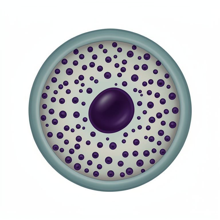
Cytological Characteristics of MCT Cells
AI-generated conceptual illustration (canine mast cell). It explains the idea and does not show study data.
View source evidence ↗Figures & tables
From the original paper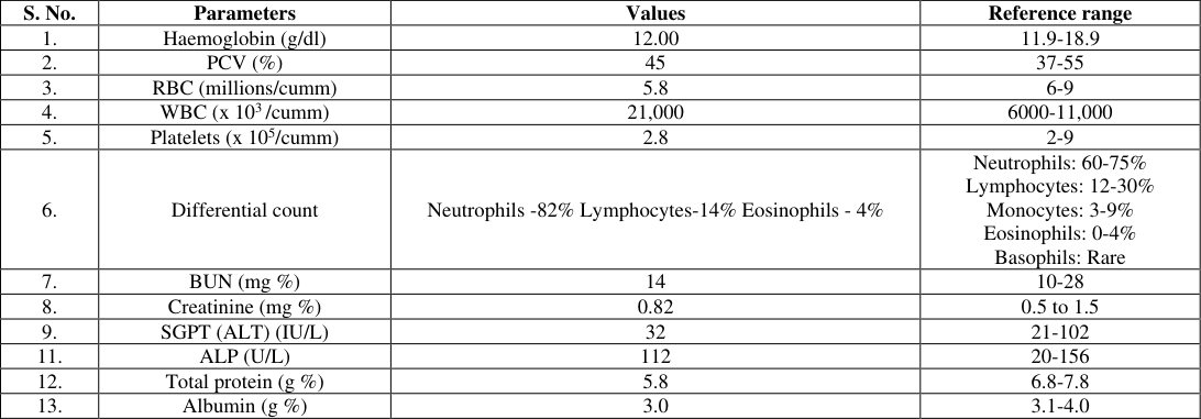 Table 1
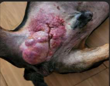 Figure 1
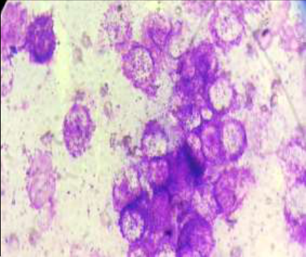 Figure 2
Introduction
Mast cell tumours (MCTs), otherwise called as mastocytoma, the most common skin tumours of dogs are highly invasive and metastatic comprising 16 to 21% of all cutaneous tumors encountered [1] . The biologic behaviours of MCTs are highly variable, ranging from solitary benign to highly malignant multiple masses which are potentially fatal. However, their true metastatic potential is not entirely known [2] . It is mostly found on the hind limbs, abdomen, perineum, and scrotum. MCTs of the preputial, inguinal, and sub inguinal regions and other mucocutaneous sites tend to be more aggressive. When these tumours are aspirated, they tend to exfoliate well, making the samples often more cellular [3] . Among different breeds affected, Boxers, Boston terriers, Beagles, and Labrador retrievers are more prone to MCTs. Some studies have shown that genetics play a role in breed predilections in which mutations in the c-kit tyrosine kinase receptor result in malignant transformation of mast cells. Although, middle-aged to older dogs are commonly affected, younger dogs as young as 3 weeks old are also sometimes affected, However, no gender predilection has been reported [4,5] .
Materials and Methods
A 9 years old male Dachshund dog was presented to the local multi-speciality hospital, Chennai with the history of presence of multiple masses on the scrotal area for the past 6 months for surgical removal. Blood sample containing EDTA as an anticoagulant, clotted blood sample, peripheral blood smears were collected and sent to the Centralised Clinical Laboratory, Madras Veterinary College, Chennai for hematobiochemical studies. In addition, fine needle aspiration procedure was performed on the mass and the aspiration smears prepared were also referred for cytological diagnosis. Haematogical and biochemical estimations were carried out using auto analyser namely Mindray BC -2800 and A15 Biosystems. The FNAC smears as well as blood smears were air dried, stained with Leishman and Giemsa cocktail stain [6] and subjected to microscopical examination.
Results and Discussion
Haematobiochemical values recorded in the present study are shown in Table 1.
| S. No. | Parameters | Values | Reference range |
|---|---|---|---|
| 1. | Haemoglobin (g/dl) | 12.00 | 11.9-18.9 |
| 2. | PCV (%) | 45 | 37-55 |
| 3. | RBC (millions/cumm) | 5.8 | 6-9 |
| 4. | WBC (x 103 /cumm) | 21,000 | 6000-11,000 |
| 5. | Platelets (x 105/cumm) | 2.8 | 2-9 |
| 6. | Differential count | Neutrophils -82% Lymphocytes-14% Eosinophils - 4% | Neutrophils: 60-75% Lymphocytes: 12-30% Monocytes: 3-9% Eosinophils: 0-4% Basophils: Rare |
| 7. | BUN (mg %) | 14 | 10-28 |
| 8. | Creatinine (mg %) | 0.82 | 0.5 to 1.5 |
| 9. | SGPT (ALT) (IU/L) | 32 | 21-102 |
| 11. | ALP (U/L) | 112 | 20-156 |
| 12. | Total protein (g %) | 5.8 | 6.8-7.8 |
| 13. | Albumin (g %) | 3.0 | 3.1-4.0 |
Table text extracted from the original PDF; superscript letters are shown inline. The original table image is below.
View original table image
Haematological parameters were found to be within normal limits except leukocytosis with left shift neutrophilia. Serum biochemistry also revealed no changes in parameters related to liver and kidney functions. Cytological examination revealed predominant population of discrete round to oval cells having moderate amounts of cytoplasm which were pale basophilic. The cytoplasm was filled with varying numbers of characteristic fine to coarse purple coloured (metachromatic) granules with eccentrically placed nucleus (Fig. 1). Only a mild anisocytosis and anisokaryosis were observed. In many areas extracellular granules released from mast cells were also noticed along with few non-degenerate neutrophils and eosinophils. Based on the cytological evaluation, the mass was confirmed as mast cell tumour (MCT). The present haematological findings were in accordance with earlier report [7] in which leukocytosis and neutrophilia in addition to mild anaemic changes in 8 year old male Pomeranian dog affected with MCT. In contrast, severe anemia was noticed in a 13 years old dog affected with mastocytoma [5] . The biochemical findings recorded in the present study were also in accordance with the earlier findings [7] who also observed no abnormalities in serum biochemical values pertaining to liver and kidney functions.
The cytological observations made in the present study correlates well with previous reports [7] who also recorded numerous discrete neoplastic cells containing purple granules in the cytoplasm, pushing the nucleus to the periphery with round to spherical nuclei in aspiration smears collected from multiple cutaneous masses located on the lower abdomen of an 8 years old male Pomeranian dog affected with mastocytoma. In addition, they also recorded extracellular granules as reported in the present study. Similar cytological observations were also reported in a 13 years old, male, halfbreed dog with a mass at the eyelid of the left eye which was confirmed as MCT [5] .
Figures and tables from the paper
References
6 in the original paper- Ehsan H, Pedram B, Bahrami AM, Moghaddam MHJ, Javanbakht J, Ghomi FE, et al. Cutaneous mast cell tumor (Mastocytoma): Cyto-histopathological and haematological investigations. Diagn Pathol. 2014;9:9. doi: 10.1186/1746-1596-9-9.
- Webster J, Yuzbasiyan GV, Miller R, Kaneene J, Kiupel M. Cellular proliferation in canine cutaneous mast cell tumors: associations with c-KIT and its role in prognostication. Vet Pathol. 2007;44:298-308. doi: 10.1354/vp.44-3-298.
- Michelle CT, Cathy AP, Goldschmidt MH, Goldschmidt KH, Rebecca ER. Scrotal tumors in dogs: A retrospective study of 676 cases (1986-2010). Can Vet J. 2014;55(1):1229-33.
- Alexandru DM, Crivineanu M, Morales I, Quintana OF. Palpebral mast cell tumour in a dog: A case report. Rev Rom Med Vet. 2020;30(3):31-34.
- Garbyal RS, Neeta A, Prachi K. Leishman-Giemsa cocktail: An effective Romanowsky stain for air-dried cytologic smears. Acta Cytol. 2006;50(4):403-406.
- Preena P, Jeny KJ, Sherin BS, Seej. Diagnosis of recurrent multiple cutaneous mast cell tumour in a dog. JIVA, 2020, 18(2).