
Research article · 2024 · Full text
International Journal of Veterinary Sciences and Animal HusbandrySurgical management of ventral hernia in a calf by hernioplasty using non-absorbable Polypropylene mesh
Volume 9(5) · Pages 638–640 · DOI: 10.22271/veterinary.2024.v9.i5j.1784 ↗
Research areasHernia repair and managementCongenital Diaphragmatic Hernia StudiesCongenital Anomalies and Fetal Surgery
Abstract
Author-writtenA two month old male crossbred Jersey calf weighing 50 kg was presented to Veterinary Polyclinic, Perambra with a history of progressive swelling on ventral abdomen since birth. The animal had normal feed and water intake, stable vital signs, and a complete blood count with no abnormalities. A large swelling on the left lateral ventral abdomen with a soft hernial ring of approximately 6-7 cm, and a smaller swelling in the caudal abdomen with a hernial ring of about 2-3 cm noticed. After confirming the congenital hernia, the hernial ring in the ventral abdomen was corrected by Hernioplasty using a non-absorbable synthetic polypropylene mesh. The second hernial ring towards caudal abdomen was corrected by Herniorrhaphy. The whole procedure was under recumbency stun anaesthesia protocol. The calf had an uneventful recovery. The hernioplasty using non-absorbable synthetic Polypropylene mesh was found effective for correcting large ventral hernia in calves.
Summary
Source-checkedPlain-language reading of the full paper, prepared with AI and checked against the text. Each point links to the authors' own words.
Why it matters
- Hernias occur when organs protrude through a tear or weakening in the diaphragm or abdominal wall, or through natural openings like the femoral or inguinal canals. View evidence ↗
- Abdominal hernias are most common in young animals and can be caused by congenital factors or abdominal trauma, such as bite wounds or accidents. View evidence ↗
- Using mesh for hernia repair is increasingly popular because it reduces the risk of the hernia recurring compared to using sutures alone. View evidence ↗
The question
- The study aims to report the successful surgical management of a ventral hernia in a 2-month-old male Jersey calf using a non-absorbable synthetic polypropylene mesh. View evidence ↗
What they did
- The calf presented with two hernias: a large one on the left lateral ventral abdomen with a 6-7 cm ring, and a smaller one in the caudal abdomen with a 2-3 cm ring. View evidence ↗
- The surgery was performed under a recumbency stun anesthesia protocol using intramuscular Butorphanol (0.1mg/kg), Xylazine (0.05mg/kg), and Ketamine (0.5mg/kg), with maintenance via intravenous Ketamine-Diazepam (1:1 ratio). View evidence ↗
- For the large hernia, a 15x10 cm non-absorbable polypropylene mesh was placed extending 2 cm beyond the defect and secured to the muscle with Polyamide 1-0 simple interrupted sutures. View evidence ↗
- The smaller caudal hernia was corrected via herniorrhaphy, using a double-breasted suture pattern with absorbable polyglycolic acid 1-0. View evidence ↗
What they found
- The calf experienced a smooth and uneventful recovery following the surgical procedures without any complications. View evidence ↗
- Following the recommended rest period, the calf showed satisfactory recovery and the suture site remained intact. View evidence ↗
What it means
- The authors conclude that polypropylene mesh is a viable option for repairing large ventral hernias in calves, whereas herniorrhaphy is better suited for smaller hernias. View evidence ↗
Visual summary
1 visualsIllustrations explain the paper; they do not show study data.
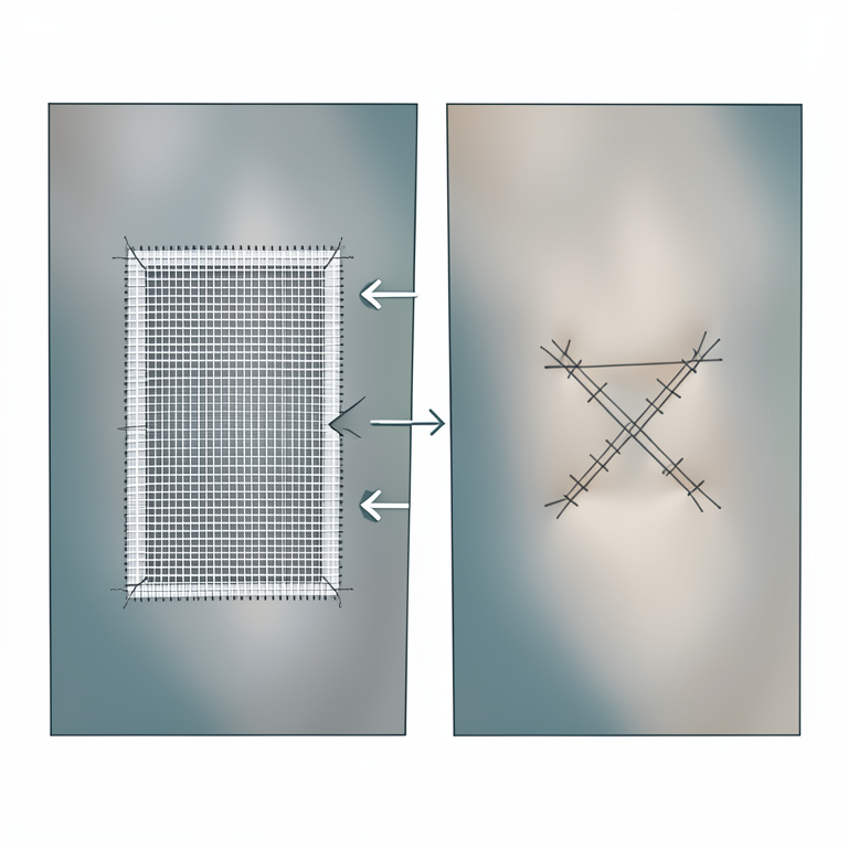
Comparison of Hernia Repair Techniques
AI-generated conceptual illustration (calf abdominal wall). It explains the idea and does not show study data.
View source evidence ↗Figures & tables
From the original paper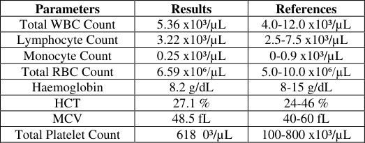 Table 1
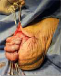 Figure 1
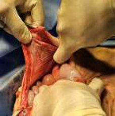 Figure 2
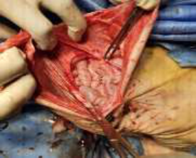 Figure 2
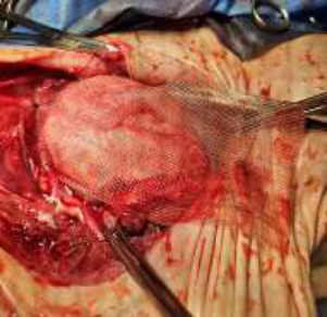 Figure 4
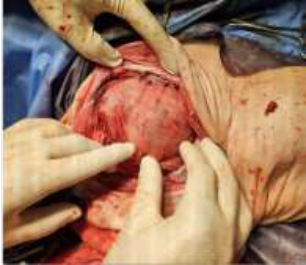 Figure 5
Introduction
Weakening or tear in the abdominal wall or diaphragm, or from natural openings such as the inguinal or femoral canal leading to the protrusion of organs resulting in hernia (Venugopalan, 1992) [14] . Hernias can be reducible or non-reducible (Farman et al., 2018) [7] . Abdominal hernias can be categorized into, external and internal hernias. External abdominal hernias can result from defects in the abdominal wall. In contrast, internal abdominal hernias occur through a hernia ring located within the abdomen or thorax. According to the location, abdominal hernias can be categorized as, ventral, prepubic, subcostal, and lateral hernias (Fossum, 2013) [8] . Trauma to the abdomen such as, kicks from cows, accidents, bite wounds, or congenital factors can increase the likelihood of developing an abdominal hernia. Young animals are mostly affected. A thorough examination of the herniated mass is important for the accurate diagnosis of the herniated contents and determining the appropriate method of correction. Hernia correction methods include the use of elastoplasts, blisters, ligatures, or clamps, as well as surgical options such as herniorrhaphy or hernioplasty (Salim et al., 2015) [13] .
Material and methods
The case presented here reports the presence of ventral hernia in a 2 month old male jersey calf and its successful surgical management using non absorbable synthetic polypropylene mesh. The herniated mass was found towards the caudo-ventral aspect of the abdomen with no external injuries over the swelling, animal showed normal physiological parameters, hematological values were normal (Table 1). The palpated mass was elastic in consistency, was reducible significantly. The mass was differentiated from abscess, hematoma, tumor and cyst through palpation.
| Parameters | Results | References |
|---|---|---|
| Total WBC Count | 5.36 x10³/µL | 4.0-12.0 x10³/µL |
| Lymphocyte Count | 3.22 x10³/µL | 2.5-7.5 x10³/µL |
| Monocyte Count | 0.25 x10³/µL | 0-0.9 x10³/µL |
| Total RBC Count | 6.59 x10⁶/µL | 5.0-10.0 x10⁶/µL |
| Haemoglobin | 8.2 g/dL | 8-15 g/dL |
| HCT | 27.1 % | 24-46 % |
| MCV | 48.5 fL | 40-60 fL |
| Total Platelet Count | 618 0³/µL | 100-800 x10³/µL |
Table text extracted from the original PDF; superscript letters are shown inline. The original table image is below.
View original table image
Management and Treatment
The animal was placed in the right lateral recumbency. Procedure was done under recumbancy stun anaethesia protocol, Inj.Butorphanol @ 0.1mg/kg body weight, Inj.Xylazine @ 0.05mg/kg body weight and Inj.Ketamine @ 0.5mg/kg body weight, intramuscularly. Anaesthesia was maintained under Ketamine-Diazepam combination @ 1:1 ratio, intravenously. The surgical site was aseptically prepared by clipping, shaving and scrubbed the site using povidone iodine solution. 2% Lignocaine was infiltered over the site of incision. A curvilinear incision of about 10-11 cm was made above the hernial swelling using BP blade of size 22. The skin was detached from subcutaneous tissue by blunt dissection, exposing hernial contents (intestine). The skin and muscular layer were grasped using tissue holding forceps (Allis tissue forceps) to get sufficient surgical field and expose hernial ring ( The second hernia towards the caudal abdomen was corrected by herniorrhaphy. By a combination of blunt and sharp dissection, the hernial ring of about 3 cm diameters was exposed and the intestinal contents were replaced into the abdominal cavity before suturing. The hernial ring was then freshened and closed with a double-breasted suture pattern using absorbable polyglycolic acid 1-0. Excess skin was removed, and the wound was finally closed with a crossmattress suture pattern using nylon 1-0. Aluspray was topically applied over the suture site. Inj.Streptopenicillin @ 2ml/50kg bodyweight and Inj.Flunixin @ 1.1mg/kg body weight were given intramuscular postoperatively and continued the same for 4 more days. Advised complete rest and restricted feeding for 2 weeks.
Result and Discussion
The congenital ventral hernia in the calf was successfully repaired using a hernioplasty procedure with non-absorbable polypropylene mesh. The mesh was securely anchored to the muscle using polyamide 1-0 sutures in a simple interrupted pattern. The second hernial ring towards caudal abdomen was corrected by herniorrhaphy in double-breasted suture pattern using absorbable polyglycolic acid 1-0. Post-operatively, the calf exhibited a smooth recovery without any complications. Ventral hernias results from the displacement of internal organs through a breach in the abdominal wall. More frequently observed in cattle and horses (Venugopalan, 1992) [14] . Congenital ventral hernia could be attributed to decreased muscle strength, which led to enlargement over time.
Excessive manipulation of young calves during calving can contribute to ventral hernias. The strain on the calf's muscles can weaken the abdominal wall, making it more susceptible to herniation (Witte et al., 2008) [16] . The use of mesh for hernia repair has gained popularity as it has been shown to reduce the risk of recurrence compared to suture repair ( [10] , [5] , [9] . Polypropylene mesh is a common prosthetic tool for reconstructing abdominal wall hernias exceeding 3 cm in length (Ober et al.,2007; Vilar et al.,2009; Sadan et al.,2019) [11] , [15] , [12] . By reducing tension on the wound, this technique effectively lowers the likelihood of hernia recurrence compared to traditional suturing (Burger et al.,2004; Finan et al.,2009; Abouelnasr et al.,2017) [4] , [6] , [1] . Polypropylene is commonly used for large hernia defect correction due to its strong and flexible nature. Its unique properties, including ease of handling, tissue compatibility, and infection resistance, make it a popular choice among surgeons. The monofilament nature of the polypropylene facilitates fibrovascular ingrowths (Bilsel and Abci, 2012) [3] . The chance of recurrence is generally higher with herniorrhaphy compared to hernioplasty, which includes the use of mesh to reinforce the repair, usually provides a more durable repair and reduces the recurrence rate. Primary closure of congenital hernias can lead to minor complications such as wound infections, abscess, seromas, recurrence and risk of strangulation, particularly in umbilical hernias (Adam and Fessler, 2000) [2] .
Conclusion
Non-absorbable synthetic polypropylene mesh proved effective in correcting large ventral hernias in calves. After the recommended rest period, the animals showed satisfactory recovery, with the suture site remaining intact. For optimal recovery, a light feeding regimen was advised for 1-2 weeks.
Our results showed that polypropylene mesh was a viable option for large ventral hernias, while herniorraphy proved to be a better choice for smaller hernias in calf.
Acknowledgements
The authors are thankful to the Senior Veterinary Surgeon, Veterinary Polyclinic, Perambra, Kerala.
Conflict of Interest
Authors declare no conflict of interest.
Financial Support
Financial Support
Figures and tables from the paper
References
13 in the original paper- Abouelnasr K, Hamed M, Lashen S, El-Adl M, Eltaysh R, Tagawa M. Enhancement of abdominal wall defect repair using allogenic platelet-rich plasma with commercial polyester/cotton fabric (Damour) in a canine model. Journal of Veterinary Medical Science. 2017;79:1301-1309.
- Bilsel Y, Abci I. The search for ideal hernia repair: mesh materials and types. International Journal of Surgery. 2012;10:317-321.
- Burger JW, Luijendijk RW, Hop WC, Halm JA, Verdaasdonk EG, Jeekel J. Long-term follow-up of a randomized controlled trial of suture versus mesh repair of incisional hernia. Annals of Surgery. 2004;204:578583.
- Christoffersen MW, Helgstrand F, Rosenberg J, Kehlet H, Strandfelt P, Bisgaard T. Long-term recurrence and chronic pain after repair for small umbilical or epigastric hernias: a regional cohort study. American Journal of Surgery. 2015;209:725-732.
- Finan KR, Kilgore ML, Hawn MT. Open suture versus mesh repair of primary incisional hernias: a cost-utility analysis. Hernia. 2009;13:173-182.
- Farman R, Al-Husseiny S, Ameer A. Surgical treatment of hernia in cattle: A review. Al-Qadisiyah Journal of Veterinary Medicine Sciences. 2018;17:61-68. doi:10.29079/vol17iss2art506.
- Kaufmann R, Halm JA, Eker HH, Klitsie PJ, Nieuwenhuizen J, van Geldere D, et al. Mesh versus suture repair of umbilical hernia in adults: a randomized, double-blind, controlled, multicenter trial. Lancet. 2018;391:860-869.
- Mosbah E, Abouelnasr KS. Commercial polyester/cotton fabric (Damour): a novel prosthetic material for hernioplasty in ruminants. International Journal of Veterinary Research. 2015;16(1):105-109.
- Ober C, Muste A, Mates N, Oana L, Beteg F, Vancea C. Techniques for implanting prosthetic meshes of polypropylene in the repair of abdominal wall defects in horses. Bulletin of University of Agricultural Sciences and Veterinary Medicine Cluj-Napoca. 2007;64:509-513.
- Sadan M, El-Sayed El Shafaey, El-Khodary S. J Vet Med Sci. 2019 May;81(5):675-681.
- Salim Md, Mirza Abul Hashim, Nasrin Sultana Juyena, Yasir Ahammad Arafat, Roney Kumar Dey, Abdus Sattar Bag M, et al. Prevalence of hernia and evaluation of herniorrhaphy in calves. International Journal of Natural and Social Sciences. 2015;2(4):35-43.
- Vilar JM, Doreste F, Spinella G, Valentin S. Double layer mesh hernioplasty for repair of incisional hernias in 15 horses. Journal of Equine Veterinary Science. 2009;29:172-176.
- Witte S, Rodgerson D, Hunt R, Spirito MA. Traumatic ventral herniation in foals as a complication of dystocia. Compendium on Equine Medicine. 2008;3:137-143.