
Research article · 2024 · Full text
International Journal of Veterinary Sciences and Animal HusbandryPolycystic kidney disease with concurrent uroabdomen in a persian cat: A case study
Volume 9(5) · Pages 431–434 · DOI: 10.22271/veterinary.2024.v9.i5g.1745 ↗
Research areasGenetic and Kidney Cyst DiseasesPediatric Urology and Nephrology StudiesUrological Disorders and Treatments
Abstract
Author-writtenA one-year old male Persian cat was presented with a history of vomiting, anorexia and anuria. The cat was dehydrated, hyperthermic, with tacky pale mucous membranes and painless abdominal distension on clinical examination. The serum biochemistry revealed azotaemia. The abdominal ultrasonography revealed intact bladder, anechoic fluid in peritoneal cavity and renomegaly with multiple well demarcated anechoic cysts (4-7 mm). Diagnostic and therapeutic peritoneocentesis and cystocentesiswere performed. Uroabdomen secondary to feline idiopathic cystitis (FIC) and concurrent polycystic kidney disease (PCKD) was diagnosed. The cat was stabilized with intravenous fluid therapy and necessary supportive medications. Iodine, glycosaminoglycan and taurine supplement were given orally for a period of two months. The haemato-biochemical values returned to normal range with no detectable abdominal fluid on the 14th day follow up. This case report highlights the likelihood of comorbidity between PCKD and FIC in un-neutered male Persian cats.
Summary
Source-checkedPlain-language reading of the full paper, prepared with AI and checked against the text. Each point links to the authors' own words.
Why it matters
- Polycystic kidney disease (PCKD) is an inherited condition common in Persian and Persian-related cat breeds, characterized by the formation of cysts in the kidney tissue. View evidence ↗
- Feline idiopathic cystitis (FIC) is identified as the primary cause of lower urinary tract disease in cats. View evidence ↗
The question
- The study aims to highlight the potential for concurrent occurrence (comorbidity) of PCKD and FIC in un-neutered male Persian cats. View evidence ↗
What they did
- The subject of the case study was a one-year-old male Persian cat presenting with vomiting, anorexia, and anuria. View evidence ↗
- Diagnostic methods included abdominal radiography and ultrasonography using a 7.5 MHz linear probe to assess the abdomen and kidneys. View evidence ↗
- The cat was sedated with ketamine (5mg/kg), midazolam (0.2 mg/kg), and butorphanol (0.2 mg/kg) to allow for transurethral catheterization and peritoneocentesis. View evidence ↗
- Treatment included a two-month oral regimen of renal supplements, glycosaminoglycan, zinc, iodine, and taurine. View evidence ↗
What they found
- Ultrasonography revealed enlarged kidneys containing multiple anechoic cysts measuring between 4 and 7 mm. View evidence ↗
- Peritoneocentesis resulted in the collection of approximately 100 ml of blood-tinged cloudy fluid from the peritoneal cavity. View evidence ↗
- By the 14th day follow-up, the cat exhibited normal urination, no detectable abdominal fluid on ultrasonography, and normal hematobiochemical values. View evidence ↗
What it means
- The authors conclude that a combination of transurethral catheterization, peritoneocentesis, fluid stabilization, and multimodal therapy effectively improved the clinical signs. View evidence ↗
- The authors recommend regular monitoring of renal function to watch for future recurrences of the condition. View evidence ↗
Visual summary
1 visualsIllustrations explain the paper; they do not show study data.
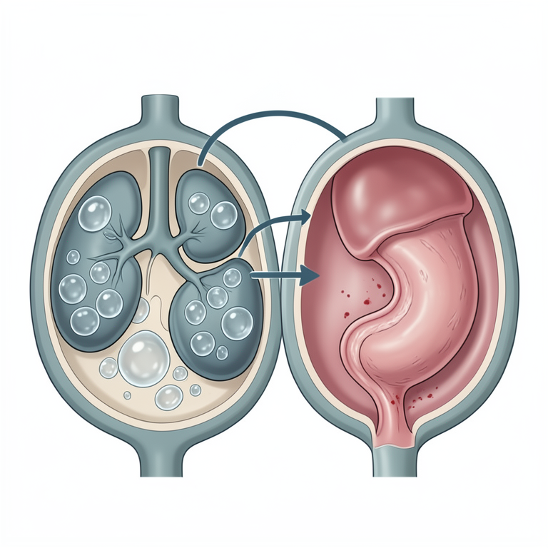
Comorbidity Concept
AI-generated conceptual illustration (Persian cat). It explains the idea and does not show study data.
View source evidence ↗Figures & tables
From the original paper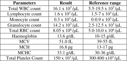 Table 1
 Table 2
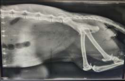 Figure 1
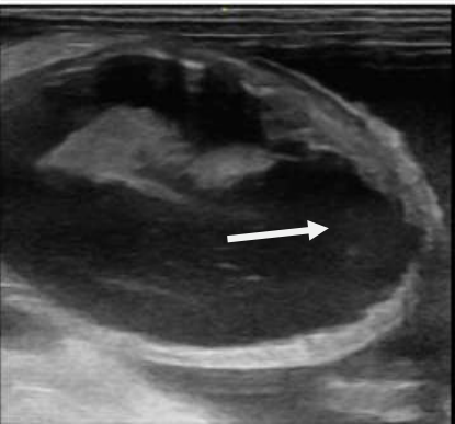 Figure 2
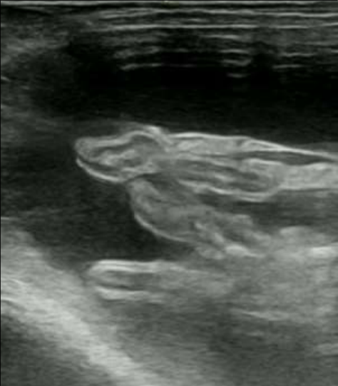 Figure 3
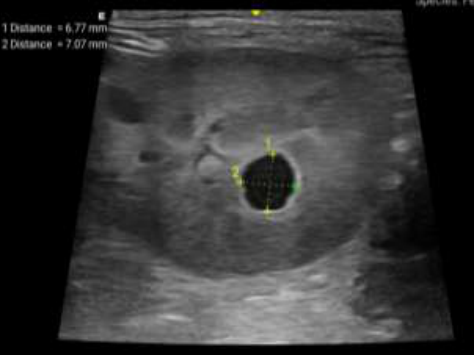 Figure 4
Introduction
The Polycystic kidney disease is autosomal inherited disease commonly seen in domestic cats especially Persian/ Persian related breeds which are characterized by typical cyst formation in renal parenchyma (Domanjko-Petrič et al., 2008) [5] . The cysts can originate in proximal tubules, distal tubules, renal cortex or medulla (Sato et al., 2019; Bilgen et al., 2020; Guerra et al., 2020) [11,2,8] . Feline idiopathic cystitis (FIC) is the leading cause of feline lower urinary tract disease (Forrester and Towell, 2015) [6] . Feline lower urinary tract diseases are characterized by periuria, hematuria, dysuria, pollakiuria, with/without urinary obstruction. Feline lower urinary tract symptoms can be attributed to a wide range of conditions, including bacterial, fungal, and parasitic urinary tract infections; different forms of uroliths and urethral plugs; neoplasia; congenital or acquired anatomical or morphological abnormalities; and factors such astrauma, neurogenic issues, or iatrogenic causes (Kruger et al., 2009) [10] . Common clinical signs of non-obstructive FIC include periuria, pollakiuria, stranguria, and gross hematuria (Westropp and Buffington, 2004) [14] . Sometimes these symptoms may improve over time without treatment, but they usually reoccur. In fact, recurrences with acute idiopathic cystitis are seen within one to two years of the disease. Considering factors such as lower urinary tract symptoms and the moisture content of the cat's food, the relative risk of the cat experiencing another episode of lower urinary tract issue within a 2-year period is about 65% (Kruger et al.,2009) [10] .
Materials and methods
A one-year old male Persian cat was presented with a history of vomiting, anorexia and anuria to the Outpatient unit of Department of Veterinary Medicine, Teaching Veterinary Clinical Complex, College of Veterinary & Animal Sciences, Pookode. The cat was subjected to systematic clinical and laboratory examinations. The cat was dehydrated with tacky pale mucus membrane, dull and depressed mentation with painless distension of abdomen. The complete blood count revealed elevated granulocyte count with thrombocytopenia (Table 1), serum biochemistry revealed elevated levels of serum creatinine, blood urea nitrogen and inorganic phosphorus (Table 2). Based on anamnesis and the imaging studies the cat was diagnosed with polycystic kidney disease and concurrent uroabdomen as a result of ongoing feline idiopathic cystitis.
| Parameters | Day 1 | Day 14 | Normal range |
|---|---|---|---|
| Creatinine | 11.28 mg/dL | 1.096 mg/dL | 0.9-2.2 mg/dL |
| Blood Urea Nitrogen | 105.62 mg/dL | 19.81 mg/dL | 19-34 mg/dL |
| Phosphorus | 12.01 mg/dL | 4.104 mg/dL | 3.0-6.1 mg/dL |
Table text extracted from the original PDF; superscript letters are shown inline. The original table image is below.
View original table image
| Parameters | Result | Reference range |
|---|---|---|
| Total WBC count | 16.1 x 10³ /μL | 5.5-19.5 x 10³ /μL |
| Lymphocyte count | 1.6 x 10³ /μL | 1.5-7 x 10³ /μL |
| Monocyte count | 0.3 x 10³ /μL | 0-0.9 x 10³ /μL |
| Granulocyte count | 14.2 x 10³ /μL | 2.5-12.5 x 10³ /μL |
| Total RBC count | 8.05 x 10⁶ /μL | 5.0-10.0 x 10⁶ /μL |
| Haemoglobin | 13.6 g/dL | 10-15 g/dL |
| MCV | 51.0 fL | 39-55 fL |
| MCH | 16.8 pg | 13-17 pg |
| MCHC | 33.1 g/dL | 30-36 g/dL |
| Total Platelet Count | 150 x 103 /μL | 300-800 x103 /μL |
Table text extracted from the original PDF; superscript letters are shown inline. The original table image is below.
View original table image
Results and Discussion
The cat was initially sedated with Inj. Ketamine @ 5mg/kg BW, Inj. Midazolam @ 0.2 mg/kg BW, Inj. Butorphanol @ 0.2 mg/kg BW intravenously for transurethral catheterization and peritoneocentesis; catheterization was done using siliconecat tube size FG 3 andturbid urine was evacuated. Peritoneocentesis was done using sterile 22G butterfly needle; about 100 ml of blood tinged cloudy fluid was collected from the peritoneum. Urinary bladder and peritoneal cavity were flushed with warm normal saline followed by metronidazole. Animal was stabilized with Inf. normal saline @ 10 ml/kg BW, Inf. ringer's lactate @ 30 ml/kg BW and Inj. sodium bicarbonate 7.5% @ 2mEq/Kg BW intravenously over 20 minutes.Treatment was initiated with Inj. amoxicillinsulbactam @ 12.5 mg/kg BW, q12h, Inj. pantoprazole @ 1mg/kg BW, q24h, and vitamin B complex intravenously. Sevelamer carbonate @ 200 mg PO q12h, bethanechol @ 5 mg per cat, TID and ethamsylate @ 125 mg per cat, BIDwere administered orally. Renalsupplements (RKleen and Nephrotec DS), glycosaminoglycan supplement (Cystopro), zinc and iodine supplement (Syr. Zincovit), taurine supplement (Syr. Catstar) were given orally for two months. The cat showed significant clinical improvement after 2 weeks of treatment. Animal showed normal urination, no abdominal fluids were noticed on ultrasonography and hematobiochemical values were well within normal range on 14 th day follow up (Table 2). Most of the clinical signs of the renal diseases could be directly related to azotemia and subsequent uremia (Breshears & Confer, 2017) [3] . Metabolic acidosis occurs when there is an imbalance of excess plasma anions over cations, leading to a decrease in plasma pH. In such cases, administering sodium bicarbonate can be beneficial for patients who have lost sodium bicarbonate due to diarrhea or renal proximal tubular acidosis (Adeva-Andany et al., 2014) [1] . Renal ultrasonography of cats was suggested as an early tool for the diagnosis of renal diseases (Debruyn et al., 2012) [4] . Sensitivity of USG in the detection of PKD was 91% (Domanjko-Petrič et al., 2008) [5] . Midazolam @ 0.3 mg/kg, butorphanol @ 0.3 mg/kg and ketamine @ 5 mg/kg when administered intramuscularly produces effective chemical restraint with minimal cardiovascular effects and is suitable for most minor procedures (Simon and Steagall, 2020) [12] . Amoxycillin/clavulanic acid @ 7 to 39 mg/kg twice daily is commonly used as an empirical antibiotic for cats with UTI (Gerber et al., 2008) [7] . Iodine when supplemented orally had anti-proliferative property and aids in cyst regression by iodination of lipids which are present in cystic fluid. Oral iodine, pre/probiotic supplementation, low glycemic index cat food and broccoli powder were found effective in treatment of PCKD in cats (Ural et al., 2021) [13] .
The glycosaminoglycan (GAG) layer on the bladder wall is vital for shielding the transitional epithelium. It enables the bladder to handle ongoing urine exposure and controls how urinary substances interact with the epithelial cells. Damage to the urothelium plays a major role in many chronic bladder disorders. Oral supplementation of GAG showed improvement in clinical signs in cats with FIC (Gunn-Moore & Shenoy, 2004) [9] . Vitamin and renal supplements help in improving immunity and in the regeneration of the functionality of the kidneys. During the course of treatment, the cat showed uneventful recovery from the condition. Hematobiochemical values were observed within the normal range during the follow-up.
Conclusion
Feline idiopathic cystitis and PCKD can lead to a variety of clinical signs and complications. Ultrasonographic diagnostic imaging was found much helpful for the early and proper diagnosis of the condition. Post renal azotemia was identified with hyperphosphatemia. Transurethral catheterization, peritoneocentesis, proper stabilization of the patient with parenteral fluids and multimodal therapy was helpful in amelioration of the clinical signs. The animal showed uneventful recovery from the condition. Regular monitoring of the renal function should be done for surveillance of the recurrences in future.
Acknowledgement
AcknowledgementsThe authors are thankful to the Dean, Director of Clinics and Head of TVCC, College of Veterinary and Animal Sciences, Pookode.
Figures and tables from the paper
References
12 in the original paper- Adeva-Andany MM, Fernández-Fernández C, MouriñoBayolo D, Castro-Quintela E, Domínguez-Montero A. Sodium bicarbonate therapy in patients with metabolic acidosis. Sci World J. 2014;2014:627673.
- Bilgen N, Bişkin Türkmen M, Çınar Kul B, Isparta S, Şen Y, Akkurt MY, et al. Prevalence of PKD1 gene mutation in cats in Turkey and pathogenesis of feline polycystic kidney disease. J Vet Diagn Invest. 2020;32(4):549-555.
- Debruyn K, Haers H, Combes A, Paepe D, Peremans K, Vanderperren K, Saunders JH. Ultrasonography of the feline kidney: technique, anatomy and changes associated with disease. J Feline Med Surg. 2012;14(11):794-803.
- Domanjko-Petrič A, Černec D, Cotman M. Polycystic kidney disease: a review and occurrence in Slovenia with comparison between ultrasound and genetic testing. J Feline Med Surg. 2008;10(2):115-119.
- Forrester SD, Towell TL. Feline idiopathic cystitis. Vet Clin North Am Small Anim Pract. 2015;45(4):783-806.
- Gerber B, Eichenberger S, Reusch CE. Guarded longterm prognosis in male cats with urethral obstruction. J Feline Med Surg. 2008;10(1):16-23.
- Guerra JM, Pellegrino A, Daniel AG, Freitas MF, Cardoso NC, Pessoa RB, et al. Cardiac structural and functional findings in Persian cats with autosomal dominant polycystic kidney disease. Ciênc Rural, 2020, 50(9).
- Gunn-Moore DA, Shenoy CM. Oral glucosamine and the management of feline idiopathic cystitis. J Feline Med Surg. 2004;6(4):219-225.
- Kruger JM, Osborne CA, Lulich JP. Changing paradigms of feline idiopathic cystitis. Vet Clin North Am Small Anim Pract. 2009;39(1):15-40.
- Sato R, Uchida N, Kawana Y, Tozuka M, Kobayashi S, Hanyu, et al. Epidemiological evaluation of cats associated with feline polycystic kidney disease caused by the feline PKD1 genetic mutation in Japan. J Vet Med Sci. 2019;81(7):1006-1011.
- Simon BT, Steagall PV. Feline procedural sedation and analgesia: when, why and how. J Feline Med Surg. 2020;22(11):1029-1045.
- Ural K, Mutlu H, Erdogan H, Erdogan S. Iodine is one of the odds for therapeutic armamentarium against feline polycystic renal disease. Int J Vet Anim Res. 2021;4(2):63-68.