
Research article · 2024 · Full text
International Journal of Veterinary Sciences and Animal HusbandryInvestigation of haematological alterations in ancylostomosis in dogs of Thrissur, Kerala
Volume 9(5) · Pages 378–380 · DOI: 10.22271/veterinary.2024.v9.i5f.1735 ↗
Research areasVeterinary Oncology ResearchVeterinary Orthopedics and NeurologyComparative Animal Anatomy Studies
Abstract
Author-writtenThe study investigated haematological alterations in dogs infected with Ancylostoma spp. in Thrissur district, Kerala, over a one-year period. Blood samples from 50 infected dogs and 10 healthy controls were analysed for complete blood count (CBC) using a five-part automatic haematological analyser. Significant changes were observed in several parameters: infected dogs exhibited elevated neutrophil and basophil counts, along with reduced red blood cell count (RBC), haemoglobin (Hb), volume of packed red cells (VPRC) and mean corpuscular haemoglobin (MCH). Platelet distribution width (PDW) was also significantly increased in infected animals. The findings indicated that dogs with ancylostomosis suffer from normocytic hypochromic regenerative anaemia, likely due to chronic blood loss and nutrient deficiencies. These results provide valuable insights into the haematological impact of canine ancylostomosis and its associated anaemia, aiding in the diagnosis and management of infected animals.
Summary
Source-checkedPlain-language reading of the full paper, prepared with AI and checked against the text. Each point links to the authors' own words.
Why it matters
- Ancylostomosis is caused by hookworms, which are the primary gastrointestinal parasites affecting dogs globally. View evidence ↗
- The parasite Ancylostoma caninum is particularly pathogenic because a single worm can consume up to 0.5 mL of blood daily. View evidence ↗
- These parasites pose a public health risk as they are zoonotic and can cause eosinophilic enteritis and cutaneous larva migrans in humans. View evidence ↗
The question
- The study aimed to examine and correlate blood parameter variations between dogs infected with hookworms and healthy control dogs in Thrissur, Kerala. View evidence ↗
What they did
- The research took place in various areas of the Thrissur district in Kerala between August 2023 and August 2024. View evidence ↗
- Blood samples were analyzed on the day of collection using an Orphee Mythic 5 vet PRO five-part automatic haematological analyzer. View evidence ↗
- The researchers used independent t-tests to statistically compare the blood parameters between the two groups. View evidence ↗
What they found
- Infected dogs had significantly higher mean neutrophil counts (12.55 ± 0.95 × 10⁹/L) than control dogs (6.59 ± 0.93 × 10⁹/L), indicating an inflammatory response. View evidence ↗
- Basophil counts were significantly higher in the infected group (0.457 ± 0.082 × 10⁹/L) compared to the control group (0.102 ± 0.054 × 10⁹/L). View evidence ↗
- The mean red blood cell (RBC) count was significantly reduced in infected dogs (4.9 ± 0.2 × 10⁶/µL) compared to controls (6.82 ± 0.24 × 10⁶/µL). View evidence ↗
- Hemoglobin (Hb) levels were significantly lower in infected dogs (10.49 ± 0.48 g/dL) than in the control group (15.26 ± 0.5 g/dL). View evidence ↗
- The volume of packed red cells (VPRC) was significantly lower in infected animals (30.6 ± 1.33%) compared to controls (45.75 ± 1.34%). View evidence ↗
- Mean corpuscular hemoglobin (MCH) was significantly lower in infected dogs (22.08 ± 0.7 pg) than in control dogs (25.65 ± 1.5 pg), indicating hypochromic anemia. View evidence ↗
- Infected dogs showed a significantly higher percentage of nucleated red blood cells (NRBC) (388.09 ± 99.44%) compared to controls (14.63 ± 3.77%), suggesting a regenerative response. View evidence ↗
- Platelet distribution width (PDW) was significantly increased in infected dogs (16.17 ± 0.72) compared to the control group (11.01 ± 1.94). View evidence ↗
What it means
- The authors concluded that dogs infected with Ancylostoma spp. exhibit normocytic hypochromic regenerative anemia. View evidence ↗
- The authors suggest that the significant increase in PDW indicates potential changes in platelet function due to the infection. View evidence ↗
Visual summary
3 visualsIllustrations explain the paper; charts are replotted from the paper's own table values.
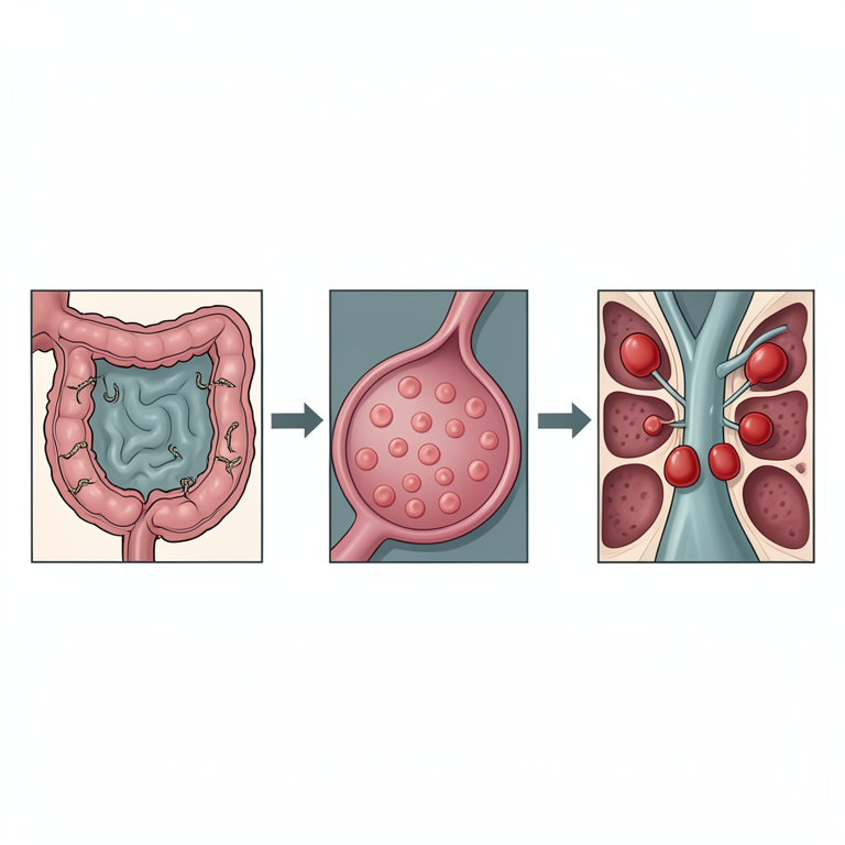
Mechanism of Regenerative Anemia in Ancylostomosis
AI-generated conceptual illustration (domestic dogs and hookworm parasites). It explains the idea and does not show study data.
View source evidence ↗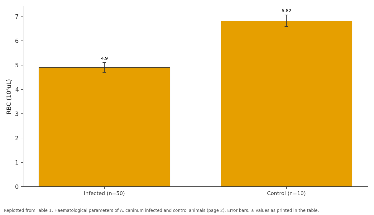
RBC (10⁶սL): Infected (n=50) vs Control (n=10)
Replotted from Table 1 (page 2) of the original paper. Every bar is the paper's own table value.
View source evidence ↗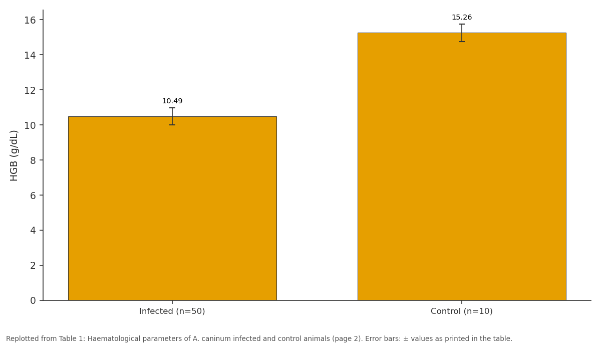
HGB (g/dL): Infected (n=50) vs Control (n=10)
Replotted from Table 1 (page 2) of the original paper. Every bar is the paper's own table value.
View source evidence ↗Figures & tables
From the original paper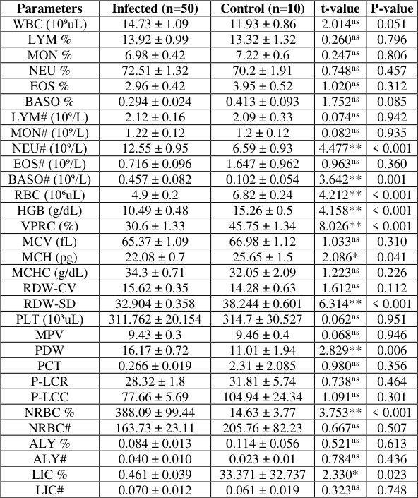 Table 1
Introduction
Hookworms are the major canine gastrointestinal parasites worldwide causing ancylostomosis. Ancylostoma caninum, Ancylostoma braziliense and Uncinaria stenocephala are common hookworm species of dogs. Transmission of ancylostomosis mainly occurs through contaminated soil and transcolostral route. Puppies under three months of age are more susceptible to infection (Dos Santos et al., 2020) [4] . Each A. caninum worm can ingest up to 0.5 mL of blood per day (Traversa, 2012) [9] . Among hookworms A. caninum is the most pathogenic (Dantas-Torres et al. 2020) [3] and have large buccal capsule with three pairs of ventral teeth and a pair of dorsal teeth (Uppal et al., 2017) [10] . Ancylostomosis in dogs causes a range of symptoms from mild enteritis to fatal haemorrhagic diarrhoea accompanied by anaemia and weight loss. The severity depends on various factors, such as the age of the host, the parasitic load and the specific species of hookworm involved (Taylor et al., 2016; Campos et al. 2017) [8,2] . Severe anaemia may develop in adult animals. However, its clinical presentation is often linked to malnutrition and the presence of concurrent chronic diseases. Ancylostoma spp. present significant public health risks due to their zoonotic potential, causing cutaneous larva migrans in humans following exposure to environmental third-stage larvae (L 3 ), as well as eosinophilic enteritis. The study on haematotological changes in canine ancylostomosis was limited in Kerala even though the prevalence was high. So the current study was conducted to examine and correlate haematological alterations between infected and control dogs of Thrissur, providing insights into infection-induced blood parameter variations.
Materials and Methods
The study was conducted on dogs in different areas of Thrissur district, Kerala from August 2023 to August 2024. Fifty dogs with ancylostomosis as well as 10 healthy animals were selected and two millilitre of blood collected aseptically using sterile syringe and needle and transferred to vacutainer tubes containing ethylene diamine tetraacetic acid (EDTA) and CBC was taken using a five part automatic haematological analyser Orphee Mythic 5 vet PRO on the same day of collection at University Veterinary hospital, Kokkalai, Thrissur for haematological analysis. Statistical analyses for comparison of each parameter was done by independent t-test.
Results
Haematological analysis revealed significant differences between infected dogs and control dogs. Among leukocytic parameters, the mean neutrophil and basophil counts were significantly elevated in infected animals. The mean neutrophil count was notably higher in the infected group (12.55 ± 0.95 × 10⁹/L) compared to the control group (6.59 ± 0.93 × 10⁹/L), with a t-value of 4.477 and a P-value of < 0.001. Similarly, the basophil count was significantly elevated in the infected group (0.457 ± 0.082 × 10⁹/L) compared to the control group (0.102 ± 0.054 × 10⁹/L), with a t-value of 3.642 and a P-value of 0.001. The mean red blood cell count (RBC) was significantly lower in the infected group (4.9 ± 0.2 × 10⁶/µL) than in the control (6.82 ± 0.24 × 10⁶/µL), with a t-value of 4.212 and a P-value of < 0.001. The mean Hb levels were also reduced in the infected group (10.49 ± 0.48 g/dL) compared to the control group (15.26 ± 0.5 g/dL), with a t-value of 4.158 and a Pvalue of < 0.001. The mean VPRC was markedly lower in the infected group (30.6 ± 1.33%) compared to the control group (45.75 ± 1.34%), with a t-value of 8.026 and a P-value of < 0.001. The mean corpuscular haemoglobin (MCH) in infected animals (22.08 ± 0.7 pg) was significantly lower than in control animals (25.65 ± 1.5 pg), with a t-value of 2.086 and a P-value of 0.041. Furthermore, the mean red cell distribution width-standard deviation (RDW-SD) was significantly reduced in the infected group (32.904 ± 0.358 fL vs. 38.244 ± 0.601 fL), with a P-value of < 0.001. The percentage of nucleated red blood cells (NRBC) was significantly higher in the infected group (388.09 ± 99.44%) compared to the control (14.63 ± 3.77%), with a t-value of 3.753 and a P-value of < 0.001. The mean value of large immature cell (LIC) was significantly decreased in infected animals (0.461 ± 0.039) with a t-value of 2.330 and a P-value of 0.023. The mean PDW was significantly increased in the infected group (16.17 ± 0.72) compared to the control group (11.01 ± 1.94), with a t-value of 2.829 and a P-value of 0.006. No significant differences were observed for the other parameters. The Comparison of haematological parameters between infected and control animals is given in Table 1.
| Parameters | Infected (n=50) | Control (n=10) | t-value | P-value |
|---|---|---|---|---|
| WBC (10⁹սL) | 14.73 ± 1.09 | 11.93 ± 0.86 | 2.014ns | 0.051 |
| LYM % | 13.92 ± 0.99 | 13.32 ± 1.32 | 0.260ns | 0.796 |
| MON % | 6.98 ± 0.42 | 7.22 ± 0.6 | 0.247ns | 0.806 |
| NEU % | 72.51 ± 1.32 | 70.2 ± 1.91 | 0.748ns | 0.457 |
| EOS % | 2.96 ± 0.42 | 3.95 ± 0.52 | 1.020ns | 0.312 |
| BASO % | 0.294 ± 0.024 | 0.413 ± 0.093 | 1.752ns | 0.085 |
| LYM# (10⁹/L) | 2.12 ± 0.16 | 2.09 ± 0.33 | 0.074ns | 0.942 |
| MON# (10⁹/L) | 1.22 ± 0.12 | 1.2 ± 0.12 | 0.082ns | 0.935 |
| NEU# (10⁹/L) | 12.55 ± 0.95 | 6.59 ± 0.93 | 4.477** | < 0.001 |
| EOS# (10⁹/L) | 0.716 ± 0.096 | 1.647 ± 0.962 | 0.963ns | 0.360 |
| BASO# (10⁹/L) | 0.457 ± 0.082 | 0.102 ± 0.054 | 3.642** | 0.001 |
| RBC (10⁶սL) | 4.9 ± 0.2 | 6.82 ± 0.24 | 4.212** | < 0.001 |
| HGB (g/dL) | 10.49 ± 0.48 | 15.26 ± 0.5 | 4.158** | < 0.001 |
| VPRC (%) | 30.6 ± 1.33 | 45.75 ± 1.34 | 8.026** | < 0.001 |
| MCV (fL) | 65.37 ± 1.09 | 66.98 ± 1.12 | 1.033ns | 0.310 |
| MCH (pg) | 22.08 ± 0.7 | 25.65 ± 1.5 | 2.086* | 0.041 |
| MCHC (g/dL) | 34.3 ± 0.71 | 32.05 ± 2.09 | 1.223ns | 0.226 |
| RDW-CV | 15.62 ± 0.35 | 14.28 ± 0.63 | 1.612ns | 0.112 |
| RDW-SD | 32.904 ± 0.358 | 38.244 ± 0.601 | 6.314** | < 0.001 |
| PLT (10³սL) | 311.762 ± 20.154 | 314.7 ± 30.527 | 0.062ns | 0.951 |
| MPV | 9.43 ± 0.3 | 9.46 ± 0.4 | 0.068ns | 0.946 |
| PDW | 16.17 ± 0.72 | 11.01 ± 1.94 | 2.829** | 0.006 |
| PCT | 0.266 ± 0.019 | 2.31 ± 2.085 | 0.980ns | 0.356 |
| P-LCR | 28.32 ± 1.8 | 31.81 ± 5.74 | 0.738ns | 0.464 |
| P-LCC | 77.66 ± 5.69 | 104.94 ± 24.34 | 1.091ns | 0.301 |
| NRBC % | 388.09 ± 99.44 | 14.63 ± 3.77 | 3.753** | < 0.001 |
| NRBC# | 163.73 ± 23.11 | 205.76 ± 82.23 | 0.667ns | 0.507 |
| ALY % | 0.084 ± 0.013 | 0.114 ± 0.056 | 0.521ns | 0.613 |
| ALY# | 0.040 ± 0.010 | 0.023 ± 0.01 | 0.784ns | 0.436 |
| LIC % | 0.461 ± 0.039 | 33.371 ± 32.737 | 2.330* | 0.023 |
| LIC# | 0.070 ± 0.012 | 0.061 ± 0.019 | 0.323ns | 0.748 |
Table text extracted from the original PDF; superscript letters are shown inline. The original table image is below.
View original table image
Discussion
In the present study, out of the 31 parameters checked 10 parameters showed significant difference between infected and control group. The mean total leucocyte (WBC) count was higher in infected animals though the difference was not statistically significant. This finding is consistent with studies by Brahmbhatt et al. (2015) [1] and Campos et al. (2017) [2] , were they reported elevated WBC counts in dogs infected with Ancylostoma spp. Brahmbhatt et al. (2015) [1] attributed this increase to the host's immune response to infection. In the present study, neutrophil counts were significantly elevated in infected dogs indicating an inflammatory response at feeding site of hookworms in the intestine. This observation aligns with Brahmbhatt et al. (2015) [1] , who similarly reported a high neutrophil percentage (74.42%) in dogs with ancylostomosis. Conversely, Nwoha and Anene (2016) [6] observed a reduction in WBC and neutrophil counts in cases of mixed infection with A. caninum and Trypanosoma brucei in Nigeria.
In the current study, no significant changes were noted in monocyte or eosinophil counts between infected and control dogs, despite eosinophilia being commonly associated with parasitic infections. Brahmbhatt et al. (2015) [1] and Campos et al. (2017) [2] reported significantly elevated eosinophil counts in infected animals. The absence of significant eosinophilia in this study could be attributed to differences in the stage of infection. Basophil counts, however, were significantly elevated, corroborating to the findings of Held and Mochizuki (2023) [5] , who found a significant association between basophilia and parasitic infections. They linked basophilia to elevated leukocyte counts in hookworm infections. Current study observed a decrease in the mean percentage of LIC in infected animals, a parameter that has not been analysed in previous researches. The reduction in LIC may be associated with recovery from infection-induced stress, as chronic blood loss and nutritional deficiencies in ancylostomosis could impair bone marrow function, leading to reduced production of immature cells. The mean RBC count was significantly lower in infected dogs indicative of anaemia. The mean Hb levels were also significantly lower in the infected group. This finding aligns with previous studies by Qadir et al. (2011) [7] , Nwoha and Anene (2016) [6] and Campos et al. (2017) [2] , who reported decreased Hb levels, further supporting the presence of anaemia. Furthermore, the mean VPRC was significantly reduced in infected dogs reflecting a marked decrease in erythrocyte count. This finding aligns with the observations of Qadir et al. (2011) [7] , Brahmbhatt et al. (2015) [1] and Nwoha and Anene (2016) [6] , who reported significant reductions in red blood cell counts in infected dogs, which are indicative of anaemia. The mean corpuscular haemoglobin (MCH) was significantly lower in infected dogs consistent with findings by Brahmbhatt et al. (2015) [1] and Campos et al. (2017) [2] , indicating hypochromic anaemia. The study also reported a significant increase in mean NRBC% in infected animals suggesting a regenerative response as the body attempted to compensate for erythrocyte loss, indicating regenerative anaemia. The marked reduction in RDW-SD in infected dogs suggests a uniform red blood cell size, while the decreased RBC count indicates the presence of normocytic anaemia. Overall, the data suggested that infected dogs exhibited normocytic hypochromic regenerative anaemia. With regard to platelet parameters, the study found a significantly higher PDW in infected dogs indicating a marked variation in platelet size, with both small and large platelets present in peripheral circulation. This finding has not been extensively explored in other studies. No significant differences were observed in platelet count (PLT) or mean platelet volume (MPV) between the groups. In conclusion, the findings of the present study on leucocyte and erythrocyte parameters largely align with previous research, particularly regarding increased neutrophils and decreased RBC count, Hb, MCH, RDW-SD and VPRC in infected dogs. Although this study did not find significant differences in eosinophil counts, which are typically elevated in parasitic infections, the overall pattern of anaemia and altered immune cell profiles is consistent with previous findings. The significant increase in PDW suggests potential alterations in platelet function in response to ancylostomosis.
Conclusion
Significant alterations in haematological parameters were observed between A. caninum infected and control groups. Infected animals exhibited reduced RBC, Hb, VPRC, MCH and RDW-SD, alongside elevated neutrophil and basophil counts, NRBC % and PDW. The anaemia in infected animals was characterised as normocytic, hypochromic and regenerative.
Acknowledgement
AcknowledgementThe authors are thankful to the Dean, College of Veterinary and Animal Sciences, Mannuthy for providing the facilities necessary to carry out the study.
References
10 in the original paper- Brahmbhatt NN, Patel PV, Hasnani JJ, Pandya SS, Patel AC. Study on haematological changes occurred in Ancylostoma spp. infected dogs at Anand district, Gujarat, India. Life Sci Leaflets. 2015;69:30-35.
- Campos DR, Perin LR, Camatta NC, Oliveira LC, de Siqueira DF, Aptekmann KP, et al. Canine hookworm: correlation between hematological disorders and serum proteins with results. Braz J Vet Med. 2017;39(3):147-151.
- Dantas-Torres F, Ketzis J, Mihalca AD, Baneth G, Otranto D, Tort GP, et al. TroCCAP recommendations for the diagnosis, prevention and treatment of parasitic infections in dogs and cats in the tropics. Vet Parasitol. 2020;283:109167.
- Dos Santos B, da Silva AN, Mora SE, Neto VA, Justo AA, de Figueiredo Pantoja JC, et al. Epidemiological aspects of Ancylostoma spp. infection in naturally infected dogs from São Paulo state, Brazil. Vet Parasitol Reg Stud Rep. 2020;22:100452.
- Held E, Mochizuki H. Hematologic abnormalities and diseases associated with moderate-to-marked basophilia in a large cohort of dogs. Vet Sci. 2023;10(12):700.
- Nwoha R, Anene B. Blood profile of dogs experimentally infected with single Trypanosoma brucei, Ancylostoma caninum and conjunct Trypanosoma brucei and Ancylostoma caninum, and treatment with diminazene aceturate and mebendazole. Br J Med Med Res. 2016;14(3):1-3.
- Qadir S, Dixit AK, Dixit P, Sharma RL. Intestinal helminths induce haematological changes in dogs from Jabalpur, India. J Helminthol. 2011;85(4):401-403.
- Taylor MA, Coop RL, Wall R. Veterinary parasitology. 4th ed. West Sussex: Wiley Blackwell; 2016. p. 343-348.
- Traversa D. Pet roundworms and hookworms: a continuing need for global worming. Parasites Vectors. 2012;5(91):1-9.
- Uppal HS, Bal MS, Singla LD, Kaur P, Sandhu BS. Morphometric and scanning electron microscopy based identification of Ancylostoma caninum parasites in dogs. J Parasit Dis. 2017;41:517-522.