
Research article · 2024 · Full text
International Journal of Advanced Chemistry ResearchThe impact of adipocytokines on thyroid function and obesity: A narrative review
Volume 6(2) · Pages 46–55 · DOI: 10.33545/26646781.2024.v6.i2a.227 ↗
Research areasCancer, Lipids, and MetabolismCardiovascular Disease and Adiposity
Abstract
Author-writtenThis review explores how adipocytokines affect thyroid function and obesity. Adipocytokines are active proteins secreted by fat tissue that play a crucial role in managing energy balance and metabolism. Recent studies suggest these substances influence thyroid function both directly and indirectly. This review focuses on how specific adipocytokines, such as leptin and adiponectin, are linked to thyroid disorders and weight gain. Leptin affects the hypothalamic-pituitary-thyroid (HPT) axis, which controls thyroid hormone release. Obesity can disrupt leptin levels, potentially exacerbating thyroid-related metabolic issues. Conversely, adiponectin, which generally benefits thyroid function, often decreases in obese individuals. The review also considers how inflammation in fat tissue- triggered by pro-inflammatory adipocytokines like TNF-α and IL-6 can impair thyroid function and contribute to a cycle of weight gain and metabolic disturbances. In obese individuals, inflammation can hinder the conversion of the thyroid hormone T4 to the more active T3, complicating their metabolic profile. By synthesizing recent research, this review highlights the significant impact of adipocytokines on the endocrine system and the interplay between thyroid disorders and obesity. Understanding these connections is essential for developing effective treatments, such as targeting adipocytokine pathways to manage obesity and thyroid dysfunction.
Summary
Source-checkedPlain-language reading of the full paper, prepared with AI and checked against the text. Each point links to the authors' own words.
Why it matters
- Thyroid hormones are essential for regulating energy balance, hunger, and the metabolism of lipids and glucose across several major organs. View evidence ↗
- There is a bidirectional relationship where thyroid hormones regulate adipose tissue, and adipose tissue influences the thyroid axis via the secretion of adipocytokines. View evidence ↗
- While obesity is often associated with normal or mildly elevated TSH levels, the exact nature of the relationship between excess body fat and thyroid dysfunction remains unclear. View evidence ↗
The question
- The review aims to explore the impact of adipocytokines on thyroid function and their role in obesity. View evidence ↗
What they did
- The authors focused their review on the links between specific adipocytokines, particularly leptin and adiponectin, and their relationship to weight gain and thyroid disorders. View evidence ↗
What they found
- Leptin can stimulate the production of TRH in the hypothalamus, helping to reverse the suppression of the thyroid axis that occurs during fasting. View evidence ↗
- Leptin promotes the conversion of the thyroid hormone T4 into the more active T3 by regulating iodothyronine deiodinase. View evidence ↗
- Individuals with hyperthyroidism typically show lower levels of ghrelin compared to those with normal thyroid function. View evidence ↗
- In patients with Graves' disease, elevated visfatin levels were found to correlate positively with increased levels of T3 and FT4. View evidence ↗
- Some findings suggest that individuals with hyperthyroidism have higher adiponectin levels than those with hypothyroidism or normal thyroid function. View evidence ↗
- Obesity can cause a shift from M2 to M1 macrophages in adipose tissue, increasing the production of reactive oxygen species (ROS) and inflammatory proteins. View evidence ↗
- A serum TSH level above 3.6 mU/l is associated with a 2.1 times higher risk of having a BMI of 30 or more compared to levels between 1 and 1.99 mU/l. View evidence ↗
- Small variations in TSH levels (e.g., 0.28 mU/l vs 4.5 mU/l) can be associated with weight differences, such as 5.5 kg. View evidence ↗
What it means
- The authors conclude that adipocytokines and thyroid disorders can create a cycle where each exacerbates the other, contributing to obesity. View evidence ↗
- The authors recommend targeting adipocytokine pathways as a potential strategy for managing both obesity and thyroid dysfunction. View evidence ↗
- The authors state that further research is needed to clarify the mechanisms of these interactions to develop targeted interventions. View evidence ↗
Visual summary
1 visualsIllustrations explain the paper; they do not show study data.
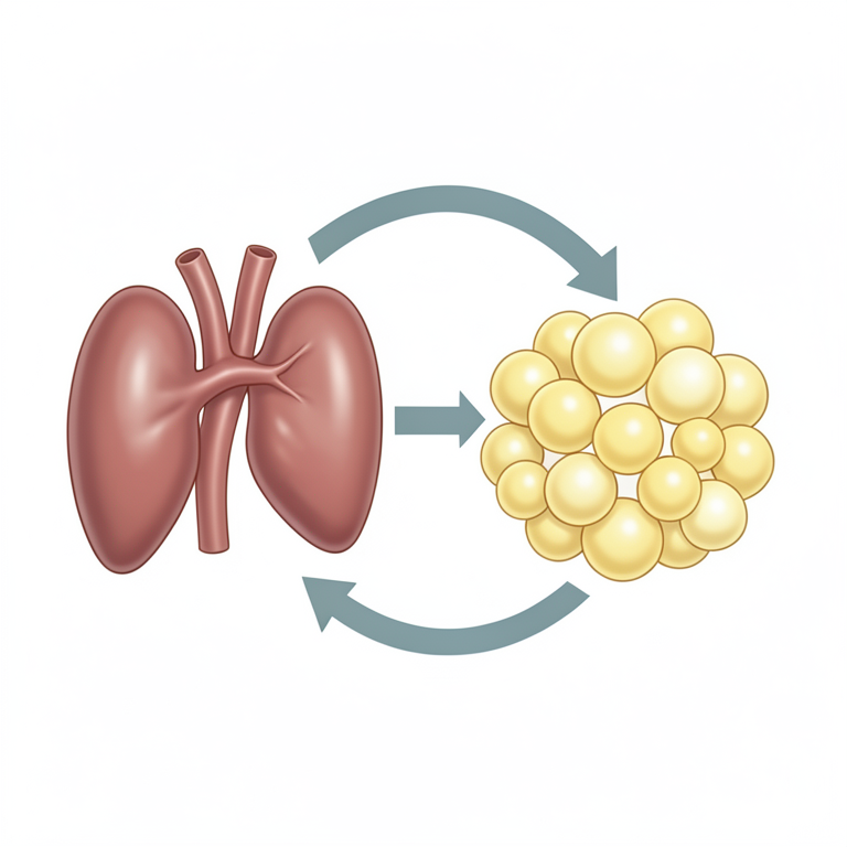
The Feedback Loop of Obesity and Thyroid Dysfunction
AI-generated conceptual illustration (Human thyroid gland and white adipose tissue). It explains the idea and does not show study data.
View source evidence ↗Figures & tables
From the original paper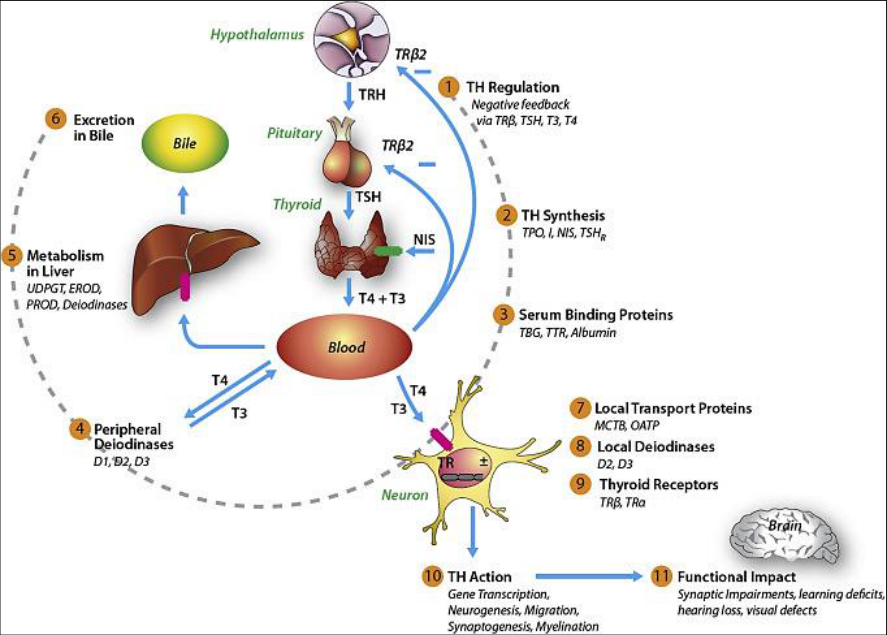 Figure 1
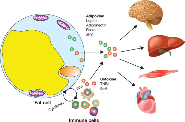 Figure 1
Introduction
Thyroid hormonal testing is commonly performed when exploring the causes of obesity [1] . Often, obesity is linked to mildly elevated or normal levels of blood thyrotropin (TSH). However, the precise relationship between thyroid dysfunction and excess body fat is still not fully understood, despite numerous studies conducted over the past decades [1,2] . Obesity is closely linked to the Hypothalamic-Pituitary-Thyroid (HPT) axis through a range of complex mechanisms. Thyroid Hormones (THs) and TSH play key roles in regulating the size and function of adipose tissue. Conversely, adipose tissue influences the HPT system by secreting adipokines, also referred to as adipocytokines [3] . Thyroid Hormones (THs) regulate various vital functions across multiple organs, including the heart, liver, brain, skeletal muscles, pancreas, and adipose tissue. They are crucial for managing energy balance, hunger, basal metabolic rate (BMR), thermogenesis, oxidation of Free Fatty Acids (FFA), and the metabolism of glucose and lipids [4] . Obesity is intricately linked to the Hypothalamic-Pituitary-Thyroid (HPT) axis through complex mechanisms. Thyroid Hormones (THs) and TSH regulate the growth and function of adipose tissue, while adipose tissue, in turn, influences the HPT system by releasing adipokines, also known as adipocytokines [5,6] . Thyroid Hormones (THs) regulate several vital organs, including the heart, liver, brain, skeletal muscles, pancreas, and adipose tissue. They are crucial for managing thermogenesis, Basal Metabolic Rate (BMR), energy balance, hunger, and the metabolism of glucose and lipids. Additionally, THs play a role in the breakdown of Free Fatty Acids (FFA) [7] . Thyrotropin primarily exerts its effects through the thyroid gland by interacting with its Gprotein-coupled receptor, TSHR.
While TSHR is well-established in thyroid tissues, it is also found in non-thyroidal tissues such as the liver, ovary, adipocytes, immune cells, ocular muscles, and erythrocytes. However, the precise role of TSHR in these non-thyroidal tissues remains unclear. The TSH/TSHR signaling pathway is thought to regulate adipose tissue function, influencing processes like thermogenesis, adipogenesis, and the balance between lipolysis and lipogenesis. Studies in mice and rats have identified TSHR in preadipocytes, with increased expression levels observed during preadipocyte development [8,9] . In rat Brown Adipose Tissue (BAT), the presence of functional Thyroid-Stimulating Hormone Receptors (TSHRs) leads to an upregulation of uncoupling protein type 1 (UCP-1) and type 2 iodothyronine deiodinase (DIO2). Additionally, research has identified TSHRs in both white and brown adipocytes in humans [10][11][12] .
Bell and his colleagues discovered that the expression of TSHR mRNA was detected in the subcutaneous adipose tissue. Additionally, they verified the presence of functional TSHR protein in human preadipocytes obtained from adipose tissue in the omentum and abdomen. Administration of TSH to preadipocytes activated the p70 S6 kinase, which is a downstream target in the TSHR pathway. This activation occurred despite the absence of cAMP involvement in the TSH signaling [13] . Crisp et al. [14] also detected TSHR transcripts in the abdominal adipose tissue of elderly individuals. Lu et al. [15] investigated the presence of TSHR protein in the subcutaneous fat tissue of 120 individuals with different body mass indexes. There is a positive correlation between the expression of the Thyroid-Stimulating Hormone Receptor (TSHR) and Body Mass Index (BMI). Janson et al. [16] demonstrated the presence of TSHR RNA in adipose tissue, as well as in distinct samples of adipose tissue obtained from infants, children, and adults. The expression appears to vary with age, since it was more elevated in the adipose tissue of neonates compared to adult tissue. A study conducted by Nannipieri et al. [17] shown that individuals who are overweight exhibit lower levels of TSHR mRNA [17] . Adipogenesis refers to the process of generating fresh adipocytes through the transformation of fibroblast-like preadipocytes into fully developed adipocytes, which is facilitated by many transcription factors. TSH-TSHR signaling in this system functions as a regulatory mechanism. During the in vitro experiment, the levels of TSHR mRNA in mature adipocytes were 100 times more than in preadipocytes. It is important to comprehend that adipogenesis might decelerate under certain circumstances. TSHR expression can be either enhanced or diminished by several factors. In the following sections, we will provide a detailed overview of the complex mechanisms that regulate TSH-TSHR signaling [18] . In Graves' disease, Thyroid-Stimulating Autoantibodies (TSAb) not only increase the levels of Thyroid-Stimulating Hormone (TSH), but also activate the Thyroid-Stimulating Hormone Receptor (TSHR) on orbital fibroblasts and T cells. In Graves' disease, the TSHR not only promotes the production of fat cells in the orbital adipose tissue but also in the white adipose tissue [19] . Researchers propose that the stimulation of TSHR by TSAb affects the distribution of body fat in individuals with Graves' illness. However, the ultimate result is determined by the functioning of the thyroid gland. Hyperthyroidism can lead to increased synthesis of thyroid hormones and activation of brown adipose tissue, resulting in heat production and weight loss through energy expenditure. Thyroid-Stimulating Hormones (TSHR) are stimulated by Thyroid-Stimulating Antibodies (TSAb), resulting in this effect. Elevated levels of Thyroid Stimulating Antibodies (TSAb) in individuals with Graves' illness and post-treatment euthyroidism stimulate the production of adipocytes in white adipose tissue, hence leading to weight gain [19] .
Fig 1:
Hypothalamic-pituitary-thyroid axis (HPT) [19] 2. Adipocytokines Adipose tissue has high activity in relation to hormones. Recent research has revealed that numerous adipocytokines have a significant role in regulating metabolism and body composition. Adipocytokines can influence several systems via autocrine, paracrine, and endocrine routes. They appear to have control over the functioning of neuroendocrine systems, thermogenesis, defense mechanisms, and nutrition. Adipokines have the potential to impact an individual's wellbeing positively or negatively. Adipokines play a crucial role in the inflammation of adipose tissue that is asymptomatic. Adipose tissue in obese individuals releases proinflammatory adipokines such as resistin, visfatin, and leptin [20] . The impact of adipokines on inflammation is also influenced by dietary factors. Their association with cardiovascular disease, metabolic syndrome, diabetes, and obesity is extensively demonstrated. Leptin has been found to function as an immunological modulator. Adipokines are linked to autoimmune disorders. Adipokines, including adiponectin, can act as prognostic markers and contribute to the prevention of cancer. Moreover, they can be utilized in the field of cancer therapy [21,22] . Thyroid hormones have a crucial role not only in appropriate growth, development, and reproduction, but also in regulating overall energy expenditure and body composition. An hypothesis has been postulated suggesting a direct association between the Body Mass Index (BMI) and the levels of Thyroid Stimulating Hormone (TSH) in the bloodstream. The reason for this is the correlation between thyroid disorders and weight fluctuations. Individuals who are obese have higher levels of serum TSH and free triiodothyronine. Higher energy levels lead to rising plasma T 3 (triiodothyronine) levels, whereas lower energy levels result in decreased plasma T 3 (triiodothyronine) levels. Among patients without an underactive thyroid, there were also documented instances of inverse relationships between FT 4 levels and body weight [23, 24] . Insulin resistance is linked to both hyperthyroidism and hypothyroidism. The methodology used to reach these results is ambiguous; yet, it is plausible that adaptive reactions played a role. There is a suggestion that there are direct relationships between BMI and Triiodothyronine (T 3 ) levels in both males and females. Roef et al. have recently found that patients without hypothyroidism but with insulin resistance exhibited higher T 3 levels [25,26] .
Leptin
Leptin, a hormone composed of 167 amino acids, induces satiety. This molecule is synthesized by adipocytes and is derived from the ob gene located on chromosome seven. Leptin is mostly synthesized in white adipose tissue, although it can also be produced in other organs such as the uterus, ovary, mammary epithelial cells, hypophysis, stomach, and liver. The db gene contains the genetic information for the leptin receptor (Ob-RL), which belongs to the class 1 family of cytokine receptors. This receptor is found in the liver and other peripheral organs, as well as being predominantly expressed in the hypothalamus [27] . Leptin's involvement in the response to fasting is widely acknowledged. Obesity and diabetes mellitus associated with obesity occur due to a hereditary lack of leptin or its receptor. Leptin affects energy consumption and appetite through mechanisms other than central brain regulation. Leptin stimulates an increase in the production of appetitesuppressing peptides and a decrease in the production of appetite-stimulating peptides [28] . In addition, leptin increases the levels of thyroid hormone [29] . The user's text is " [23] ". Leptin exerts both rapid and wellestablished long-term effects on metabolism. Additionally, it can exert a direct influence on the thyroid axis, so indirectly affecting thyroid metabolism. Leptin administration stimulates the production of TRH in the hypothalamus, which helps to reverse the suppression of the hypothalamuspituitary-thyroid axis caused by fasting at the central level [24] . Ortiga-Carvalho et al. provided evidence demonstrating that leptin enhances the release of TSH in living beings [25] . Furthermore, there is a conjecture that leptin has a direct stimulating effect on the synthesis of Thyroxine (T 4 ) by the thyroid gland [26] . Leptin regulates the function of iodothyronine deiodinase and promotes the transformation of T 4 into T 3 in both the central and peripheral areas [27] . Studies have shown that administering leptin can counteract the changes in blood T 3 and T 4 levels caused by fasting. Araujo et al. [28] revealed that the administration of leptin restores the activity of deiodinase 1 in rodents. Boelen et al. discovered that fasting led to an elevation in liver D3 mRNA levels in animals. In addition, the infusion of leptin reversed the rise in hepatic D3 expression that was produced by fasting, regardless of the levels of thyroid hormones in the blood [29] . Leptin additionally boosts D2 function in the central nervous system, leading to an elevation in T 3 levels [30] . Therefore, it is plausible that leptin plays a role in the development of the euthyroid ill condition [31] . In addition, TSH has a direct impact on adipocytes, possibly via binding to TSH-receptors located on the surface of adipocytes, which in turn triggers the secretion of leptin. The release of leptin by fat cells can be positively correlated with Thyroid-Stimulating Hormone (TSH) due to the direct action of TSH on leptin secretion [31] .
Ghrelin
Ghrelin, a hormone, consists of 28 amino acids and acts as a ligand for the Growth Hormone Secretagogue-Receptor (GSH-R). It was initially extracted from the intestines of rats. Ghrelin is primarily secreted by neuroendocrine cells located in the stomach wall. However, it can also be synthesized in various other tissues such as the hypothalamus, hypophysis, pancreas, kidneys, intestines, placenta, and others [32] . Ghrelin is recognized as the regulator of energy balance, appetite, and thermogenesis. Additionally, it stimulates the release of GH at the arcuate nucleus of the brain, where NPY-and AgRP are also located. NPY stimulation is believed to regulate the impact of ghrelin on appetite. Ghrelin levels increase during periods of fasting and other catabolic states. Administering ghrelin intravenously can induce hunger. Ghrelin hormone antagonizes leptin hormone in the body. Excessive fat accumulation and obesity result in a decrease in ghrelin levels in individuals who are overweight [33,34] . The presence of GHS-R mRNA and ghrelin in the thyroid gland has been demonstrated. Several studies indicate that individuals with hyperthyroidism experience a decrease in ghrelin levels in comparison to individuals who have normal thyroid function. Ghrelin levels normalize after hyperthyroidism therapy. There was no association seen between alterations in ghrelin levels and subclinical hyperthyroidism. A study conducted on rats found a correlation between hyperthyroidism and decreased levels of ghrelin in the bloodstream, as well as increased levels of ghrelin immunoreactivity in the stomach mucosa. The mechanism behind the decrease in ghrelin levels is not fully understood, however it is plausible that elevated insulin levels in hyperthyroidism or excessive thyroid hormone may contribute to this phenomenon [35,36] . There is a correlation between decreased thyroid function and elevated levels of ghrelin. A recent study discovered that ghrelin stimulates the synthesis of fT 4 in organisms. Conversely, certain researchers discovered that individuals with hypothyroidism exhibited typical levels of ghrelin in their bloodstream. Altinova et al. demonstrated a significant decrease in ghrelin levels in individuals with Hashimoto's thyroiditis in comparison to those who are in good condition. There was a correlation between decreased levels of ghrelin and increased titers of thyroid peroxidase antibodies. The coexistence of autoimmune thyroid illness in individuals with atrophic gastritis is believed to be the cause of reduced ghrelin production in their stomach cells [37,38] . Administering recombinant TSH to individuals who underwent thyroidectomy due to thyroid cancer resulted in a reduction of their blood ghrelin levels independently. The researchers proposed that TSH may induce the release of ghrelin in the gastric mucosa. However, none of the research accurately quantify the amount of food individuals consume or the impact it has on ghrelin levels [39] .
Obestatin
Obestatin is a newly discovered adipocytokine that is linked to ghrelin. The stomach is the main location where it is expressed. It is suggested that obestatin does not possess any inherent action. However, the effect of it on body composition is still not well understood. There is a lack of extensive study on the correlation between thyroid disorders and obestatin levels. Kosowicz and his colleagues discovered a positive correlation between hypothyroidism and elevated obestatin levels, while hyperthyroidism was associated with reduced obestatin levels. Gurgul et al. identified a direct relationship between ghrelin, obestatin, and TSH levels in individuals with hypothyroidism and suggested that obestatin might serve as a regulatory molecule [40,41] .
Visfatin
Visfatin is an adipocytokine derived from adipose tissue in the abdomen. It is alternatively referred to as pre-B cell colony-enhancing factor. This protein exhibits a somewhat substantial magnitude, and its corresponding gene is located on chromosome 7. The first source for the identification of pre-B cell colony-enhancing factor was peripheral blood leukocytes. Furthermore, it was discovered that adipocytes contained the molecule [42] . Visfatin is an insulin-like molecule that significantly contributes to the process of adipocyte differentiation. Previous research has demonstrated a correlation between levels of visfatin and obesity. The presence of elevated levels of visfatin in obese individuals is believed to be attributed to the activation of macrophages in adipose tissue. However, recent evidence indicates that visfatin can also be synthesized by macrophages in adipose tissue, hepatocytes, and skeletal muscles [43] . Visfatin is believed to induce inflammation, and elevated levels have been observed in autoimmune disorders. The immune system is responsible for the alterations in visfatin levels observed in thyroid disorders. IL-6 has the ability to enhance the activity of Visfatin genes [44] , while Visfatin is believed to exert a contrary impact by improving the functionality of IL-6. Patients diagnosed with graves disease exhibited elevated levels of visfatin, which were positively correlated with increased levels of T 3 and FT 4 . A study conducted by MacLaren and his colleagues shown that T 3 has the ability to inhibit the production of visfatin mRNA in adipocytes. Han et al. discovered that individuals with hyperthyroidism or hypothyroidism exhibited elevated levels of visfatin compared to individuals with normal thyroid function (euthyroid). Visceral fat accumulation is considered an indication of insulin resistance. Additionally, there is a potential correlation between elevated levels of visfatin in the bloodstream and increased visfatin expression in visceral fat. Another hypothesis suggests that visfatin is synthesized by the circulatory system as a mechanism to safeguard itself in cases of thyroid hyperactivity or hypoactivity [45,46] . Ozkaya et al. found that hyperthyroid individuals had lower levels of visfatin compared to both the hypothyroid and control groups. Visfatin levels shown a substantial decrease after treatment for hypothyroidism, but they demonstrated a notable increase following antithyroid therapy for hyperthyroidism. Caixàs et al. found that restoring normal thyroid function led to elevated plasma visfatin levels in individuals with both hypo-and hyperthyroidism. Furthermore, in both hyperthyroidism and hypothyroidism, the levels of visfatin in the blood were higher compared to the control group. Their findings were in direct opposition to the prior research, since they concluded that there was no correlation between visfatin levels and insulin resistance and inflammatory markers [47,48] .
Adiponectin
Adiponectin, a peptide consisting of 244 amino acids, is secreted by the adipose tissue. The primary mechanism of action involves two distinct receptors: adiponectin receptor 2 (AdipoR2), predominantly situated in the liver, and adiponectin receptor 1 (AdipoR1), primarily present in muscle [49]. Individuals who are obese and have insulin resistance see a decrease in their serum adiponectin levels. Adiponectin levels also rise after weight loss or medicines aimed at enhancing insulin sensitivity. Furthermore, a hypothesis proposing a correlation between decreased adiponectin levels and coronary artery disease [50] is presented. Thyroid hormones and adiponectin, despite their distinct effects on metabolism, are believed to be associated in all weight conditions. The findings indicate that individuals with hyperthyroidism have higher levels of adiponectin compared to those with hypothyroidism or normal thyroid function (euthyroidism). On the other hand, Santini et al. and Iglesias et al. found that the levels of adiponectin in individuals with hyperthyroidism were not significantly different from those in the control groups. Altinova et al. failed to show a link between adiponectin levels and thyroid function. Furthermore, there is a hypothesis suggesting a correlation between thyroid hormone levels and adiponectin levels in persons who are in a state of excellent health [51,52] . Adiponectin is believed to have the ability to inhibit the growth of new blood vessels and to prevent the development of cancer. Adiponectin levels have been found to be below the normal range in many forms of cancer. In a recent study by Mitsiades et al., it was found that individuals diagnosed with thyroid cancer have markedly reduced levels of adiponectin compared to the control group. The researchers demonstrated the presence of AdipoR1 and AdipoR2 in thyroid cancer cell lines. The therapy, which included recombinant adiponectin, did not have any impact on the in vitro proliferation and viability of cells. Empirical investigations have established a connection between insulin resistance and the development of thyroid cancer [53] .
Resistin
Resistin is primarily synthesized in the pancreas, adipose tissue, muscle, and macrophages. The structure consists of a polypeptide chain with 114 amino acids. The association between obesity and insulin resistance is widely regarded to be interconnected, and resistin partially explains this connection. Resistin is believed to be associated with inflammatory disorders that are not influenced by insulin resistance due to its pro-inflammatory properties. The exact correlation between resistin levels and thyroid disorders remains uncertain. Ziora et al. [54] found a favorable correlation between FT 4 and serum resistin levels in persons diagnosed with anorexia nervosa. Iglesias et al. found that the blood resistin levels of individuals with hypothyroidism and individuals with normal thyroid function were substantially similar. Unlike the euthyroid state, Kaplan et al. found that hypothyroidism caused by thyroidectomy did not cause significant temporary alterations in adiponectin, leptin, or resistin levels. An independent investigation establishes a conclusive link between resistin levels and thyroid hormones. Discrepancies in thyroid function data may arise due to fluctuations in resistin levels caused by various adipocytokines. In instances of thyroid malfunction, it is hypothesized that alterations in resistin levels serve as adaptive mechanism [55] .
Metabolic inflammation and adipocytokines
In 1993, Hotamisligil and his colleagues made the discovery that adipose tissue in obese mice produces TNFα, a proinflammatory cytokine that is usually generated by immune cells. Furthermore, it has been found that adipocytes release TNFα directly, leading to the emergence of insulin resistance commonly observed in obesity. The idea of metabolic inflammation, recognized as a crucial mechanistic link between obesity and its related consequences, has evolved from the original correlation between obesity and inflammation [56] . Evidence demonstrated that adipose tissue produces a diverse range of cytokines and chemokines, such as IL-6 and MCP-1, after the administration of TNFα. Cytokines can exert either beneficial or detrimental effects on the body's overall metabolism of glucose and fats. Several adipokines share similarities with cytokines and have the ability to regulate inflammatory responses, which is fascinating. The name "adipocytokines" is commonly used to denote these two chemicals that are produced by adipose tissue. In 2003, two studies were released at the same time, showing that obesity causes the entry of macrophages into adipose tissue in both people and animals. This finding not only clarifies the source of cytokines produced by adipose tissue, but also establishes a unique link between immune and metabolic cells in a metabolic organ for the first time [57,58] .
Adipose tissue contains two distinct types of macrophages: M1 and M2. M1 macrophages are activated through classical pathways and are known for releasing proinflammatory cytokines such as TNFα and IL-6. They also produce Reactive Oxygen Species (ROS) and inducible Nitric Oxide Synthase (iNOS), which contribute to the development of insulin resistance. In contrast, M2 macrophages are activated by different mechanisms and secrete substances like IL-10, arginase-1, and blockers of the IL-1 receptor, which are involved in tissue remodeling.
Excessive adipose tissue can alter macrophage function, leading to a shift from M2 to M1 macrophages. This shift, driven by insulin resistance, results in increased production of inflammatory proteins and ROS [59,60] . During this time, the lack of particular beneficial effects linked to M2 macrophages may potentially contribute to the decrease in metabolic function in obesity. For example, M2 macrophages produce catecholamines that promote adaptive thermogenesis. During fasting, lipolysis triggers the recruitment of macrophages, which helps prevent fat buildup in some areas and protects the functionality of adipose tissue. Over the past few years, almost all major categories of immune cells have been discovered in adipose tissue, after the discovery of macrophages. These cells play a crucial role in regulating metabolic processes in the body by contributing to the endocrine function of adipose tissue.
In obese persons, the liver, muscle, and pancreas serve as the main metabolic organs. They have close physical and signaling connections with cells involved in metabolism and the immune system. These findings indicate that metabolic inflammation is a common and fundamental factor contributing to the metabolic dysfunction associated with obesity [61,62] . Obesity-induced adipose inflammation has multiple underlying mechanisms. Hypoxia can occur due to the enlargement of adipose tissue during the progression of obesity, leading to the stimulation of compensatory angiogenesis. The process of vascularization is enhanced by the recruitment of macrophages to the specified area. Immune cells in other metabolic tissues, such as the liver, also perform a similar job. Kupffer cells in the mouse liver secrete TNF-a and IL-6, which are crucial for the liver's regeneration. Macrophages that have entered adipose tissues have also been proposed as a means of eliminating apoptotic cells. Furthermore, the progression of endotoxemia, which is associated with obesity and changes in the permeability of the intestines, might worsen inflammation in adipose tissue [63] . While there is increasing data suggesting that inflammation in adipose tissue negatively affects energy metabolism, it is important to note that not all metabolic inflammation is harmful to metabolic balance. In order to adapt to extra energy and maintain a stable metabolic state, the body may need to undergo inflammation that is linked to the growth or regeneration of adipose tissue. Similarly, some cytokines can aid in the prevention of obesity by boosting energy expenditure and reducing food consumption. Therefore, it is essential to consider the physiological basis when examining the metabolic effects of adipose inflammation. Further investigation is necessary to fully comprehend the scope and process of the beneficial inflammatory reactions linked to different phases of obesity [64, 65] . Adipocytokines and metabolic inflammation in adipose tissue [65]
Thyroid function and obesity
The Resting Energy Expenditure (REE) is increased in overt hyperthyroidism, whereas it is decreased in overt hypothyroidism. REE, or Resting Energy Expenditure, constitutes almost 60% of the overall energy used by adult males. However, there have been cases where there are noticeable variations in Resting Energy Expenditure (REE) that are linked to a decrease in the range of thyroid function. In this particular animal, a decrease in blood TSH levels from 4.0 to 0.4 mU/l leads to a significant rise (about 40%) in the amount of L-T 4 that is absorbed [66,67] . Approximately 10 grams of fat are metabolized daily when there is a 7-8% alteration in Resting Energy Expenditure (REE). If there are no changes in your energy intake or expenditure, or in your diet or body's temperature regulation, then variations in thyroid function tests within the normal range in the laboratory may be associated with a yearly weight loss or gain of a few kilograms [68] . It is crucial to bear in mind that the fact that a person's thyroid function test results fall within the "normal" range established by the laboratory does not necessarily imply that they are indeed normal. The variability in individuals' thyroid function test findings is significantly narrower compared to the standard range employed in laboratory settings. There exist disparities between the individual's perceived "normal" state and around half of the accepted range of "normal" values [69] . Figure 11 Despite including several significant variables that could influence BMI, this correlation persisted [70] . Furthermore, a correlation was observed between serum fT 4 levels and BMI, indicating a negative link. However, no correlation was found between serum fT 3 levels and BMI [14] . The selected individuals had fT 4 and fT 3 values that fell within the established laboratory ranges. The group with a serum TSH level greater than 3.6 mU/l had a 2.1 times increased risk of becoming overweight with a BMI of 30 or more compared to the group with a serum TSH level between 1 and 1.99 mU/l. A difference of 5.5 kg distinguished a lady with a serum TSH level of 0.28 mU/l from another woman with a serum TSH level of 4.5 mU/l. The blood levels of thyroid-stimulating hormone (TSH) [71] were directly associated with the increase in body weight over the past five years. Individuals with moderate hypothyroidism typically have decreased levels of free Thyroxine (fT 4 ), elevated levels of Thyroid-Stimulating Hormone (TSH), and minimal fluctuations in free Triiodothyronine (fT 3 ). The findings of the DanThyr study indicate that variations in individuals' energy expenditure, influenced by their thyroid hormones, are the main factor that accounts for the relationship between thyroid function variables and BMI. Prior research has consistently shown a detrimental association between blood fT 4 levels and indicators of the metabolic syndrome, as well as BMI [72,73] .
Obesity and thyroid dysfunction
There appears to be a moderate association between thyroid hormones and body composition. Thyroid hormones regulate various physiological processes including food intake, fat burning, heat production, basal metabolic rate, and the metabolism of lipids and glucose. Thyroid dysfunction is characterized by changes in body weight and composition, body temperature, total and Resting Energy Expenditure (REE), all of which are not influenced by physical activity [74] . Decreased thermogenesis, decreased metabolic rate, and hypothyroidism have also been associated with higher Body Mass Index (BMI) and increased prevalence of obesity. While the topic is not yet fully understood, there is clinical data that suggests a connection between even slight thyroid dysfunction, known as subclinical hypothyroidism, and significant increases in body weight. This indicates that subclinical hypothyroidism may be a risk factor for overweight and obesity. Additionally, there is an observed association between significant alterations in Resting Energy Expenditure (REE) in individuals with hypothyroidism and minor variations in blood Thyroid-Stimulating Hormone (TSH) levels caused by moderate adjustments in L-T 4 dosage during replacement therapy.
There is less knowledge regarding the actual extent of weight gain and weight loss associated with L-T 4 therapy in hypothyroidism [75,76] .
Materials and Methods
The data in the analysis was altered in November 2023. We did a comprehensive analysis of national guidelines and conducted a systematic evaluation of many renowned medical databases, including PubMed Central, and Google Scholar) applied similar methodologies that utilized identical language. At first, the criteria for deciding which papers to include were based on the titles and abstracts. Afterwards, thorough textual studies of relevant papers were conducted. The search was refined using specific terms such as "Echinococcus granulosus", "E. granulosus", "echinococcosis", "cystic echinococcosis", "prevalence", "incidence", "diagnosis", "clinical presentation", "treatment", and "prevention". We employed these terms alone as well as in conjunction with Boolean operators. The database search yielded additional CE-related terms such as "hydatid disease" and "hydatid cyst," however none of these terms generated any additional outcomes. The scope of our evaluation encompassed scientific articles on cystic echinococcosis, with a focus on taxonomic modifications, rates of occurrence and prevalence in humans, available treatment options, and current advancements in disease management and prevention, such as the utilization of novel biomarkers for diagnostic purposes. The following criteria were used to eliminate some studies: those that did not involve human subjects, those that were not written in English, review articles that did not include original data, editorials or letters to the editor that did not include original data, and papers that did not provide an estimate of Incidence Rate (IR) or Prevalence Rate (PR).
Conclusion
The relationship between adipocytokines, thyroid function, and obesity is intricate and interdependent. These consequences have significant implications for our comprehension and management of metabolic disorders. This study examines how adipocytokines, particularly leptin and adiponectin, affect thyroid function and, consequently, energy balance and body weight. Evidence suggests that changes in the levels and activities of these adipocytokines can contribute to thyroid disorders, which, in turn, exacerbate obesity, creating a challenging cycle. Leptin plays a crucial role in regulating thyroid hormone synthesis, especially in obese individuals, who often struggle with leptin regulation. This not only affects thyroid function but also compounds the metabolic issues associated with obesity. Conversely, adiponectin, which has an inverse relationship with body fat percentage and protects thyroid function, indicates that targeting adiponectin pathways might be beneficial in treatment strategies. Additionally, chronic inflammation in adipose tissue, driven by proinflammatory adipocytokines like TNF-α and IL-6, disrupts thyroid hormone metabolism and prolongs metabolic disorders. This highlights the need for a comprehensive approach to addressing both obesity and thyroid-related conditions. A thorough understanding of the interactions between adipose tissue and the endocrine system is essential for developing effective treatments. Future research should aim to clarify these mechanisms and create targeted interventions to improve outcomes for individuals facing these interconnected health issues.
Figures and tables from the paper
References
76 in the original paper- Pasquali R, Casanueva F, Haluzik M, van Hulsteijn L, Ledoux S, Monteiro MP, et al. European Society of Endocrinology Clinical Practice Guideline: Endocrine work-up in obesity. Eur J Endocrinol. 2020;182-G32. DOI: 10.1530/EJE-19-0893.
- Knudsen N, Laurberg P, Rasmussen LB, Bülow I, Perrild H, Ovesen L, et al. Small differences in thyroid function may be important for body mass index and the occurrence of obesity in the population. J Clin Endocrinol Metab. 2005;90:4019-4024. DOI: 10.1210/jc.2004-2225.
- Lai Y, Wang J, Jiang F, Wang B, Chen Y, Li M, et al. The relationship between serum thyrotropin and components of metabolic syndrome. Endocr J. 2011;58:23-30. DOI: 10.1507/endocrj.K10E-272.
- Santini F, Marzullo P, Rotondi M, Ceccarini G, Pagano L, Ippolito S, et al. Mechanisms in endocrinology: The crosstalk between thyroid gland and adipose tissue: Signal integration in health and disease. Eur J Endocrinol. 2014;171:137-152. DOI: 10.1530/EJE-140067.
- Ortiga-Carvalho TM, Sidhaye AR, Wondisford FE. Thyroid hormone receptors and resistance to thyroid hormone disorders. Nat Rev Endocrinol. 2014;10:582591. DOI: 10.1038/nrendo.2014.143.
- Mullur R, Liu YY, Brent GA. Thyroid hormone regulation of metabolism. Physiol Rev. 2014;94:355382. DOI: 10.1152/physrev.00030.2013.
- Senese R, Cioffi F, de Lange P, Goglia F, Lanni A. Thyroid: Biological actions of 'nonclassical' thyroid hormones. J Endocrinol. 2014;221-R12. DOI: 10.1530/JOE-13-0573.
- Lu M, Lin RY. TSH stimulates adipogenesis in mouse embryonic stem cells. J Endocrinol. 2008;196:159-169. DOI: 10.1677/JOE-07-0452.
- Haraguchi K, Shimura H, Lin L, Endo T, Onaya T. Differentiation of rat pre-adipocytes is accompanied by expression of thyrotropin receptors. Endocrinology. 1996;137:3200-3205. DOI: 10.1210/endo.137.8.8754740.
- Lu S, Guan Q, Liu Y, Wang H, Xu W, Li X, et al. Role of extra-thyroidal TSHR expression in adipocyte differentiation and its association with obesity. Lipids Health Dis. 2012;11:17. DOI: 10.1186/1476-511X-1117.
- Bell A, Gagnon A, Grunder L, Parikh SJ, Smith TJ, Sorisky A. Functional TSH receptor in human abdominal preadipocytes and orbital fibroblasts. Am J Physiol Cell Physiol.; c2000, p. 279-C340. doi: 10.1152/ajpcell.2000.279.2.C335.
- Janson A, Karlsson FA, Micha-Johansson G, Bolme P, Brönnegård M, Marcus C. Effects of stimulatory and inhibitory thyrotropin receptor antibodies on lipolysis in infant adipocytes. J Clin Endocrinol Metab. 1995;80:1712-1716. DOI: 10.1210/jcem.80.5.7745024.
- Bell A, Gagnon A, Grunder L, Parikh SJ, Smith TJ, Sorisky A. Functional TSH receptor in human abdominal preadipocytes and orbital fibroblasts. Am J Physiol Cell Physiol.; c2000. p. 279-C340. DOI: 10.1152/ajpcell.2000.279.2.C335.
- Crisp MS, Lane C, Halliwell M, Wynford-Thomas D, Ludgate M. Thyrotropin receptor transcripts in human adipose tissue. J Clin Endocrinol Metab. 1997;82:20032005. DOI: 10.1210/jcem.82.6.2003.
- Lu S, Guan Q, Liu Y, Wang H, Xu W, Li X, et al. Role of extrathyroidal TSHR expression in adipocyte differentiation and its association with obesity. Lipids Health Dis. 2012;11:17. DOI: 10.1186/1476-511X-1117.
- Janson A, Karlsson FA, Micha-Johansson G, Bolme P, Brönnegård M, Marcus C. Effects of stimulatory and inhibitory thyrotropin receptor antibodies on lipolysis in infant adipocytes. J Clin Endocrinol Metab. 1995;80:1712-1716. DOI: 10.1210/jcem.80.5.7745024.
- Nannipieri M, Cecchetti F, Anselmino M, Camastra S, Niccolini P, Lamacchia M, et al. Expression of thyrotropin and thyroid hormone receptors in adipose tissue of patients with morbid obesity and/or type 2 diabetes: Effects of weight loss. Int J Obes. 2009;33:1001-1006. DOI: 10.1038/ijo.2009.140.
- Moreno-Navarrete JM, Moreno M, Ortega F, Xifra G, Hong S, Asara JM, et al. TSH mRNA is linked to cholesterol metabolism in adipose tissue. FASEB J. 2017;31:4482-4491. DOI: 10.1096/fj.201700161R.
- Draman MS, Stechman M, Scott-Coombes D, Dayan CM, Rees DA, Ludgate M, et al. The role of thyrotropin receptor activation in adipogenesis and modulation of fat phenotype. Front Endocrinol. 2017;8:83. DOI: 10.3389/fendo.2017.00083.
- Hotamisligil GS. Inflammation and metabolic disorders. Nature. 2006;444:860-867. DOI: 10.1038/nature05485.
- Piya MK, McTernan PG, Kumar S. Adipokine inflammation and insulin resistance: The role of glucose, lipids and endotoxin. J Endocrinol. 2013;216T15.
- Mattu HS, Randeva HS. Role of adipokines in cardiovascular disease. J Endocrinol. 2013;216-T 3 6.
- Knudsen N, Laurberg P, Rasmussen LB, Bülow I, Perrild H, Ovesen L, et al. Small differences in thyroid function may be important for body mass index and the occurrence of obesity in the population. J Clin Endocrinol Metab. 2005;90:4019-4024. DOI: 10.1210/jc.2004-2225.
- Hoogwerf BJ, Nutall FQ. Long-term weight regulation in treated hyperthyroid and hypothyroid subjects. Am J Med. 1984;76:963-970.
- Alevizaki M, Saltiki K, Voidonikola P, Mantzou E, Papamichael C, Stamatelopoulos K. Free thyroxine is an independent predictor of subcutaneous fat in euthyroid individuals. Eur J Endocrinol. 2009;161:459465. DOI: 10.1530/EJE-09-0441.
- Roef G, Lapauw B, Goemaere S, Zmierczak HG, Toye K, Kaufman JM, et al. Body composition and metabolic parameters are associated with variation in thyroid hormone levels among euthyroid young men. Eur J Endocrinol. 2012;167:719-726.
- Fei H, Okano HJ, Li C, Lee GH, Zhao C, Darnell R, et al. Anatomic localization of alternatively spliced leptin receptors (Ob-R) in mouse brain and other tissues. Proc Natl Acad Sci U S A. 1997;94:7001-7005. DOI: 10.1073/pnas.94.13.7001.
- Chelikani PK, Glimm DR, Kennelly JJ. Short communication: Tissue distribution of leptin and leptin receptor mRNA in the bovine. J Dairy Sci. 2003;86:2369-2372.
- Montague CT, Farooqi IS, Whitehead JP, Soos MA, Rau H, Wareham NJ, et al. Congenital leptin deficiency is associated with severe early-onset obesity in humans. Nature. 1997;387:903-908. DOI: 10.1038/43185.
- Légràdi G, Emerson CH, Ahima RS, Flier JS, Lechan RM. Leptin prevents fasting-induced suppression of prothyrotropin-releasing hormone messenger ribonucleic acid in neurons of the hypothalamic paraventricular nucleus. Endocrinology. 1997;138:2569-2576. DOI: 10.1210/en.138.6.2569.
- Ortiga-Carvalho TM, Oliveira KJ, Soares BA, PazosMoura CC. The role of leptin in the regulation of TSH secretion in the fed state: in vivo and in vitro studies. J Endocrinol. 2002;174(1):121-125. DOI: 10.1677/joe.0.1740121.
- Kojima M, Hosoda H, Date Y, Nakazato M, Matsuo H, Kangawa K. Ghrelin is a growth-hormone-releasing acylated peptide from stomach. Nature. 1999;402(6762):656-660. DOI: 10.1038/45230.
- Date Y, Kojima M, Hosoda H, Sawaguchi A, Mondal MS, Suganuma T, et al. Ghrelin, a novel growth hormone-releasing acylated peptide, is synthesized in a distinct endocrine cell type in the gastrointestinal tracts of rats and humans. Endocrinology. 2000;141(11):4255-4261.
- Hewson AK, Dickson SL. Systemic administration of ghrelin induces Fos and Egr-1 proteins in the hypothalamic arcuate nucleus of fasted and fed rats. J Neuroendocrinol. 2000;12(11):1047-1049. DOI: 10.1046/j.1365-2826.2000.00584.x.
- Tanda ML, Lombardi V, Genovesi M, Ultimieri F, Lai A, Gandolfo M, et al. Plasma total and acylated ghrelin concentrations in patients with clinical and subclinical thyroid dysfunction. J Endocrinol Invest. 2009;32(1):74-78.
- Dadan J, Zbucki RL, Sawicki B, Winnicka MM. Estimation of gastric ghrelin-positive cells activity in hyperthyroid rats. Folia Histochem Cytobiol. 2008;46(4):511-517. DOI: 10.2478/v10042-008-00610.
- Altinova AE, Toruner F, Karakoc A, Yetkin I, Ayvaz G, Cakir N, et al. Serum ghrelin levels in patients with Hashimoto's thyroiditis. Thyroid. 2006;16(12):12591264. DOI: 10.1089/thy.2006.16.1259.
- Checchi S, Montanaro A, Pasqui L, Ciuoli C, Cevenini G, Sestini F, et al. Serum ghrelin as a marker of atrophic body gastritis in patients with parietal cell antibodies. J Clin Endocrinol Metab. 2007;92(11):43464351. DOI: 10.1210/jc.2007-0988.
- Ciuoli C, Brusco L, Theodoropoulou A, Castagna MG, Neri O, Pasqui L, et al. Effects of acute recombinant human TSH on serum ghrelin levels. Front Endocrinol. 2011;2:94. DOI: 10.3389/fendo.2011.00094.
- Gourcerol G, St-Pierre DH, Taché Y. Lack of obestatin effects on food intake: should obestatin be renamed ghrelin-associated peptide (GAP)? Regul Pept. 2007;141(1-3):1-7. DOI: 10.1016/j.regpep.2006.12.008.
- Kosowicz J, Baumann-Antczak A, Ruchala M, Gryczyńska M, Gurgul E, Sowiński J. Thyroid hormones affect plasma ghrelin and obestatin levels. Horm Metab Res. 2011;43(2):121-125. DOI: 10.1055/s0031-1274725.
- Jia SH, Li Y, Parodo J, Kapus A, Fan L, Holstein OD, et al. Pre-B cell colony-enhancing factor inhibits neutrophil apoptosis in experimental inflammation and clinical sepsis. J Clin Invest. 2004;113(9):1318-1327. DOI: 10.1172/JCI200419930.
- Costford SR, Bajpeyi S, Pasarica M, Albarado DC, Thomas SC, Xie H, et al. Skeletal muscle NAMPT is induced by exercise in humans. Am J Physiol Endocrinol Metab. 2010;298(1)-E126. DOI: 10.1152/ajpendo.00318.2009.
- Moschen AR, Kaser A, Enrich B, Mosheimer B, Theurl M, Niederegger H, et al. Visfatin, an adipocytokine with proinflammatory and immunomodulating properties. J Immunol. 2007;178(3):1748-1758. DOI: 10.4049/jimmunol.178.3.1748.
- Chu CH, Lee JK, Wang MC, Lu CC, Sun CC, Chuang MJ, et al. Change of visfatin, C-reactive protein concentrations, and insulin sensitivity in patients with hyperthyroidism. Metabolism. 2008;57(10):1380-1383. doi: 10.1016/j.metabol.2008.05.013.
- Han J, Zhang TO, Xiao WH, Chang CQ, Ai H. Upregulation of visfatin expression in subjects with hyperthyroidism and hypothyroidism is partially relevant to a nonlinear regulation mechanism between visfatin and tri-iodothyronine with various concentrations. Chin Med J. 2012;125(5):874-881. DOI: 10.3760/cma.j.issn.0366-6999.2012.05.026.
- Ozkaya M, Sahin M, Cakal E, Yuzbasioglu F, Sezer K, Kilinc M, et al. Visfatin plasma concentrations in patients with hyperthyroidism and hypothyroidism before and after control of thyroid function. J Endocrinol Invest. 2009;32(5):435-439. DOI: 10.3275/6296.
- Caixàs A, Tirado R, Vendrell J, Gallart L, Megía A, Simón I, et al. Plasma visfatin concentrations increase in both hyper-and hypothyroid subjects after normalization of thyroid function and are not related to insulin resistance, anthropometric or inflammatory parameters. Clin Endocrinol (Oxf). 2009;71(5):733738. DOI: 10.1111/j.1365-2265.2009.03546.x.
- Kamon J, Tobe K, Kita S, Kadowaki T. Cloning of adiponectin receptors that mediate antidiabetic metabolic effects. Nature. 2003;423(6941):762-769. DOI: 10.1038/nature01705.
- Kumada M, Kihara S, Sumitsuji S, Kawamoto T, Matsumoto S, Ouchi N, et al. Association of hypoadiponectinemia with coronary artery disease in men. Arterioscler Thromb Vasc Biol. 2003;23(1):85-89. DOI: 10.1161/01.ATV.0000048.
- Yaturu S, Prado S, Grimes SR. Changes in adipocyte hormones leptin, resistin, and adiponectin in thyroid dysfunction. J Cell Biochem. 2004;93(3):491-496. DOI: 10.1002/jcb.20188.
- Altinova AE, Törüner FB, Aktürk M, Bukan N, Cakir N, Ayvaz G, et al. Adiponectin levels and cardiovascular risk factors in hypothyroidism and hyperthyroidism. Clin Endocrinol (Oxf). 2006;65(4):530-535.
- Mantzoros CS, Petridou E, Dessypris N, Chavelas C, Dalamaga M, Alexe DM, et al. Adiponectin and breast cancer risk. J Clin Endocrinol Metab. 2004;89(3):11021107. DOI: 10.1210/jc.2003-031804.
- Steppan CM, Bailey ST, Bhat S, Brown EJ, Banerjee RR, Wright CM, et al. The hormone resistin links obesity to diabetes. Nature. 2001;409(6818):307-312. DOI: 10.1038/35053000.
- Iglesias P, Alvarez Fidalgo P, Codoceo R, Díez JJ. Serum concentrations of adipocytokines in patients with hyperthyroidism and hypothyroidism before and after control of thyroid function. Clin Endocrinol (Oxf). 2003;59(5):621-629. DOI: 10.1046/j.1365-2265.2003.01897.x.
- Hotamisligil GS. Inflammation and metabolic disorders. Nature. 2006;444:860-867.
- Weisberg SP, McCann D, Desai M, Rosenbaum M, Leibel RL, Ferrante AW Jr. Obesity is associated with macrophage accumulation in adipose tissue. J Clin Invest. 2003;112(12):1796-1808.
- Xu H, Barnes GT, Yang Q, Tan G, Yang D, Chou CJ, et al. Chronic inflammation in fat plays a crucial role in the development of obesity-related insulin resistance. J Clin Invest. 2003;112(12):1821-1830.
- Gordon S. Alternative activation of macrophages. Nat Rev Immunol. 2003;3(1):23-35.
- Lumeng CN, Bodzin JL, Saltiel AR. Obesity induces a phenotypic switch in adipose tissue macrophage polarization. J Clin Invest. 2007;117(1):175-184.
- Winer S, Chan Y, Paltser G, Truong D, Tsui H, Bahrami J, et al. Normalization of obesity-associated insulin resistance through immunotherapy. Nat Med. 2009;15(9):921-929.
- Feuerer M, Herrero L, Cipolletta D, Naaz A, Wong J, Nayer A, et al. Lean, but not obese, fat is enriched for a unique population of regulatory T cells that affect metabolic parameters. Nat Med. 2009;15:930-939. DOI: 10.1038/nm.2000.
- Pang C, Gao Z, Yin J, Zhang J, Jia W, Ye J. Macrophage infiltration into adipose tissue may promote angiogenesis for adipose tissue remodeling in obesity. Am J Physiol Endocrinol Metab. 2008;295E322. DOI: 10.1152/ajpendo.00181.2008.
- Ye J, Keller JN. Regulation of energy metabolism by inflammation: A feedback response in obesity and calorie restriction. Aging (Albany NY). 2010;2:361368. DOI: 10.18632/aging.100092.
- Ye J, McGuinness OP. Inflammation during obesity is not all bad: evidence from animal and human studies. Am J Physiol Endocrinol Metab. 2013;304-E477. DOI: 10.1152/ajpendo.00423.2012.
- Kim B. Thyroid hormone as a determinant of energy expenditure and the basal metabolic rate. Thyroid. 2008;18:141-144. DOI: 10.1089/thy.2007.0263.
- al-Adsani H, Hoffer LJ, Silva JE. Resting energy expenditure is sensitive to small dose changes in patients on chronic thyroid hormone replacement. J Clin Endocrinol Metab. 1997;82:1118-1125. DOI: 10.1210/jcem.82.4.3874.
- Eisenberg M, Distefano JJ. TSH-based protocol, tablet instability, and absorption effects on L-T 4 bioequivalence. Thyroid. 2009;19:103-110. DOI: 10.1089/thy.2008.0198.
- Andersen S, Pedersen KM, Bruun NH, Laurberg P. Narrow individual variations in serum T(4) and T(3) in normal subjects: A clue to the understanding of subclinical thyroid disease. J Clin Endocrinol Metab. 2002;87:1068-1072. DOI: 10.1210/jc.87.3.1068.
- Laurberg P, Jørgensen T, Perrild H, Ovesen L, Knudsen N, Pedersen IB, et al. The Danish investigation on iodine intake and thyroid disease, DanThyr: status and perspectives. Eur J Endocrinol. 2006;155:219-228. DOI: 10.1530/eje.1.02283.
- Knudsen N, Laurberg P, Rasmussen LB, Bülow I, Perrild H, Ovesen L, et al. Small differences in thyroid function may be important for body mass index and the occurrence of obesity in the population. J Clin Endocrinol Metab. 2005;90:4019-4024. DOI: 10.1210/jc.2005-0151.
- Shon HS, Jung ED, Kim SH, Lee JH. Free T 4 is negatively correlated with body mass index in euthyroid women. Korean J Int Med. 2008;23:53-57. DOI: 10.3904/kjim.2008.23.1.53.
- Makepeace AE, Bremnert AP, O'Leary P, Leedman PJ, Feddema P, Michelangeli V, et al. Significant inverse relationship between serum free T 4 concentration and body mass index in euthyroid subjects: differences between smokers and nonsmokers. Clin Endocrinol (Oxf). 2008;69:648-652. DOI: 10.1111/j.13652265.2008.03270.x.
- Rosenbaum M, Hirsch J, Murphy E, Leibel RL. Effects of changes in body weight on carbohydrate metabolism, catecholamine excretion, and thyroid function. Am J Clin Nutr. 2000;71:1421-1432. DOI: 10.1093/ajcn/71.5.1421.
- Danforth E Jr, Horton ES, O'Connell M, Sims EA, Burger AG, Ingbar SH, et al. Dietary-induced alterations in thyroid hormone metabolism during overnutrition. J Clin Invest. 1979;64:1336-1347. DOI: 10.1172/JCI109504.
- Knudsen N, Laurberg P, Rasmussen LB, Bülow I, Perrild H, Ovesen L, et al. Small differences in thyroid function may be important for body mass index and the occurrence of obesity in the population. J Clin Endocrinol Metab. 2005;90:4019-4024. DOI: 10.1210/jc.2005-0151.
Cited by 3
From OpenAlex, OpenCitations and reference lists read by Scinovex
- Biochemical markers in sepsis: Advances in early diagnosis and prognosis ↗
Semiu Adebayo Kareem, Sajjad Ahmed, Suha F Mohammed AL-Ma’aroof, Mohamed A. Naser · International Journal of Chemical Studies · 2025
Sepsis is a life-threatening condition characterized by a dysregulated immune response to infection, leading to systemic inflammation and organ dysfunction. Early diagnosis and prognosis assessment are critical for improving patient outcomes. In recent years, biochemical markers have emerged as valuable tools in identifying sepsis at an early stage and predicting disease severity. Among these markers, C-reactive protein (CRP) and procalcitonin
- The relationship between insulin and obesity molecular and physiological insights ↗
Priyanka Saini · Journal of Entomology and Zoology Studies · 2025
Obesity is a global health crisis intricately linked with insulin dysfunction, which plays a pivotal role in metabolic regulation. This paper explores the molecular and physiological mechanisms underpinning the relationship between insulin and obesity, focusing on how insulin resistance develops in key tissues such as muscle, liver, and adipose tissue. Chronic low-grade inflammation, adipokine imbalance, and elevated free fatty acids
- First trimester screening for thyroid disorders in pregnancy ↗
Simon Gregori, Gian Luca Garnotel · International Journal of Gynaecology Research · 2024
Thyroid disorders are common during pregnancy and can have significant implications for both maternal and fetal health. Early detection and management are crucial to prevent adverse outcomes. This review explores the importance of screening for thyroid disorders in the first trimester, the current guidelines and practices, and the potential impact on pregnancy outcomes.