
Research article · 2024 · Full text
International Journal of Veterinary Sciences and Animal HusbandryQ fever in small ruminants and its public health significance
Volume 9(5S) · Pages 160–165 · DOI: 10.22271/veterinary.2024.v9.i5sc.1697 ↗
Research areasVector-borne infectious diseasesVector-Borne Animal DiseasesMicrobial infections and disease research
Abstract
Author-writtenCoxiellosis, or Q fever, is a significant global zoonotic disease caused by Coxiella burnetii. It ranks among the top 13 global priority zoonoses and poses an increasing threat to high-risk individuals worldwide. Coxiella burnetii is frequently detected in animal-derived foods (such as bulk milk, eggs, and meat) and tick vectors. The disease can lead to spontaneous abortions in humans and reproductive issues in livestock, causing production losses. In developing countries, Q fever is prevalent but underreported and neglected, especially in regions like the Indian subcontinent (including Sri Lanka, Bhutan, Pakistan, Nepal, Maldives, and Myanmar). Policymakers and funding agencies often do not prioritize this disease, despite its public health significance. This review emphasizes the importance of recognizing and addressing the occurrence and epidemiology of Q fever.
Summary
Source-checkedPlain-language reading of the full paper, prepared with AI and checked against the text. Each point links to the authors' own words.
Why it matters
- Q fever, caused by the bacterium Coxiella burnetii, is a major global zoonotic disease and is ranked among the top 13 priority zoonoses. View evidence ↗
- The disease is significant because it causes production losses in livestock through reproductive issues and can cause spontaneous abortions in humans. View evidence ↗
- Q fever is often neglected and underreported in developing countries, particularly within the Indian subcontinent. View evidence ↗
The question
- The aim of this review is to highlight the importance of recognizing and addressing the epidemiology and occurrence of Q fever. View evidence ↗
What they did
- The pathogen is described as an obligate intracellular gram-negative bacterium with pleomorphic rods measuring 0.2-0.4 µm in diameter and 0.4-1.0 µm in length. View evidence ↗
- The bacterium exists in two forms: a metabolically active large cell variant (LCV) and an environmentally resistant, spore-like small cell variant (SCV). View evidence ↗
- The Immunofluorescence Assay (IFA) is the reference diagnostic test used to detect phase I and II IgG and IgM antibodies. View evidence ↗
- Polymerase Chain Reaction (PCR) is used for rapid and specific detection by targeting genes such as IS1111, Com1, and 16s rRNA. View evidence ↗
What they found
- One cited study found a slightly higher prevalence of C. burnetii in cattle (20%) than in small ruminants (15%). View evidence ↗
- In a study from Yemen involving 250 staff and 263 goats, the seroprevalence of Q fever was 5.2% in staff and 8.57% in goats. View evidence ↗
- A study of 1,350 ruminants in Ethiopia found seroprevalence rates of 37.6% in cattle and 28.7% in small ruminants. View evidence ↗
- In Egyptian sheep, the overall seroprevalence was 37.5%, with significantly higher rates in females (39.5% vs 8.3% in males), pregnant ewes (47.7%), and sheep older than 3 years (71.2%). View evidence ↗
- In a Mexican study, PCR identified C. burnetii in 61.1% of vaginal samples from aborting sheep and 60% of ram semen samples. View evidence ↗
- A biosensor using SMR structure detected C. burnetii within 20 minutes with 81-93% sensitivity and a detection limit ten times higher than PCR. View evidence ↗
What it means
- The authors conclude that effective management of Q fever requires increased surveillance, improved diagnostics, and better awareness among healthcare providers. View evidence ↗
- The authors state that further research is needed to understand the bacterium's pathogenesis and transmission, and to develop better guidelines and preventive measures. View evidence ↗
Visual summary
1 visualsIllustrations explain the paper; they do not show study data.
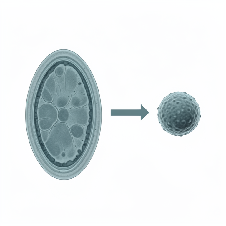
C. burnetii Life Cycle Variants
AI-generated conceptual illustration (Coxiella burnetii bacteria). It explains the idea and does not show study data.
View source evidence ↗Introduction
Coxiella burnetii is an obligate intracellular bacterium causing Q fever in humans and coxiellosis in animals, reported globally except in New. It belongs to the class Gammaproteobacteria, order Legionellales, family Coxiellaceae, and is the sole species in the genus Coxiella. Humans typically acquire infection through airborne transmission from animal reservoirs, especially domestic ruminants (OIE, 2010) [35] . Q fever is considered a neglected zoonotic disease in many developing countries and is notifiable by the OIE. However, it remains non-notifiable in some nations (Njeru et al., 2016) [33] . The International Livestock Research Institute (ILRI) identifies it as "the most contagious disease" due to its extremely low infectious dose (ILRI, 2012) [25] . Classified as a Category B bioterrorism agent by the CDC, Coxiella burnetii primarily infects ruminants, with small ruminants being significant reservoirs. Infected sheep and goats release millions of bacteria through placenta, amniotic fluid, fetal membranes, and lochia following birth or abortion. (Bauer et al., 2016) [33] . In India, the Indian Council 1of Medical Research (ICMR) established a Task Force on Rickettsioses in 2002 to enhance research on Q fever diagnosis, prevention, and control. However, limited research has been conducted on C. burnetii in sheep, goats, and humans.
Transmission of Coxiella burnetii
The organism is secreted in milk, making the ingestion of contaminated raw milk and dairy products a source of infection for humans. Individuals in close contact with farm animals, such as veterinarians, farm workers, butchers, and laboratory personnel, are at heightened risk (Hatchette et al., 2001). Goats typically contract the infection through inhalation of aerosols or environmental exposure, often showing no symptoms. Sheep and goats serve as primary reservoirs of the pathogen. Recent research indicates that goats primarily excrete C. burnetii through milk, whereas sheep predominantly do so via vaginal mucus or feces (Rodolakis, 2009) [38] . High-risk groups for Q fever include livestock farmers, shepherds, sheep shearers, animal by-product processors, meat plant employees, abattoir workers, veterinarians, and researchers handling the pathogen or samples.
While the bacterium can infect various animals, birds, and arthropods, ruminants are considered the major reservoirs (VandenBrom et al., 2015) [45] . Guatteo et al. (2011) [22] found that the prevalence of C. burnetii was slightly higher in cattle (20%) compared to small ruminants (15%), though this may be influenced by the design of existing studies
Morphology
Coxiella burnetii is an obligate intracellular gram-negative bacterium with pleomorphic rods measuring approximately 0.2-0.4 µm in diameter and 0.4-1.0 µm in length. The bacterium exists in two forms the large cell variant (LCV) and the small cell variant (SCV). The LCV is metabolically active and replicative, while the SCV, a spore-52 like form, is highly resistant to environmental stresses such as high temperatures, UV radiation, and osmotic pressure (McCaul and Williams, 1981) [28] . The SCV is associated with the presence of lipopolysaccharide (LPS), which is also found in the LCV (Coleman et al., 2007) [12] . The two LPS phenotypes that are displayed by organism are phase 1 (smooth) and phase 2 (rough), the latter lacking the O-antigenic region (Toman et al., 2009) [43] . Phylogenetically, C. burnetii has been reclassified from the Rickettsiales order to the Legionellales order.
History
The term "Q fever" was coined by Edward Holbrook Derrick in 1935, following investigations of febrile illness in abattoir workers in Brisbane, Australia (Derrick, 1973) [14] . The bacterium was first observed by Macfarlane Burnet in infected guinea pigs' spleens, with Burnet and Mavis Freeman initially hypothesizing it as a Rickettsial agent. Concurrently, Gordon Davis isolated the pathogen from Dermacentor andersoni ticks, and in 1938, Cox propagated the agent in embryonated eggs.
Epidemiology
Q fever outbreaks occurred during World War II among troops in Europe, with over 20,000 cases reported in Italy (Babudieri, 1959) [7] . In farm settings, individuals involved in birth assistance, handling dead animals and cleaning are exposed to C. burnetii. Slaughterhouse workers and veterinarians, who frequently handle infected animals, are at higher risk, with field veterinarians showing a high seroprevalence (22.2%) (Whitney et al., 2009) [48] . Coxiella burnetii is endemic in over 51 countries, including India, and remains a significant public health issue due to poor disease surveillance (Malik et al., 2014; Maurin and Raoult, 1999) [26,27] . The infection is often underestimated due to its pleomorphic clinical presentation and reliance on physician awareness and diagnostic capabilities (Fournier et al., 1998) [17] . Infected ruminants, particularly during parturition, are major sources of infection for other livestock and humans. Transmission is facilitated by animal trade and milk products. The pathogen can survive for over a year at temperatures below -20 °C and requires fewer than 10 organisms to establish an infection (Bauer et al., 2016; Meadows et al., 2015) [33,30] .
Pathogenesis
Coxiella burnetii exists in two phases: phase I (virulent, smooth) and phase II (avirulent, rough). Phase variation involves differences in surface antigens, primarily lipopolysaccharides (LPS). Phase I bacteria have full-length LPS O-chains, crucial for cell internalization and survival in the phagosome, while phase II bacteria lack these chains (Thompson et al., 2003; Coleman et al., 2007) [42,12] . Upon entering the host cell by phagocytosis, phase I small cell variants (SCVs) transform into large cell variants (LCVs), multiply by binary fission, and prevent host cell apoptosis, persisting for a long time (Arricau-Bouvery, 2005) [6] . As the organism is highly infectious, with an infectious dose as low as one organism (McQuiston et al., 2002) [29] , primary multiplication occurs in regional lymph nodes, leading to bacteraemia lasting 5-7 days, then localizing in mammary glands and the placenta of pregnant animals (Babudieri, 1959) [7] . Acute human infections often involve atypical pneumonia and hepatitis, with the bacteria spreading to various organs and forming granulomatous lesions, especially in the liver and bone marrow (Maurin and Raoult, 1999) [27] . Chronic infections can lead to endocarditis. The immune response involves both humoral and cellular immunity. Antibodies against phase II bacteria develop within three weeks postinfection (Dupuis et al., 1985) [15] . Cellular immunity, particularly involving T-cells and cytokines like interferon gamma (IFN-γ), plays a key role in clearing the infection (Honstettre et al., 2004) [24] . In animals, C. burnetii is often contracted from the environment or ticks, leading to primary infections with minimal clinical signs. The bacteria persist in infected females, shedding large numbers during pregnancy. Experimental studies show the organism can be recovered from various organs during early pregnancy, with high multiplication rates in the placenta at term (Freick et al., 2017) [18] . Infected pregnant goats exhibit severe inflammation in the allantochorion, with aborted fetuses appearing normal but the placenta showing significant pathological changes (Palmer et al., 1983) [36] .
Clinical Signs Humans
Q fever in humans can be either acute or chronic, often presenting sub clinically or with mild symptoms. Approximately 50% of those infected develop clinical disease, with an incubation period ranging from 14 to 39 days (Dupuis et al., 1985) [15] . Acute symptoms include prolonged fever, abdominal pain, nausea, vomiting, weight loss, lower respiratory infection, chills, sweating, coughing, chest pain, pharyngitis, myalgia, disorientation, and severe headache (Graves and Islam, 2016) [21] . Pneumonia occurs in 30-50% of cases, and some may develop granulomatous hepatitis. Common acute manifestations are flu-like symptoms, highgrade fever, fatigue, headache, and myalgia (Njeru et al., 2016) [33] . Atypical pneumonia is characterized by a nonproductive cough, fever, and minimal auscultatory abnormalities. Hepatitis in Q fever presents in three forms: infectious hepatitis with hepatomegaly, asymptomatic hepatitis, and prolonged fever of unknown origin with granulomas on liver biopsy. Myocarditis, accounting for 0.5-1% of cases, can be life-threatening (Urrutia et al., 1991) [44] . Rarely, Q fever presents with pericarditis, skin rash, encephalitis, meningoencephalitis, or encephalomyelitis. Other less common symptoms include hemolytic anemia, thyroiditis, pancreatitis, and neurological diseases. Chronic Q fever, persisting for six months or more, can develop years after initial infection, often resulting in endocarditis in highrisk patients (Njeru et al., 2016) [33] . Pregnant women may experience reactivation of the infection, leading to complications such as abortion and low birth weight (Zeman et al., 1989) [49] .
Animals
In ruminants, Q fever is usually asymptomatic but can cause abortion and stillbirth in small ruminants. Approximately 50% of pregnant animals may abort within a flock or herd (Van den Brom et al., 2015) [45] . Other signs include uterine infection, premature births, and weak offspring. Coxiella burnetii also causes abortion in wild ruminants, with 27.3% of North African gazelles with a history of abortion testing positive for the bacterium (McQuiston et al., 2002) [29] .
Diagnosis Identification by Microscopy
Gimenez's stain is used to identify C. burnetii in smears from infected chicken embryos, liver, and spleen, showing pinkstained coccobacillary bodies against a blue or green background (OIE, 2004) [34] . Electron microscopy distinguishes between SCV and LCV forms of the bacterium.
Serological Diagnosis
Antibody levels in serum, detectable from five days postinfection, help identify Q fever. Phase II antibodies appear 7-15 days after symptom onset, subsiding after 3-6 months. Primary is indicated by a 4-fold increase in phase II IgG or IgM antibodies, with significant titers being IgG ≥200 and IgM ≥50 (Fournier et al., 1998) [17] . Chronic Q fever shows elevated phase I IgG titers (≥1:800) (Freick et al., 2017) [18] .
Complement Fixation Test (CFT)
CFT detects phase I and II antibodies, used for diagnosing chronic Q fever. A titer of >1/10 is positive, while >1/80 indicates progressive infection. CFT has been largely replaced by ELISA and IFA due to its poor sensitivity (OIE, 2004; Emery et al., 2012) [34,16] .
ELISA
ELISA is more sensitive than CFT and quicker than IFA. It serves as an epidemiological screening tool (Mohan et al., 2017). Studies show varying prevalence rates in cattle, sheep, goats, and farm workers using ELISA (Dhaka et al., 2019) [32] . AlShaibani et al. (2024) [3] conducted a study on 250 staff workers and 263 goats in slaughterhouses in Dhamar city, Yemen. Using Immuno DOT and ELISA tests, they found a seroprevalence of 5.2% in staff workers and 8.57% in goats. Higher seroprevalence in staff was noted among those aged 31-40, with 6-10 years of experience, and butchers. In goats, significant associations were found with sex, season, source, and tick presence. Getachew et al. (2024) [20] examined 1350 ruminants (450 cattle, 450 goats, and 450 sheep) across three 1districts in the South Omo zone, Ethiopia. They used ELISA testing to determine seroprevalence rates, finding 37.6% in cattle and 28.7% in small ruminants. The study also identified 2720 ticks (1299 from cattle, 1020 from goats, and 401 from sheep) as potential vectors for C. burnetii. The research highlighted a significant gap in community awareness, with only 43% of animal owners able to recognize Q fever symptoms, compared to 76.2% of animal health professionals who were aware 1of the causative agent. These findings underscore the widespread presence of and the urgent need for improved awareness and control measures for Q fever. Kamaly et al. (2024) in his study investigated the seroprevalence of C. burnetii using ELISA among sheep in Egypt. The overall seroprevalence of Q fever in sheep was 37.5%, with higher prevalence in females (39.5%) compared to males (8.3%). Pregnant ewes had a higher seroprevalence (47.7%) compared to non-pregnant (31.4%) and abortive ewes (43.8%). Sheep older than 3 years showed significantly higher seroprevalence (71.2%).
Immunofluorescence Assay (IFA)
IFA is the reference test for Q fever, detecting phase I and II IgG and IgM antibodies. It can differentiate between acute and chronic infections. Studies indicate its effectiveness in various samples from animals and humans.
Other Serological Assays
New methods like Coxiella Immunospot assay and automated immunofluorescence assays have been developed, showing varied sensitivity and specificity (Bizzini et al., 2015) [9 ].
Molecular Diagnosis
Hybridization Technique: Fluorescent in-situ hybridization (FISH) uses specific probes to detect C. burnetii at the tissue level, targeting genes like 16s rRNA (Melenotte et al., 2016) [31] .
Polymerase Chain Reaction (PCR)
PCR is a rapid, sensitive, and specific method for detecting C. burnetii. It targets genes such as IS1111, Com1, and 16s rRNA. Studies show PCR's effectiveness in various samples, including blood, milk, and placental tissue. Sánchez-Rodríguez et al. (2024) [39] conducted a study to determine the presence of C. burnetii DNA in vaginal samples from sheep that presented abortion and ram semen in five Central and Southern States of Mexico. A total of 180 vaginal exudate samples and 20 semen samples were tested. PCR amplification and sequencing of the IS1111 insertion sequence identified C. burnetii in 110 (61.1%) vaginal samples and 12 (60%) semen samples. This marks the first report of C. burnetii in sheep that aborted and in ram semen in Mexico. In Egypt, Kamaly et al. (2024) used PCR for molecular detection of the IS1111 gene in vaginal swabs of sheep and revealed the presence of the IS1111 gene in 20% of the vaginal swabs tested, with no significant statistical difference between aborted and non-aborted ewes. PCR results showed no significant association with age, pregnancy status, abortion history, or farm location.
Genome Sequencing
Sequencing genes like IS1111 and 16s rRNA helps differentiate C. burnetii strains and understand their spread. Whole genome sequencing has identified closely related strains in human and animal samples (Schets et al., 2013) [41] .
Biosensors
The first biosensor for detecting the agent of Q fever, developed by Koo et al., utilizes a silicon micro ring resonator (SMR) that is optically interrogated by a feed wave-guide. The SMR detects changes in the refractive index near the wave-guide caused by the binding reaction of biomolecules immobilized on its surface alters the resonant wavelength. The assay employs DNA amplification (IDA) using recombines polymerase amplification (RPA). This highly sensitive DNA amplification combined with advanced SMR detection addresses issues like false-negative reactions and low sensitivity due to inhibitors in blood plasma, such as hemoglobin, IgG fraction, and heparin. A recently described electrochemical biosensor for Q fever is based on the immobilization of the CBU_1718 (GroEL) protein (UniProtKB-P19421) onto a gold electrode modified by a self-assembled monolayer. The immobilization of the antigen on the gold electrode was optimized by adjusting parameters such as protein concentration and incubation time to achieve optimal performance. GroEL was selected for immobilization due to its higher expression yield (0.65 mg/mL) compared to the genetic region Com1. The sensor was tested with PBS, sera from blood donors, and patients with acute and chronic Q fever infections. It demonstrated the necessary discrimination ability for accurate serological detection of chronic Q fever, which is expected given the period of seroconversion. Koo et al. functionalized the sensing chip with an amine group to immobilize C. burnetii DNA primers. DNA elongation and detection occurred on the SMR structure, addressing low pathogen concentration and inhibitor effects in clinical samples. Using samples from formaldehyde-fixed paraffin-embedded tissue and frozen blood plasma, C. burnetii was detected within 20 minutes with a sensitivity of 81-93% and a detection limit ten times higher than PCR methods.
Prevention and Control
Screening and vaccination are key strategies. Pasteurization of milk and proper heat treatment of milk products prevent infection. Vaccination reduces bacterial shedding and subsequent transmission. Separation of pregnant animals after abortion helps control the spread. Antibiotics like doxycycline are effective, especially when combined with hydroxychloroquine for chronic cases. Pregnant women are treated with co-trimoxazole. High-risk groups must adopt safety measures to prevent exposure (Alonso et al., 2015) [2] .
Future Prospects
Research is needed to better understand the bacterium, its pathogenesis, and its transmission. Improved diagnostic methods, guidelines for practitioners, and preventive measures for medical staff are essential. Veterinary aspects, including the effectiveness of antibiotic treatment and the impact of Q fever on various animal species, require further investigation. Developing new vaccines and better management practices will reduce the zoonotic risk. Developing a "one health" strategy, establishing surveillance networks, and raising awareness are crucial.
Conclusion
Q fever remains a significant public health concern, particularly in developing countries. Increased surveillance, better diagnostic capabilities, and awareness among healthcare providers are essential for effective management and prevention of this zoonotic disease. Collaborative efforts between veterinarians, healthcare professionals, and public health officials can help mitigate the risks associated with Q fever and protect at-risk populations
Conflict of Interest Not available
Conflict of Interest Not available
Financial Support
Financial Support
References
44 in the original paper- Panda BB, Gaur K, Kori ML, Tyagi LK, Nema RK, Sharma CS, et al. Anti-inflammatory and analgesic activity of Jatropha gossypifolia in experimental animal models. Global Journal of Pharmacology. 2009;3(1):1-5.
- Alonso E, Etxaniz LI, Hurtado A, Liendo P, Urbaneja F, Aspiritxaga I, et al. Q fever outbreak among workers at a waste-sorting plant. PLOS One. 2015;10(9).
- AlShaibani IRMS, Al-Houthi AAM, Al-Dhurani MYA, et al. Q fever among staff workers and goats in slaughterhouses at Dhamar city, Yemen. Yemeni Journal of Agriculture and Veterinary Sciences. 2024;5:1.
- Alonso AR, Zendoia II, Barandika JF, Jado I, Hurtado A, López CM, et al. Monitoring Coxiella burnetii infection in naturally infected dairy sheep flocks throughout four lambing seasons and investigation of viable bacteria. Frontiers in Veterinary Science. 2020;7:352.
- Angelakis E, Raoult D, et al. Q fever. Veterinary Microbiology. 2010;140(3-4):297-309.
- Bouvery AN, Rodolakis A, et al. Is Q fever an emerging or reemerging zoonosis? Veterinary Research. 2005;36(3):327-349.
- Babudieri B. Q fever: A zoonosis. Advances in Animal and Veterinary Sciences. 1959;5:81.
- Bauer AE, Hubbard KR, Johnson AJ, Messick JB, Weng HY, Pogranichniy RM, et al. A cross-sectional study evaluating the prevalence of Coxiella burnetii, potential risk factors for infection, and agreement between diagnostic methods in goats in Indiana. Preventive Veterinary Medicine. 2016;126:131-137.
- Bizzini A, Peter O, Baud D, Edouard S, Meylan P, Greub G, et al. Evaluation of a new serological test for the detection of anti-Coxiella and anti-Rickettsia antibodies. Microbes and Infection. 2015;17:811-816.
- Camacho MT, Outschoorn I, Kovacova E, Tellez A, et al. Distribution of immunoglobulin G (IgG) and A (IgA) subclasses following Q fever vaccination with soluble phase I C. burnetii extract. Vaccine. 2000;18:1773-1777.
- Coleman SA, Fischer ER, Cockrell DC, Voth DE, Howe D, Mead DJ, et al. Proteome and antigen profiling of C. burnetii developmental forms. Infection and Immunology. 2007;75:290-298.
- Cox HR. A filter-passing infectious agent isolated from ticks III. Description of organism and cultivation experiments. Public Health Reports. 1938;27:53-70.
- Derrick EH. Q fever, a new fever entity: clinical features, diagnosis and laboratory investigation. The Medical Journal of Australia. 1937;2:281-299.
- Dupuis G, Peter O, Peacock M, Burgdorfer W, Haller E, et al. Immunoglobulin responses in acute Q fever. Journal of Clinical Microbiology. 1985;22:484-487.
- Emery MP, Ostlund EN, Schmitt BJ, et al. Comparison of Q fever serology methods in cattle, goats, and sheep. Journal of Veterinary Diagnostic Investigation. 2012;24:379-382.
- Fournier PE, Marrie TJ, Raoult D, et al. Diagnosis of Q fever. Journal of Clinical Microbiology. 1998;36:18231834.
- Freick M, Enbergs H, Walraph J, Diller R, Weber J, Konrath A, et al. Serological reactions and bacterial shedding in primiparous dairy cows in an endemically infected herd-impact on milk yield and fertility. Reproduction in Domestic Animals. 2017;52:160-169.
- Kalou GP, Dyankov G, Krumova S, Simeonov K, Valkov T, Baymakova M, et al. Applicability of biosensor technologies in the detection of Coxiella burnetii infection in clinical samples. Biotechnology & Biotechnological Equipments. 2024;38:2350163.
- Getachew S, Kumsa B, Getachew Y, Kinfe G, Gumi B, Rufael T, et al. Seroprevalence of Coxiella burnetii and potential tick vectors infesting domestic ruminants and community perception of the disease in pastoral areas of south Omo zone, southern Ethiopia. Parasitology Epidemiology and Control. 2024;26:00369.
- Graves SR, Islam A. Endemic Q fever in New South Wales, Australia: A case series (2005-2013). American Journal of Tropical Medicine and Hygiene. 2016;95:5559.
- Guatteo R, Seegers H, Taurel AF, Joly A, Beaudeau F, et al. Prevalence of C. burnetii infection in domestic ruminants: a critical review. Veterinary Microbiology. 2011;149:1-16.
- Hatchette T, Hudson R, Schezch WF, et al. Goat associated-Q fever: A new disease in Newfoundland. Emerging Infectious Diseases. 2001;7:413-419.
- Honstettre A, Ghigo E, Moynault A, Capo C, Toman R, Akira S, et al. Lipopolysaccharide from C. burnetii is involved in bacterial phagocytosis, filamentous actin reorganization, and inflammatory responses through Tolllike receptor 4. Journal of Immunology Research. 2004;172:3695-3703.
- Malik SS, Das DP, Rawool D, Kumar A, Suryawanshi D, Negi M, Barbuddhe SB, et al. Screening of foods of animal origin for C. burnetii in India. International Journal of Epidemiology. 2014;34:8-27.
- Maurin M, Raoult D. Q fever. Journal of Clinical Microbiology. 1999;12:518-553.
- McCaul TF, Williams JC. Developmental cycle of C. burnetii: structure and morphogenesis of vegetative and sporogenic differentiations. Journal of Bacteriology. 1981;147:1063-1076.
- McQuiston JH, Childs JE, Thompson HA, et al. Q fever. Journal of American Veterinary Medical Association. 2002;221:796-799.
- Meadows S, Bitton JA, McEwen S, Jansen J, Menzies P, et al. C. burnetii seropositivity and associated risk factors in goats in Ontario, Canada. Preventive Veterinary Medicine. 2015;121:199-205.
- Melenotte C, Million M, Audoly G, Gorse A, Dutronc H, Roland G, et al. B-cell non-Hodgkin lymphoma linked to C. burnetii. The Journal of American Society of Hematology. 2016;127:113-121.
- Mohan V, Nair A, Kumar M, Dhaka P, Vergis J, Rawool DB, et al. Seropositivity of goats for coxiellosis in Bareilly region of UP India. Advances in Animal and Veterinary Sciences. 2017;5:226-228.
- Njeru J, Henning K, Pletz M, Heller R, Neubauer H, et al. Q fever is an old and neglected zoonotic disease in Kenya: A systematic review. BMC Public Health. 2016;16:1-20.
- Q fever. In: Manual of Diagnostic Tests and Vaccines for Terrestrial Animals. 2004;2:565-572.
- Palmer NC, Kierstead M, Key DW, Williams JC, Peacock MG, Velland H, et al. Placentitis and abortion in goats and sheep in Ontario caused by C. burnetii. The Canadian Veterinary Journal. 1983;24:60-61.
- Raoult D, Marrie TJ, Mege JL, et al. Natural history and pathophysiology of Q fever. The Lancet Infectious Diseases. 2000;5:219-226.
- Rodolakis A. Q fever in dairy animals. Annals of the New York Academy of Sciences. 2009;1166:90-93.
- Rodríguez SO, Resendiz PEG, Aparicio DE, Reynoso AB, Chavarría MLC, Serrano MG, et al. Molecular Detection of Coxiella burnetii in Vaginal Swab Samples from Sheep That Aborted. Journal of Vector Borne Diseases. 2024;35:145-162.
- Sandoz KM, Sturdevant DE, Hansen B, Heinzen RA, et al. Developmental transitions of C. burnetii grown in axenic media. Journal of Microbiological Methods. 2014;96:104-110.
- Schets FM, Heer DL, Husman DRAM, et al. C. burnetii in sewage water at sewage water treatment plants in a Q fever epidemic area. International Journal of Hygiene and Environmental Health. 2013;216:698-702.
- Thompson HA, Hoover TA, Vodkin MH, Shaw EI, et al. Do chromosomal deletions in the lipopolysaccharide biosynthetic regions explain all cases of phase variation in C. burnetii strains? An update. Annals of the New York Academy of Sciences. 2003;990:664-670.
- Toman R, Skultety L, Ihnatko R, et al. C. burnetii glycomics and proteomics tools for linking structure to function. Annals of the New York Academy of Sciences. 2009;1166:67-78.
- Urrutia A, García JP, Collazos J, et al. Acute myopericarditis as a manifestation of Q fever. Medicina Clinica. 1991;96:319-329.
- Brom VDR, Berends SI, Luttikholt S, Moll L, Engelen VE, Vellema P, et al. Bulk tank milk surveillance as a measure to detect C. burnetii shedding in dairy goat herds in the Netherlands between 2009 and 2014. Journal of Dairy Science. 2015;98:1-12.
- Whitney EAS, Massung RF, Candee AJ, Ailes EC, Myers LM, Patterson NE, Berkelman RL, et al. Seroepidemiologic and occupational risk survey for C. burnetii antibodies among US veterinarians. Clinical Infectious Diseases. 2009;48:550-557.
- Zeman DH, Kirkbride CA, Leslie-Steen P, Duimstra JR, et al. Ovine abortion due to C. burnetii infection. Journal of Veterinary Diagnostic Investigation. 1989;1:178-180.