
Research article · 2024 · Full text
International Journal of Veterinary Sciences and Animal HusbandryThe fragility of rabbits red blood cells under different anticoagulants and temperatures
Volume 9(5) · Pages 215–218 · DOI: 10.22271/veterinary.2024.v9.i5c.1688 ↗
Research areasAdipose Tissue and MetabolismThermoregulation and physiological responsesErythrocyte Function and Pathophysiology
Abstract
Author-writtenThe goal of this study was to distinguish between the anticoagulant heparin (antithrombin) and Calcium chelating anticoagulants (EDTA, citrate and oxalate) for assessing the health condition of specimens by the osmotic fragility of rabbit red blood cells at room temperature 25 °C - 18 °C and 50 °C for 1 hr and for long time (1 week) at 25 °C. Anticoagulants (heparin, EDTA, citrate, and oxalate) were added directly to four tubes containing four millilitres of blood samples obtained from the hearts of five male rabbits. Throughout November and December 2022, five male rabbits weighing between 800-1000 g were intermittently housed in cages of 60 cm-40 cm in College of Veterinary Medicine 12:12 dark light cycle, the osmotic fragility test were done for each type to record the zero time and ensure the activity of anticoagulants; The blood samples were stored at ambient temperature 25 °C, under a water bath at 50 °C, and in a refrigerator at -18 °C, for 1hr followed by examination of red blood cell fragility of each type of anticoagulant under different temperature; finally the blood samples of room temperature 25 °C were tested after 24 hr, 48 hr,72 hr and a week to point the long term anticoagulant (keeping the red blood cell membrane). The osmotic fragility of erythrocyte was determined by adding few drops of blood to tubes contain 5ml of normal saline 0.85%, the mixtures put in centrifuge at 3000 rpm for 15 min then the supernatant color indicate hemolysis while the clear mean not. 32 blood samples were analysed for each kind of anticoagulant: EDTA, citrate oxalate, and heparin, both at zero time and after 1 hour at 25 °C, -18 °C and 50 °C. The hemolytic percent at zero time and under 25 °C were zero% for all types but the hemolytic percent of heprinized blood sample after 1 hr revealed 100% and 50% under -18 °C and 50 °C respectively; while the rest anticoagulants types appear hemolytic percent 50% under -18c and 25% under 50 °C. on the other hand the hemolytic percent of blood samples containing EDTA, citrate, oxalate and heparin for long period (1week) under 25 °C room temperature were zero % after 24 hr and 48 hr while the hemolytic percent after 72 hr were only zero in heprinized blood samples with 100% to the rest of anticoagulants blood samples; with time progressing 4th, 5th, 6th day the hemolytic percent were zero %; from 7th day the heprinized blood samples became complete hemolysis. In conclusion; Ca chelating anticoagulants (EDTA, citrate and oxalate) are more effective than heparin in preserve blood samples under extreme cool and hot condition while heparin was best in preserve blood samples for long time under room temperature 25 °C.
Summary
Source-checkedPlain-language reading of the full paper, prepared with AI and checked against the text. Each point links to the authors' own words.
Why it matters
- Anticoagulants are critical in veterinary medicine for maintaining blood samples in a stable condition for hematology and biochemistry analysis. View evidence ↗
- The erythrocyte osmotic fragility test (EOFT) is used to precisely determine the resistance of red blood cell membranes when exposed to different osmotic fluids. View evidence ↗
What they did
- Samples were stored for 1 hour at three different temperatures: 25 °C (room temperature), 50 °C (water bath), and -18 °C (refrigerator) before fragility testing. View evidence ↗
- Osmotic fragility was measured by adding blood to 5 ml of 0.85% normal saline, centrifuging at 3000 rpm for 15 minutes, and observing the supernatant for hemolysis. View evidence ↗
What they found
- After 1 hour, heparinized blood samples showed 100% hemolysis at -18 °C and 50% hemolysis at 50 °C. View evidence ↗
What it means
- The authors concluded that calcium-chelating anticoagulants (EDTA, citrate, and oxalate) are more effective than heparin for preserving blood samples under extreme heat and cold. View evidence ↗
Visual summary
3 visualsIllustrations explain the paper; charts are replotted from the paper's own table values.
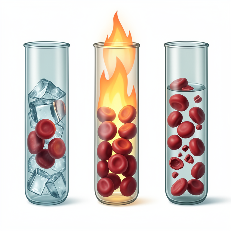
Anticoagulant Performance Comparison
AI-generated conceptual illustration (rabbit red blood cells in test tubes). It explains the idea and does not show study data.
View source evidence ↗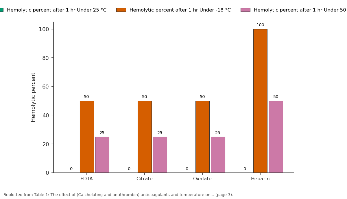
Hemolytic percent after 1 hr Under 25 °C and Hemolytic percent after 1 hr Under -18 °C and more: EDTA vs Citrate vs Oxalate vs Heparin
Replotted from Table 1 (page 3) of the original paper. Every bar is the paper's own table value.
View source evidence ↗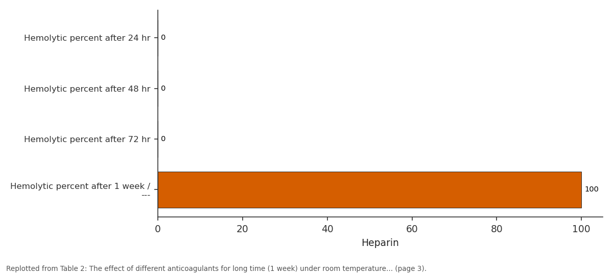
Heparin: Hemolytic percent after... vs Hemolytic percent after... vs Hemolytic percent after... vs Hemolytic percent after...
Replotted from Table 2 (page 3) of the original paper. Every bar is the paper's own table value.
View source evidence ↗Figures & tables
From the original paper Table 1
 Table 2
Introduction
In veterinary medicine the evaluation of animals healthy status usually need hematology and biochemistry analysis for the collected blood samples within mints or several hours; therefore the anticoagulants act important factor in keeping blood samples in save condition (BuffEnstein, et al., 2001) [3] The characteristic features of mammalian red blood cell it contain respiratory pigment called hemoglobin, without nucleus and life span about 120 days, they travel thousand of miles through various size of vessels in order to transport O2 to tissues and become attachment with endogenous and exogenous chemicals like anticoagulants, the erythrocytes activity limited by its intact and flexible membrane (Smith, 1987) [20] . The erythrocyte osmotic fragility test EOFT represent the precise determination to the red blood cell membrane resistance when exposed to hyper or hypo osmotic fluid and challenges associated with cyclic dehydration and rehydration; beside abnormality membrane detection (Ryall et al., 1978; Pacheco, et al., 2020) [17,15] . The blood clot formation impaired by a medical dominant chemical anticoagulants (heparin, ethylene diamine tetraacetic acid EDTA, citrate, oxalate) either in vivo and in vitro (Hugo, et al; 2020) [8] .
The aims of this study 1. Evaluating the efficacy of chelating calcium anticoagulants (EDTA, citrate, and oxalate) and antithrombin (heparin) anticoagulants for routine blood analysis at room temperature and severe temperatures (-18 °C & 50 °C). 2. To investigate the efficient anticoagulant conserve red blood cell membrane during storage of blood samples for long term at room temperature 25 °C.
The driving of water molecule from area of high concentration to area of low concentration through semipermeable membrane until reach to equilibrium is called osmosis (Jaio et al, 2015) [11] .
When the red blood cell suffer from stress it tends to hemolyse this is called fragility while the resistance of red blood cell to hemolysis when put in different dilute saline is called osmotic fragility (Pagano and Faggio, 2015) [16] .
Hemolysis:
The distraction of erythrocyte membrane lead to liberate hemoglobin (Hb), as the hemolysis occurs the incline of osmotic fragility of the cells; when the red blood cell put in isotonic fluid the number of water molecules influx is equal to the number of water molecules outflux; while in hypotonic solution the number water molecules influx more than outflux leading to swelling and rupture of red blood cell and visaversa in hypertonic the RBC become shrinkage; the size and shape of red blood cells remain intact inside the normal saline 0.9% NaCl (Samuel and Sowemimo 2002) [18] .
Anticoagulants:
The maintains of blood in fluid form for hematological analysis or get plasma for chemical analysis the anticoagulants are added to collecting tubes; the prevention of blood clot formation within the test tubes is occur due to irreversible remove of calcium ion which is responsible for almost coagulate formation (Farias et al., 2016; Cedrone, et al., 2018) [5,4] . The Ca chelating anticoagulants are EDTA, citrate and oxalate prevent the conversion of prothrombin into thrombin and fibrinogen into fibrin resulting Ca EDTA, Ca citrate and Ca oxalate (Shanker, 2017) [19] . The chelating activity occur when organic molecule make abound with a metal ion resulting of new complex molecule highly soluble and inactive ion that could not form precipitation (Guder,, 2002; Oliveira et al., 2015; Gros et al., 2023) [7,12,6] . Antithrombin anticoagulants represented mainly by heparin which is a monosaccharid chain which bind to lysine sites on antithrombin resulting in conformational changes at the arginine binding site that cause rapid inhibitor (Hirsh et [9,14,22] .
Materials and Methods
Experimental animals: Five male rabbits weighing between 800-1000 g were housed at the animal section of the College of Veterinary Medicine at the University of Diyala. They were subjected to a 12:12 dark light cycle and provided with free food and water in separate cages measuring 60 cm and 40 cm. This arrangement was maintained throughout November and December of 2022.
Blood collection: Four milliliters of blood samples were collected by heart puncture using a 23-gauge needle. These samples were placed into four tubes containing anticoagulants: EDTA, citrate, oxalate, and heparin. One milliliter of blood was added to each tube. The osmotic fragility test was conducted for each formulation to determine the zero time and verify the effectiveness of the anticoagulants. The blood samples were then stored at room temperature of 25 degrees Celsius and water bath temperature of 50 degrees Celsius (MeMMert 854 Schwabach), and refrigerator -18 °C (Arecilik Turkey), for 1hr followed by examination of red blood cell fragility in each type of anticoagulant under different temperature; finally the blood samples of room temperature 25 °C were tested after 24 hr, 48 hr, 72 hr and a week to point the long term anticoagulant (keeping the red blood cell membrane).
Determination of osmotic fragility test:
The osmotic fragility of erythrocyte was determined by adding few drops of blood to tubes contain 5ml of normal saline 0.85%, the mixtures put in centrifuge (EBA20 Hettich Zentrifu Gen; Germany) at 3000 rpm for 15min then the supernatant color indicate hemolysis while the clear mean not.
Preparation of physiological normal saline 0.85% 8.5 gm of NaCl dissolved in 1000 ml of distilled water. Determination of results: occur by percent % (Steel and Torrie 1980) [21] .
Preparation of anticoagulants
Preparation of anticoagulants
Results and Discussion
Hemolytic percent of male rabbits blood samples under some different types of anticoagulants and temperature According to table (1): A total of 32 blood samples were analysed for each kind of anticoagulant -EDTA, citrate, oxalate, and heparin -at zero time and after 1 hour at room temperature of 25 °C, -18 °C and 50 °C. The hemolytic percent at zero time and under 25 °C were zero% for all types but the hemolytic percent of heprinized blood sample after 1 hr revealed 100% and 50% under -18 °C and 50 °C respectively; while the Ca chelating anticoagulants types appear hemolytic percent 50% under -18c and 25% under 50 °C.
In several synthetic compound the EDTA were used as Ca ++ detector, it is unique by acidic COOH group which increase the interaction that lower erythrocyte hemolysis or may be it act as diluent resulted in red blood cell hemolysis (Monfared, et al., 2022) [13] . The synthetic anticoagulants heparin extract mainly from porcine intestinal mucosa, and it is prevent clot formation by inhibiting formation of fibrin from fibrinogen through impair thrombin activation, the heparin also modulate the electrolytes bound concentration and free ion beside antigen-antibody reactions during precipitation (Bowen and Ley, 2014) [2] .
Figures and tables from the paper
References
20 in the original paper- Banfi G, Salvagno L, Lippi G. The role of ethylenediamine tetraacetic acid (EDTA) as in vitro anticoagulant for diagnostic purposes. Clin Chem Lab Med. 2007;45(5):565-576.
- Bowen R, Andley R. Interferences from blood collection tube components on clinical chemistry assays. Biochem Med (Zagreb). 2014;24(1):31-44.
- Buffenstein R, McCarron H, Dawson T. Erythrocyte fragility of red (Macropus rufus) and grey (Macropus fuliginosus and Macropus giganteus) kangaroos and freeranging sheep of Australia. Comp Physiol B. 2001;171:41-47.
- Cedrome E, Neun B, Rodriguez J, Vermilya A, Clogeston J. Anticoagulants influence the performance of in vitro assays intended for characterization of nanotechnology-based formulations. Molecules. 2018;23(12):2926. doi:10.3390/molecules23122926.
- Farias V, Pereira L, Pádua B, Alves V, Sakabe R, Belo A, et al. Na2EDTA anticoagulant impaired blood samples from the teleost Piaractus mesopotamicus. Pesq Vet Bras. 2016;36(5):431-5.
- Gros N, Klobucar T, Gaber K. Accuracy of citrate anticoagulant amount, volume, and concentration in evacuated blood collection tubes evaluated with UV molecular absorption spectrometry on a purified water model. Molecules. 2023;28(2):486.
- Hugo Y, Nogueira N, Santos V, Boas F, Paulo J. Anticoagulant treatment for subsegmental pulmonary embolism. Cochrane Database Syst Rev. 2020;2
- Hirsh J, Anand S, Halperin J, Fuster V. Guide to anticoagulant therapy: heparin. Circulation. 2001;103:2994-3018.
- American Heart Association. A statement for healthcare professionals from the American Heart Association. Circulation. 2001;103:2994-3018.
- Oliveira A, Santos M, Lemos J, Dias M, Marcon J. Comparison of the effects of anticoagulants used in blood collection to determine blood parameters of free-living stingrays from the Potamotrygon genus (Elasmobranchii: Potamotrygonidae). Biota Amazônia. 2015;5(3):55-58.
- Monfared K, Mahmoudian M, Hoti G, Bisericaru M, Caldera F, Cavalli R, et al. Hyper-branched cyclodextrinbased polymers as anticoagulant agents: in vitro and in vivo studies. Bioengineering. 2022;9:765.
- Onishi A, Ange K, Dordick J, Linhardt R. Heparin and anticoagulation. Front Biosci. 2016;21:445-461.
- Pacheco AP, Salazar LB, Garcia AV, Mendoza ER, Martin A, Carrillo IO, et al. Erythrocytes osmotic fragility test with a standard Abb-refractometer. Rev Med Hosp Gen Mex. 2020;83(1):20-25.
- Pagano M, Faggio C. The use of erythrocytes fragility to assess xenobiotic cytotoxicity. J Cell Biochem Funct. 2015;33(6):351-355.
- Ryall RG, Chittleborough AJ, Kane A. A microtechnique for measuring red cell osmotic fragility of infants. J Clin Pathol. 1978;31:878-882.
- Samuel O, Sowemimo C. Red blood cell hemolysis during processing. Transfusion Med Rev. 2002;16(1):4660.
- Shankar V. Anticoagulants in hematologic investigation. Clin Pathol. 2017;10:263-269.
- Smith JE. Review: Erythrocyte membrane, structure, function and pathophysiology. Vet Pathol. 1987;24:471476.
- Steel RG, Torrie JH. Principles and procedures of statistics: a biometrical approach. 2nd ed. New York: McGraw-Hill; 1980. p. 693-696.
- Zang L, Zhu H, Wang K, Lui Y, Yu F, Zhao X. Review: Not just anticoagulation-new and old applications of heparin. Molecules. 2022;27:6968. doi:10.3390/molecules27206968.