
Research article · 2024 · Full text
International Journal of Veterinary Sciences and Animal HusbandryFrom lab to animal facility: A complete guide for tumor xenograft model creation
Volume 9(5) · Pages 149–155 · DOI: 10.22271/veterinary.2024.v9.i5c.1668 ↗
Research areasCancer Research and Treatments
Abstract
Author-writtenThis article presents a comprehensive and accessible protocol for establishing cell-derived xenograft (CDX) models in immunocompromised mice, specifically tailored to align with the practices at IISER Bhopal. Designed for beginners in cancer research, this protocol not only serves as a guide to studying human tumor biology and evaluating new therapeutic strategies but also provides an integrated approach that connects the laboratory and the animal care facility.The protocol covers the entire process from selecting and preparing human tumor cells to their implantation and monitoring in various strains of immunocompromised mice. It also includes detailed methodology addressing key aspects such as hygiene, housing, and pain management, ensuring that ethical standards are maintained while optimizing experimental outcomes.Importantly, this guide extends beyond the laboratory procedures to include practices followed in the animal experimentation room and the post-experiment care of animals. By offering clear instructions and practical tips, this protocol bridges the knowledge gap between researchers and veterinary staff, fostering a collaborative environment that enhances both the quality of research and animal welfare.By following this protocol, researchers at IISER Bhopal and beyond can establish robust tumor models with confidence, ensuring that their cancer research is both impactful and ethically responsible.
Summary
Source-checkedPlain-language reading of the full paper, prepared with AI and checked against the text. Each point links to the authors' own words.
Why it matters
- Cell-derived xenograft (CDX) models are essential in cancer research because they allow for the study of human tumors within living immunocompromised mice. View evidence ↗
- These models are valuable for understanding tumor growth, metastasis, and therapeutic responses because they closely mimic human cancer dynamics. View evidence ↗
- CDX models are specifically useful for testing drugs in preclinical stages and developing personalized medicine. View evidence ↗
The question
- The aim of this work is to provide a comprehensive and accessible protocol for establishing CDX models in immunocompromised mice, based on practices at IISER Bhopal. View evidence ↗
What they did
- The protocol was demonstrated using the MCF-7 breast cancer cell line. View evidence ↗
- The animal model used for the demonstration consisted of 4-week-old female NOD SCID mice. View evidence ↗
- The cell preparation involved mixing 1 x 10^7 cells with a 1:1 ratio of Matrigel and PBS to support cell survival and tumor formation. View evidence ↗
- Anesthesia was induced with 5% isoflurane and maintained at 2% during the implantation procedure. View evidence ↗
- To protect the immunocompromised mice from contaminants, they were housed in Individually Ventilated Cages (IVC) with HEPA filters. View evidence ↗
- Post-surgical pain management included the administration of meloxicam at 5 mg/kg body weight once daily for up to three days. View evidence ↗
- The ethical protocol defines the maximum allowable subcutaneous tumor size as 20 mm for mice and 40 mm for rats. View evidence ↗
What they found
- By day 28, tumors reached approximately 20 mm in size at both the flank and mammary gland implantation sites. View evidence ↗
- Histopathological analysis confirmed successful engraftment and invasive growth, characterized by a densely packed cellular architecture. View evidence ↗
What it means
- The authors conclude that this guide serves as a holistic tool to bridge the gap between laboratory procedures and animal care facility practices. View evidence ↗
- The authors recommend this detailed approach to foster collaboration between researchers and veterinary staff to prioritize animal welfare alongside scientific goals. View evidence ↗
Visual summary
1 visualsIllustrations explain the paper; they do not show study data.
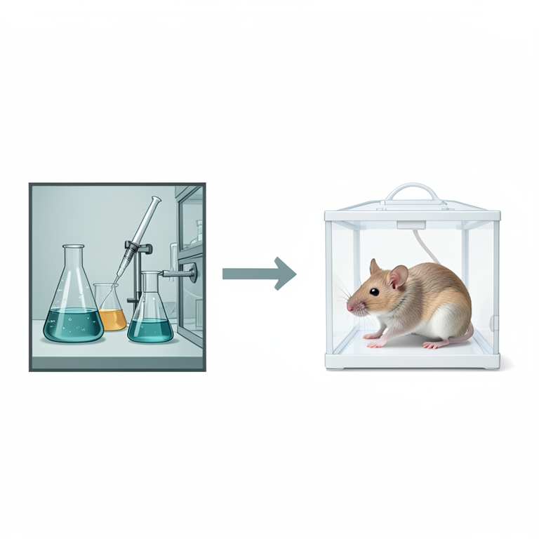
The Lab-to-Facility Workflow
AI-generated conceptual illustration (immunocompromised mice and laboratory cell culture equipment). It explains the idea and does not show study data.
View source evidence ↗Figures & tables
From the original paper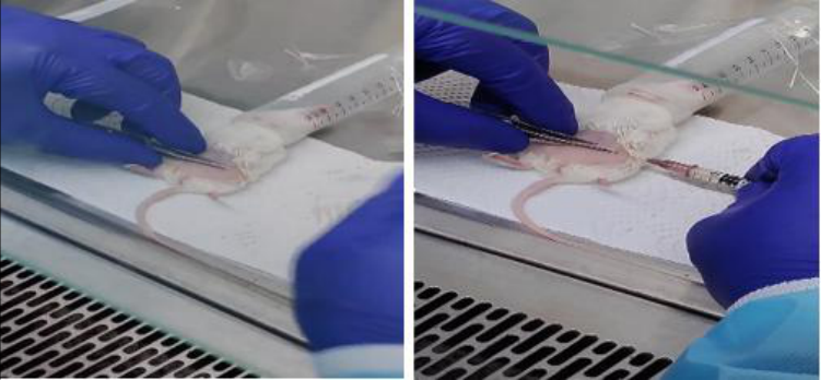 Figure 1
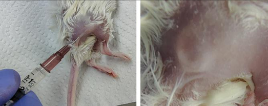 Figure 2
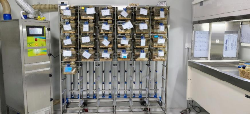 Figure 3
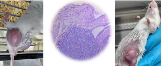 Figure 3
Introduction
Cell-derived xenograft (CDX) models are indispensable tools in cancer research, providing a platform to study human tumors in vivo within immunocompromised mouse models. These models are crucial for understanding tumor biology, including tumor growth, metastasis, and responses to therapeutic interventions, as they closely replicate human cancer dynamics. CDX models are particularly valuable for preclinical drug testing and the development of personalized medicine approaches. This protocol not only streamlines the process of establishing CDX models across different mouse strains but also integrates the comprehensive practices followed at IISER Bhopal, ensuring reproducibility, reliability, and ethical integrity in research outcomes. By adhering to standardized procedures, researchers can generate robust and consistent data, which is critical for advancing cancer research and the development of new therapies. Moreover, this protocol emphasizes the importance of collaboration between researchers and veterinary staff, detailing the entire process from cell preparation to post-experiment animal care. This holistic approach ensures that all aspects of the experiment are conducted with the highest standards of precision and animal welfare, ultimately leading to more meaningful and impactful research outcomes. By following this protocol, researchers can not only achieve reproducible results but also contribute to the ethical and responsible conduct of cancer research, thereby playing a vital role in the ongoing fight against cancer.
Materials and Equipment
Human Tumor Cells: Prepared and ready for implantation. Immunocompromised Mice: Nude or SCID mice.
Surgical Instruments: Needles, syringes, and forceps. Anaesthesia: Isoflurane or another approved anesthetic. Cell Culture Media: For preparing and resuspending cells. Sterile Equipment: Gloves, sterile tubes, and cell culture dishes. The following procedure is a part of IAEC approved study.
Procedure Cell Preparation
Proper preparation of human tumor cells is crucial for the successful establishment of cell-derived xenografts (CDXs), ensuring high-quality and reproducible results. We begin this process with careful cell line selection, choosing a wellcharacterized human tumor cell line that aligns with our research objectives. It is essential to authenticate the selected cell line and test for contamination, such as mycoplasma, which could compromise the integrity of our research (Clarke, 2018) [9] . We also confirm the cell line's growth characteristics, genetic stability, and suitability for implantation to ensure that the cells will perform as expected in vivo (MacLeod & Reddel, 2007) [35] .
In this experiment, we utilized the MCF-7 cell line, a breast cancer cell line isolated in 1970 from a 69-year-old White woman. MCF-7, an acronym for Michigan Cancer Foundation-7, refers to the institute in Detroit where the cell line was established in 1973 by Herbert Soule and his team (Soule et al., 1973) [52] . We cultured these tumor cells in appropriate media supplemented with necessary growth factors (Freshney, 2010) [18] . Specifically, we used RPMI-1640 or DMEM, supplemented with 10% fetal bovine serum and 1% antibiotic and antimycotic, tailored to the cell type. We maintained the cells at 37°C in a humidified incubator with 5% CO 2 , mimicking physiological conditions to promote optimal growth. During cell harvesting, we detached adherent cells using a trypsin-EDTA solution or a non-enzymatic cell dissociation reagent, carefully monitoring under a microscope to avoid over-trypsinization, which could damage the cells (Vandesompele & De Paepe, 2002) [56] . After harvesting, we neutralized the trypsin with complete culture media containing serum to provide necessary nutrients for cell recovery.
Cell counting is another critical step in our preparation process. We transferred the cells to a sterile centrifuge tube and centrifuged at 300-400 x g for 5-10 minutes to pellet the cells. After discarding the supernatant, we resuspended the cell pellet in a suitable volume of fresh medium or PBS. Using a hemocytometer or an automated cell counter, we determined cell concentration and performed trypan blue exclusion staining to assess cell viability (McCoy & Murphy, 2016) [38] . Viable cells excluded the dye and appeared clear, while dead cells took up the dye and appeared blue (Liu & Li, 2015) [34] . We then adjusted the cell concentration to the desired level for implantation, ensuring reproducibility and effectiveness in our experimental models.
For this experiment, we mixed 1 x 10^7 cells with Matrigel and PBS in a 1:1 ratio to resuspend the cell pellet. This combination enhances cell viability by providing a supportive matrix that mimics the extracellular environment, essential for cell survival and potential tumor formation (Kleinman & Philp, 2003) [30] . Matrigel also improves the efficiency of cell engraftment by promoting adhesion and growth, which is critical for establishing a robust tumor model. The 1:1 ratio of Matrigel to PBS allowed us to control and consistently deliver cells, reducing the risk of clumping or uneven distribution during injection. This method ensures the implantation of a sufficient number of cells, optimizing the chances of successful engraftment and reliable experimental outcomes. Quality control is paramount before implantation. We ensured high cell viability (Typically greater than 80%) and confirmed the sterility of the cell suspension by checking for contamination. This step is critical, as low viability can lead to poor engraftment and unreliable results. For implantation preparation, we aliquoted the cell suspension into sterile syringes or tubes, keeping them on ice or at 4 °C to maintain cell viability until use. We labeled all aliquots with relevant information, including cell line, concentration, date, and other pertinent details to ensure proper tracking. If not used immediately, we stored the cells at 4°C for short periods of up to 4 hours, avoiding extended storage to prevent cell stress or death. For long-term storage, we froze cells in aliquots using cryoprotectants like DMSO in a freezing medium, storing them in liquid nitrogen to preserve cell lines for future use (Balan & Tang, 2019) [4] . Throughout the entire process, we strictly adhered to safety and handling considerations. We wore appropriate personal protective equipment (PPE), including gloves, lab coats, and eye protection, to prevent contamination and exposure to hazardous materials. We also ensured the proper disposal of all cell culture materials and contaminated waste according to biohazard safety protocols, maintaining a safe working environment. By following these comprehensive guidelines for the preparation and handling of human tumor cells, we enhance the reliability and reproducibility of our CDX studies, ultimately contributing to more meaningful scientific insights in cancer research.
Animal Preparation Animal Strain:
We have used NODSCID Females of 4 week of age in our experiment. There are other strains like Nude or NSG Mice which can also be used based on the researcher need.
Subcutaneous Implantation at the Mammary Gland
Begin by inducing anesthesia with 5% isoflurane, followed by maintaining it at 2% to keep the mouse immobilized and painfree during the procedure (Flecknell, 2009) [17] . Once the mouse is fully anesthetized, identify the 4th nipple in the lower abdominal region. Disinfect the injection site with 70% ethanol (Hargraves & O'Neill, 2020) [21] . Using forceps, gently lift the skin near the nipple to create a tent-like structure, forming a tunnel for the injection. Insert the syringe needle just parallel to the skin surface with the bevel facing upwards. Guide the needle carefully into the tunnel, ensuring it moves under the skin without penetrating deeper tissues (Cuthbertson & Clarke, 2013) [10] . Align the bevel so it is positioned just below the nipple, then slowly release the cells while retracting the syringe from the tunnel. Successful injection will be indicated by the formation of a small bleb or swelling at the injection site, confirming that the cells have been properly deposited subcutaneously. It is essential to maintain the needle's angle throughout to avoid accidental intraperitoneal (IP) injections, which could affect the outcome of the procedure (De Matteis & Schultz, 2015) [11] . Immobilize the mouse by using anesthesia-starting with 5% induction followed by 2% maintenance-or by manual restraint if anesthesia is not required (Flecknell, 2009) [17] . Disinfect the injection site (e.g., the flank of the mouse) with 70% ethanol (Hargraves & O'Neill, 2020) [21] . Prepare a 26gauge needle for injecting the cells. Gently elevate the skin at the flank site and insert the needle with the bevel side facing upwards. Once inserted, move the needle in a fan-like fashion to create more space under the skin, allowing for a larger area to accommodate the tumor cells (Cuthbertson & Clarke, 2013) [10] . After injecting the cells, wait for a moment before retracting the syringe to ensure the cells are deposited properly. Upon removing the syringe, apply gentle pressure to the injection site with a tissue to prevent any cell solution from oozing out. Successful injection will be indicated by the formation of a small bleb or swelling at the injection site, confirming that the cells have been properly deposited subcutaneously (De Matteis & Schultz, 2015) [11] [55] . Prompt identification of any concerning changes is crucial for minimizing suffering and ensuring animal welfare throughout the study (Flecknell, 2009) [17] .
Effective pain management is a critical component of postoperative care in CDX research. Researchers must adhere to established anesthesia and analgesia protocols to minimize pain and discomfort during and after surgical procedures (Morton & Griffiths, 1985) [41] . Approved anesthetic agents should be used, with careful monitoring of anesthesia depth during surgery to ensure adequate pain relief (Flecknell, 2009) [17] . Postoperatively, analgesics such as buprenorphine or meloxicam should be administered based on the animals' weight and condition, with ongoing assessments of pain to adjust protocols as needed (Roughan & Flecknell, 2000) [42] . By implementing comprehensive monitoring and effective pain management strategies, researchers can promote animal welfare and enhance the reliability of their findings while upholding ethical research practices (Balthazar & Goh, 2012) [6] .
Monitoring
In our daily routine, we conduct thorough inspections of our mice to identify any signs of distress, health issues, or complications. Each day, we carefully monitor changes in behavior, weight, and physical appearance. We keep an eye on key health indicators, such as signs of infection, including redness, swelling, or discharge. We also observe for abnormal tumor growth, which might present as excessive swelling or necrosis. When it comes to general health, we document any instances of lethargy or weight loss. For tumors, we take precise measurements of subcutaneous implants using calipers or other measurement tools to assess size. For orthotopic implants, we employ imaging techniques like ultrasound or MRI to effectively monitor tumor growth and position (Van den Broek & Thompson, 2015) [55] . By following this comprehensive approach, we ensure timely intervention and contribute to the overall welfare of our mice (Flecknell, 2009) [17] .
Pain and Stress Management
In managing pain and discomfort for our mice undergoing tumor assessments and treatments, we administer analgesics as needed, adhering to institutional guidelines for dosing and frequency. We commonly use meloxicam at a dosage of 5 mg/kg body weight, administering it subcutaneously or orally once daily for up to three days post-surgery, or as needed based on the animal's pain assessment (Morton & Griffiths, 1985) [41] . We also consider buprenorphine as an effective option, administering it at a dosage ranging from 0.1 to 0.5 mg/kg body weight, given subcutaneously or intravenously every 6 to 12 hours as required for pain management (Roughan & Flecknell, 2000) [42] . Additionally, for any procedures requiring anesthesia, we ensure proper administration and monitoring to minimize stress and pain, safeguarding the welfare of our mice throughout their tumor evaluation and treatment process (Balthazar & Goh, 2012) [6] .
Hygiene and Housing
We house NOD SCID mice which lack adaptive immunity as well as our other strains, in Individually Ventilated Cages (IVC) equipped with inbuilt HEPA filters to protect them from potential contaminants, as recommended by Dutton & Garner (2018) [13] . Due to their high susceptibility to environmental pathogens, these mice cannot be housed in traditional cages.
To maintain a sterile environment, we handle the mice with utmost care, always wearing gloves and appropriate personal protective equipment (PPE). All procedures are conducted in a laminar flow hood or safety cabinet, following the guidelines of Whitford & Kline (2019) [57] . We change the cages in a dedicated cage-changing station with laminar flow and filters, ensuring that each step is taken to minimize the risk of infection.
We also ensure that the cages are kept clean and provide fresh bedding regularly, using wood chips from Himachal pine trees, which are autoclaved along with the cages. The housing environment is meticulously maintained to be free from contaminants, with cages properly sanitized, in line with the practices outlined by Kohn & Wills (2012) [31] .
Feeding and Hydration
We provide a balanced, commercial rodent diet specifically formulated for our strain of mice. To ensure their well-being, we make sure that food is always available and regularly monitor for any changes in their eating habits. To minimize the risk of contamination, we offer autoclaved water. Although gamma-irradiated feed is more expensive, we use it to maintain a pathogen-free environment, as recommended by Lee & Zhang (2018) [33] . We also regularly check that the mice have easy access to water and ensure that the water delivery system is functioning properly, following guidelines from the National Research Council (1995).
For bedding, we use wood chips from Himachal pine trees, which are incorporated along with Individually Ventilated Cages (IVC) and autoclaved to maintain a sterile environment. We provide Reverse Osmosis (RO) water with a Total Dissolved Solids (TDS) level of 100, and this water is also autoclaved, along with the autoclave-safe water bottles, to ensure purity.For our immunocompromised animals, we provide a diet formulated to meet their specific nutritional needs. The feed contains 14% fat, 27% protein, and 59% carbohydrates, offering a comprehensive nutrient profile. The crude nutrient analysis includes 11.1% moisture, 6.1% crude ash, 4.5% crude fiber, 5.1% crude fat, 22.5% crude protein, and 50.7% nitrogen-free extract, as outlined by the American Association of Feed Control Officials (2021). [52] in the characterization of the MCF-7 cell line. The dense cellular infiltrate observed in the dermis is likely the result of tumor formation from the injected MCF-7 cells, consistent with findings from Amin and Biliciler (2015) [2] , who reported similar histopathological features in MCF-7induced tumors. The irregular, densely packed cells observed are consistent with tumor tissue, as described in various studies on MCF-7 xenografts. The lack of clear demarcation between the dense cellular infiltrate and the surrounding tissue suggests possible invasion of the tumor into adjacent areas, which is typical of malignant growths and has been similarly observed in in vivo models of MCF-7 tumor formation.
Data Collection and Analysis
Equally important is the need for consistency in measurement techniques. Utilizing the same methods and tools throughout the study ensures that the data collected is comparable, minimizing variability that could arise from differences in measurement approaches. This uniformity enhances the reliability of the results, allowing researchers to draw meaningful conclusions about the efficacy of treatments based on robust and consistent data (Houghton & Houghton, 2009) [25] . By adhering to these data collection protocols, researchers can ensure a systematic and thorough investigation of tumor behavior in response to various therapeutic strategies. The analysis of tumor growth is crucial in evaluating the efficacy of therapeutic interventions in cancer research. The volume of preclinical tumor are measured by using V = 0.5 × L × W 2 , where V is the tumor volume, L is the tumor length, and W is the tumor width .Researchers begin by plotting tumor volume over time to generate growth curves, which serve as visual representations that help identify trends in tumor progression and assess the effects of various treatments (LaBree & Rogers, 2015) [32] . By comparing growth curves across different experimental groups, researchers can effectively evaluate the relative efficacy of the therapeutic approaches being studied. Statistical analysis further enhances the robustness of the findings. Descriptive statistics, including mean, median, standard deviation, and range of tumor volumes for each group, provide foundational insights into the data distribution (LaBree & Rogers, 2015) [32] . For comparative analysis, appropriate statistical tests such as t-tests or ANOVA are employed to assess differences in tumor volumes between treatment groups and controls, ensuring that the assumptions of these tests, such as normality and homogeneity of variances, are satisfied. Additionally, survival analysis can be conducted to examine the time taken for tumors to reach specific sizes or to assess overall animal survival, providing valuable context to the treatment effects observed (Klein & Moeschberger, 2003) [29] .
Interpreting the data requires careful consideration of both efficacy and biological relevance. Researchers assess treatment effectiveness by comparing tumor volume reductions or growth inhibition between treated and control groups. However, it is essential to interpret the results within the context of their biological significance, not solely their statistical significance (Roth & Kwon, 2012) [46] . This involves considering how the findings contribute to the broader understanding of tumor biology and their potential therapeutic implications, ultimately guiding future research and treatment strategies in oncology. access to food and water (Hawkins, 2014) [22] . Daily health monitoring is necessary to detect signs of distress, illness, or discomfort, allowing for prompt intervention and veterinary care when needed. Implementing humane endpoints is crucial for minimizing suffering, with predefined criteria for euthanasia if animals exhibit severe distress, excessive tumor burden, or other health complications (International Council for Laboratory Animal Science [ICLAS], 2012). Humane treatment practices are fundamental in all procedures involving the mice. Anesthesia should be administered to minimize pain during surgical interventions, using approved anesthetics and ensuring proper monitoring of anesthesia depth for effective pain relief (Flecknell, 2009) [17] . Postsurgery, analgesics should be provided as needed throughout the study to manage discomfort effectively. Surgical procedures must be conducted using sterile techniques to prevent infections, and all personnel involved should be adequately trained in surgical techniques and animal handling (Kilkenny et al., 2010) [28] . By adhering to these guidelines, researchers can uphold ethical standards and ensure the wellbeing of the animals involved in their studies, ultimately contributing to the integrity and validity of the research findings.
Safety and Ethical Considerations in
Safety and Ethical Considerations in
Guidelines for Tumor Burden and Ethical Considerations for Xenograft Models
We must prioritize the responsible management of human cells and biological materials when conducting research involving cell-derived xenografts. Adhering to biosafety guidelines ensures safety and compliance with ethical standards. We use appropriate personal protective equipment (PPE) to prevent contamination and exposure while handling human tumor cells (National Institutes of Health [NIH], 2016). We also follow strict decontamination procedures, including fumigation after injecting cells, to maintain a safe working environment and protect both ourselves and the integrity of the biological materials being studied (Centers for Disease Control and Prevention [CDC], 2020). When determining the humane endpoint for an experiment, the size of the tumor is only one factor. We must prioritize the overall well-being of the animal in both oncological experiments and cases of spontaneous tumors, as outlined by the guidelines of the Institute for Laboratory Animal Research (ILAR) and other ethical standards in animal research (Workman et al., 2010) [60] . To minimize harm to surrounding normal tissues, we carefully select the site for tumor implantation, avoiding areas that could impact critical senses (Hendriksen & Morton, 1999) [23] . We have outlined specific endpoints clearly in the Institutional Animal Ethics Committee (IAEC) protocol. For subcutaneous tumors, the maximum size should not exceed 20 mm in diameter for mice and 40 mm for rats (Hendriksen & Morton, 1999) [23] . If an animal hosts multiple tumors, this size limit applies to the total combined size of all tumors. Measurements are taken with calipers at least twice a week once they become visible. When a tumor reaches 10 mm in mice or 20 mm in rats, the frequency of measurements is increased to every other day. For rapidly growing tumors, daily measurements are advisable. All measurements are recorded properly, ensuring they are accessible to husbandry and veterinary staff for health or welfare concerns (Workman et al., 2010) [60] . If the skin over or near a tumor becomes ulcerated, we withdraw the animal from the study and euthanize it. If the protocol allows for exceptions, we explicitly state this in the IAEC-approved animal use protocol. We inject tumors in areas that do not hinder the animal's movement or physiological and social behaviors. If the tumor impairs these functions, we remove the animal from the study and euthanize it humanely (Roughan & Flecknell, 2003) [48] . All these practices have received approval from our institutional ethics committee, which follows IAEC guidelines.
In conclusion, adhering to safety and ethical considerations is essential for conducting responsible and humane research with cell-derived xenografts. By following these guidelines, researchers can ensure that their work not only yields valuable scientific insights but also upholds the highest standards of animal welfare and research ethics, ultimately contributing to the advancement of knowledge while maintaining the integrity of the research process.
Conclusion
The establishment of cell-derived xenograft (CDX) models in immunocompromised mice is a cornerstone of contemporary cancer research, and this protocol reflects the comprehensive practices followed at the IISER Bhopal animal facility. This guide is not just a procedural manual; it is a holistic tool designed to bridge the gap between the laboratory and the animal care facility, providing an eagle-eye view of the entire process-from the initial stages of cell culture in the lab to the meticulous care of animals post-experimentation.
We have endeavored to include every critical step, ensuring that researchers are well-informed about the procedures taking place both within the laboratory and the animal experimentation room. Beginning with the careful preparation of cell suspensions and extending to the precision required in subcutaneous injections, our protocol ensures that experiments are conducted with the highest level of accuracy and reproducibility. This methodical approach not only validates preclinical findings but also enhances communication between researchers and veterinary staff. Additionally, we have emphasized the protocols for the humane treatment and care of the animals, both during the experimental phases and afterward. This aspect is often overlooked, with researchers sometimes unaware of the conditions within the animal holding rooms and veterinarians not fully informed of the laboratory procedures. By detailing these practices, we aim to foster a deeper understanding and collaboration between the lab and veterinary teams, ensuring that the welfare of the animals is prioritized alongside the pursuit of scientific knowledge. In summary, this protocol is a testament to the commitment of IISER Bhopal to advancing cancer research while maintaining the highest ethical standards in animal care. It is a comprehensive guide that empowers researchers to achieve breakthrough findings, all while ensuring the responsible and humane treatment of the animals involved in this critical research.
Figures and tables from the paper
References
47 in the original paper- Amin R, Biliciler S. Histopathological and immunohistochemical study of MCF-7 cell line-induced tumors in a xenograft model. Appl Immunohistochem Mol Morphol. 2015;23(4):271-278.
- Baek NH, et al. In vivo tumor formation of MCF-7 cells in nude mice: Characterization of MCF-7 derived breast carcinoma tumor. Cancer Res Treat. 2013;45(1):43-51.
- Balan V, Tang J. Cryopreservation of cell lines: Techniques and protocols. Methods Mol Biol. 2019;1928:1-12.
- Balleine BW, Killcross AS. The role of the basolateral amygdala in learning and memory. J Neurosci. 2006;26(11):2981-2987.
- Balthazar J, Goh J. Effective postoperative care strategies for laboratory animals. J Am Assoc Lab Anim Sci. 2012;51(5):620-626.
- Carbone L, Austin J. Pain and laboratory animals: Publication practices for better data reproducibility and better animal welfare. PLoS ONE. 2016, 11(5).
- Centers for Disease Control and Prevention (CDC). Guidelines for Disinfection and Sterilization in Healthcare Facilities, 2008. CDC; c2020. Available from: CDC Disinfection Guidelines
- Clarke C. Importance of cell line authentication and mycoplasma testing. J Cell Biol. 2018;217(11):40104018.
- Cuthbertson L, Clarke S. Techniques for subcutaneous injection in laboratory mice. J Lab Anim Sci. 2013;53(6):673-678.
- De Matteis F, Schultz R. Subcutaneous tumor cell implantation in mice: Techniques and considerations. Cancer Res. 2015;75(7):1479-1487.
- Dittmer DP, Karakousis G. A375 cell line: A valuable tool for melanoma research. Melanoma Res. 2012;22(2):147-155.
- Fan W, Zhang J. HCT116 cell line: Applications and utility in colorectal cancer research. J Cell Biochem. 2015;116(8):1825-1832.
- Fenton C. Subcutaneous injection techniques and outcomes in mouse models. J Biomed Sci. 2017;24(1):39.
- Friedman A, Nelson T. U87-MG cell line: A model for glioblastoma research. Neuro-Oncology. 2009;11(3):242251.
- Gibson RL, Tyler AA. MDA-MB-231 and MCF-7 cell lines: A comparative study of breast cancer model systems. J Cancer Res Ther. 2013;9(2):225-231.
- Hargraves L, O'Neill K. Best practices for injection site preparation and handling in laboratory animals. Lab Anim. 2020;54(1):65-76.
- Hawkins P. Recognizing and assessing pain, suffering and distress in laboratory animals: A survey of current practice in the UK with recommendations. Lab Anim. 2014;48(1):1-18.
- Hendriksen C, Morton D, editors. Humane Endpoints in Animal Experiments for Biomedical Research. Proceedings of the International Conference, 22-25 November 1998, Zeist, The Netherlands. Royal Society of Medicine Press; c1999.
- Holliday DL, Speirs V. Choosing the right cell line for breast cancer research. Breast Cancer Res. 2011;13(4):215.
- Houghton PJ, Houghton JA. Nude mice and the study of human tumors: The legacy of basic and applied cancer research. Cancer Res. 2007;67(16):7530-7533.
- Kaighn ME, Narayan KS. PC-3 cell line: A new model for prostate cancer research. Prostate. 1979;3(1):87-99.
- Kilkenny C, Browne WJ, Cuthill IC, Emerson M, Altman DG. Improving bioscience research reporting: The ARRIVE guidelines for reporting animal research. PLoS Biol. 2010, 8(6).
- Kleinman HK, Philp D. Matrigel: A review of the use of Matrigel in cancer research and tumor modeling. J Cell Biochem. 2003;88(1):28-36.
- LaBree RT, Rogers J. Analyzing tumor growth data: Statistical methods and best practices. J Stat Comput Simul. 2015;85(10):1924-1937.
- Lee CC, Zhang J. Autoclaving and gamma-irradiation of feed for laboratory rodents: Benefits and risks. J Anim Sci Biotechnol. 2018;9(1):67.
- Liu Y, Li X. Trypan blue exclusion test and its applications in cell viability assessment. J Biomed Sci. 2015;22(1):60.
- MacLeod RA, Reddel RR. Mycoplasma contamination in cell culture: A problem revisited. J Cell Biochem. 2007;102(6):1221-1230.
- Markman M, Gold M. SKOV-3 cell line: Utility in ovarian cancer research. Gynecol Oncol. 2004;94(3):771779.
- Martin L, Wagner R. Monitoring injection sites and postoperative care for laboratory mice. J Exp Anim Sci. 2018;25(3):209-219.
- McCoy JC, Murphy CJ. Cell counting and viability assays: A comprehensive guide. J Cell Sci. 2016;129(12):2353-2360.
- Miller RE, Liu H. A549 cell line: Model of lung cancer in preclinical research. Am J Cancer Res. 2016;6(8):16271638.
- Mombaerts P, Iacomini J. RAG-1-deficient mice have severe combined immunodeficiency and lack functional T and B lymphocytes. Cell. 1992;68(5):855-867.
- Morton DB, Griffiths PH. Guidelines on the recognition of pain, distress and discomfort in experimental animals and a hypothesis for assessment. Vet Rec. 1985;116(16):431-436.
- Roth JA, Kwon K. Interpreting data from cancer research studies: A focus on biological significance. Cancer Res. 2012;72(14):3618-3625.
- Roughan JV, Flecknell PA. The assessment of postoperative pain in laboratory animals: A review. Lab Anim. 2000;34(2):129-145.
- Roughan JV, Flecknell PA. Behavioural effects of laparotomy and analgesic effects of ketoprofen and carprofen in rats. Pain. 2003;104(3):489-498.
- Sato Y, Matsui Y. Guidelines for monitoring tumor growth and tumor-related effects in mice and rats. J Toxicol Pathol. 2017;30(2):105-114.
- Shultz LD, Lang P. Humanized mice in cancer research. J Clin Invest. 2008;118(1):101-105.
- Shultz LD, Schweitzer I. NOD/SCID mice: A model of human disease. J Immunol Methods. 1995;193(1):111123.
- Soule HD, et al. A human cell line from a pleural effusion derived from a breast carcinoma. J Natl Cancer Inst. 1973;51(5):1409-1416.
- Ullman-Culleré MH, Foltz CJ. Body condition scoring: A rapid and accurate method for assessing health status in mice. Lab Anim Sci. 1999;49(3):319-323.
- Van den Broek J, Thompson D. Postoperative care and monitoring in animal research. Lab Anim Sci. 2015;65(4):552-556.
- Vandesompele J, De Paepe A. Efficient cell harvesting: Techniques and protocols. Cell Res. 2002;12(1):11-16.
- Whitford WG, Kline K. Guidelines for the use of individually ventilated cages in laboratory animal facilities. Lab Anim. 2019;53(2):139-153.
- Wilentz RE, Yeo CJ. PANC-1 cell line: Characteristics and use in pancreatic cancer research. Cancer Res. 2003;63(1):290-297.
- Workman P, Aboagye EO, Balkwill F, Balmain A, Bruder G, Chaplin DJ, et al. Guidelines for the welfare and use of animals in cancer research. Br J Cancer. 2010;102(11):1555-1577.
- Workman P, et al. Guidelines for the welfare and use of animals in cancer research. Br J Cancer. 2010;102(11):1555-1577.