
Research article · 2024 · Full text
International Journal of Veterinary Sciences and Animal HusbandryTherapeutic management of fetal mummification in a bitch: A case report
Volume 9(5) · Pages 104–105 · DOI: 10.22271/veterinary.2024.v9.i5b.1660 ↗
Research areasUrological Disorders and TreatmentsVeterinary Medicine and SurgeryGenital Health and Disease
Abstract
Author-writtenAn 18-month-old bitch was presented with dystocia due to uterine inertia at the Referral Veterinary Polyclinic, IVRI, Izatnagar. Clinical examination revealed a foul-smelling vaginal discharge, systemic illness, and a lodged fetal mass in the reproductive tract. Following treatment, the mummified fetus and a normal-sized dead fetus were delivered. The dog recovered uneventfully after three days of supportive care.
Summary
Source-checkedPlain-language reading of the full paper, prepared with AI and checked against the text. Each point links to the authors' own words.
Why it matters
- Fetal mummification occurs when a fetus dies after its bones have ossified, provided there is no bacterial infection. View evidence ↗
- During mummification, the fetus and uterus shrink as fetal fluids and the placenta are absorbed by the body. View evidence ↗
- In dogs, fetal death leading to mummification can be caused by various factors, including infections, chromosomal abnormalities, or maternal endocrine disorders. View evidence ↗
The question
- The case aimed to manage a 18-month-old bitch presenting with dystocia caused by uterine inertia. View evidence ↗
What they did
- Clinical assessment of the patient showed systemic illness, anorexia, a rectal temperature of 104.2°F, and foul-smelling brownish vaginal discharge. View evidence ↗
- A per-vaginal examination was used to identify a fetal mass lodged in the reproductive tract. View evidence ↗
- Initial therapeutic intervention consisted of 250 ml of 25% Dextrose saline, 15 IU of Oxytocin, and 10 ml of Calcium borogluconate administered intravenously. View evidence ↗
- Post-delivery care included Ceftriaxone (20 mg/kg IV), Meloxicam (0.2 mg/kg IM), and 2.5 ml of Eldervit multivitamin injection. View evidence ↗
What they found
- The dog fully recovered without complications after three days of supportive therapy and follow-up. View evidence ↗
What it means
- The authors conclude that uterine inertia was likely the primary cause for the failure to deliver the mummified fetus. View evidence ↗
- The authors recommend standard vaccination, reproductive monitoring, and proper hygiene to prevent fetal mummification. View evidence ↗
- A limitation of this case was that radiography and ultrasonography, which are effective for diagnosing mummification, were not performed. View evidence ↗
Figures & tables
From the original paper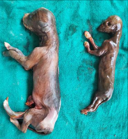 Figure 1
Introduction
Fetal death following ossification of the bones without bacterial infection typically results in fetal mummification (Noakes,1986) [3]. In this condition, the uterus and fetus gradually shrink as the placenta and fetal fluids are absorbed, leading to the formation of a mummy within the uterus. The cervix remains tightly closed, and the fetus becomes sterile (Purohit and Gaur, 2011) [7]. In polytocous species, the occurrence of mummification in some fetuses typically does not disrupt the continuation of pregnancy for the remaining fetuses. (Noakes et al., 2001) [4].
In canines, fetal death can result from developmental or chromosomal abnormalities, infections, maternal endocrine disorders, contraceptive drugs, uterine torsion, and dystocia. These factors can disrupt the uterine environment, causing fetal death and eventual mummification. (Planellas et al., 2012) [6]. Canine Herpes Virus infection may cause fetal mummification in dogs (Arthur et al., 1996) [1]. It is uncommon and sporadic in canines (Roberts, 2004) [8].
Case History and Clinical Observations An 18-month-old female dog, in its first parity, was presented to the ‘Referral Veterinary Polyclinic’ at IVRI, Izatnagar, following the completion of its gestation period. As per the owner, the dog had already delivered nine puppies; however, six had died, and only three were alive. Clinical examination revealed the presence of a foul-smelling, brownish vaginal discharge from the vagina. The dog appeared weak, anorexic and showed signs of systemic illness with an elevated rectal temperature of 104.2°F.
Additionally, the conjunctival mucous membranes were congested. A per-vaginal examination was performed, which revealed the presence of a fetal mass lodged within the reproductive tract. Based on history and findings, a tentative diagnosis of dystocia, due to uterine inertia, was made. Treatment and Discussion Initial treatment included 250 ml of 25% Dextrose saline intravenously to address potential hypoglycemia, Inj.
Oxytocin @ 15 IU intravenously to stimulate uterine contractions, and Inj. Calcium borogluconate @ 10 ml intravenously (slowly, diluted with normal saline) to support muscle contractions and prevent hypocalcemia. Approximately 40 minutes post-treatment, the bitch Surprisingly delivered a mummified fetus (Figure 1), followed by a normal-sized dead VET 2024; 9(5): 104-105 Chinmay Warghat M.V.Sc. Student, Division of Animal Reproduction, ICAR-Indian Veterinary Research Institute, Izatnagar, Uttar Pradesh, India Poornima Dwivedi Division of Animal Reproduction, ICAR-Indian Veterinary Research Institute, Izatnagar, Bareilly, Uttar Pradesh, India Mohan Gawai Division of Animal Reproduction, ICAR-Indian Veterinary Research Institute, Izatnagar, Bareilly, Uttar Pradesh, India Manoj Donadkar Division of Animal Reproduction, ICAR-Indian Veterinary Research Institute, Izatnagar, Bareilly, Uttar Pradesh, India Amala Jackson Division of Animal Reproduction, ICAR-Indian Veterinary Research Institute, Izatnagar, Bareilly, Uttar Pradesh, India Saurabh Nistane Division of Animal Reproduction, ICAR-Indian Veterinary Research Institute, Izatnagar, Bareilly, Uttar Pradesh, India Vishnu Vadera Division of Animal Reproduction, ICAR-Indian Veterinary Research Institute, Izatnagar, Bareilly, Uttar Pradesh, India Chinmay Warghat M.V.Sc.
Student, Division of Animal Reproduction, ICAR- Indian Veterinary Research Institute, Izatnagar, Uttar Pradesh, India https://www.veterinarypaper.com 2. Baruti M, Ahuja AK, Singh R, Dhindsa SS, Singh P. Fetal mummification in German Spitz bitch: case report; c2018. 3. Noakes D.
Fertility and obstetrics in cattle; c1986. p. xi- 139. 4. Noakes DE, Parkinson TJ, England GCW. Maternal fetus 20 minutes later. Post-whelping, female received Ceftriaxone @ 20 mg/kg body weight intravenously with normal saline to prevent or treat bacterial infections, Meloxicam @ 0.2mg/kg body weight intramuscularly for inflammation and pain relief, and Eldervit (a multivitamin injection) @ 2.5 ml intravenously for overall health support.
After continuous follow-up and supportive therapy over three days the female showed steady improvement and ultimately recovered without any complications. dystocia: causes and treatment. In: Arthur’s Veterinary Reproduction and Obstetrics. 8th ed. London: Saunders; c2001. p. 229-44. 5. Perumal P, Srivastava SK.
Mummified fetus and a pup with eviscerated intestines in normal litter. Indian Pet J. 2011;10:122. 6. Planellas M, Martin N, Pons C, Font J, Cairo J. Mummified fetus in the thoracic cavity of a domestic short-haired cat.
Top Companion Anim Med. 2012;27(1):36-37. 7. Purohit GN, Gaur M. Etiology, antenatal diagnosis and therapy of fetal complications of gestation in large and small domestic ruminants. Therio Insight. 2011;1(1):43- 62. 8.
Roberts SJ. Veterinary obstetrics and genital diseases. 2nd ed. New Delhi: CBS Publishers & Distributors Pvt. Limited; c2004.
Fig 1: Mummified fetus (right side) along with fully developed dead How to Cite This Article Warghat C, Dwivedi P, Gawai M, Donadkar M, Jackson A, Nistane S, et al. Therapeutic management of fetal mummification in a bitch: A case report. International Journal of Veterinary Sciences and Animal Husbandry. 2024;9(5):104-105. Creative Commons (CC) License This is an open access journal, and articles are distributed under the terms of the Creative Commons Attribution-NonCommercial- ShareAlike 4.0 International (CC BY-NC-SA 4.0) License, which allows others to remix, tweak, and build upon the work non- commercially, as long as appropriate credit is given and the new creations are licensed under the identical terms. fetus Fetal mummification is frequent issue in polytocous species and rare in monotocous species (Perumal and Srivastava, 2011) [5].
In this case, the primary reason for the failure to deliver the mummified fetus may be uterine inertia, which is commonly observed in canine species. Mummified fetus along with normal live fetuses at the time of whelping have been reported in canines by Baruti et al. (2018) [2] which was also observed in the present study. Radiography and ultrasonography (USG) are effective clinical methods for diagnosing mummification. However, in this case, radiography and USG were not done.
Fetal mummification can be prevented by maintaining proper hygiene and sanitation, adhering to standard vaccination schedules, and regularly implementing recommended reproductive monitoring programs.
Acknowledgement
The authors express their gratitude to the Director of the Indian Veterinary Research Institute (IVRI), Izatnagar, the head of the Division of Animal Reproduction, and the In- charge of the Referral Veterinary Polyclinic for their support and facilities provided for the treatment of animals. Conflict of interest No conflict of interest to declare. Financial Support Not available
References
1 in the original paper- Arthur GH, Noakes DE, Pearson H, Parkinson TJ. Veterinary reproduction and obstetrics. 8th ed. London: WB Saunders Company Ltd; c1996.