
Research article · 2024 · Full text
International Journal of Veterinary Sciences and Animal HusbandrySurgical management of bilateral cryptorchidism with congenital agenesis of scrotum in a mongrel breed dog: A rare case report
Volume 9(5) · Pages 98–100 · DOI: 10.22271/veterinary.2024.v9.i5b.1658 ↗
Research areasUrological Disorders and TreatmentsTesticular diseases and treatmentsVeterinary Medicine and Surgery
Abstract
Author-writtenThe case report discusses the surgical management of bilateral cryptorchidism with congenital agenesis of scrotum in a dog, which is a rare condition. The report details the case presentation of a nine-month-old male mongrel breed dog with no scrotal sac and the surgical procedure performed to remove both testicles. The dog underwent orchiectomy under general anesthesia, and post-operative treatment was also outlined. The report highlights the rarity of bilateral cryptorchidism with congenital agenesis of scrotum in dogs and provides valuable insights into the surgical and post-operative aspects of the condition.
Summary
Source-checkedPlain-language reading of the full paper, prepared with AI and checked against the text. Each point links to the authors' own words.
Why it matters
- Cryptorchidism is a congenital urological condition where one or both testicles fail to descend fully or at all. View evidence ↗
- In dogs, testicles normally move through the inguinal canal 3–4 days after birth and reach the scrotum by day 35. View evidence ↗
- Dogs with bilateral cryptorchidism are always sterile due to the high temperature at which sperm is produced in the abdomen. View evidence ↗
The question
- The authors aimed to report a rare case of bilateral cryptorchidism combined with the congenital absence of the scrotum in a dog, as no such cases had been published in India. View evidence ↗
What they did
- The subject was a nine-month-old male mongrel dog presented for castration at the West Bengal University of Animal and Fishery Sciences. View evidence ↗
- Physical examination and digital manipulation confirmed the absence of a scrotal sac and the absence of testicles under the abdominal skin. View evidence ↗
- The dog underwent an orchiectomy under general anesthesia, starting with a 4-centimeter incision parallel to the preputial sheath on the right side. View evidence ↗
- Both testicles were located within the abdomen, exteriorized, and removed using Vicryl no 1 sutures for the spermatic cord stumps. View evidence ↗
- Post-operative care included intramuscular Ceftriaxone/Tazobactam (20mg/kg for 5 days), intramuscular Ranitidine (1mg/kg for 3 days), and oral systemic enzymes (twice daily for 7 days). View evidence ↗
What they found
- The surgical procedure successfully located and removed both testicles from within the abdominal cavity. View evidence ↗
- The authors found that orchiectomy was the most appropriate surgical choice for this case, as orchiopexy is generally avoided due to the risk of testicular neoplasia. View evidence ↗
What it means
- The authors conclude that the case of a nine-month-old mongrel dog with both bilateral cryptorchidism and congenital agenesis of the scrotum is a rare occurrence. View evidence ↗
Visual summary
1 visualsIllustrations explain the paper; they do not show study data.
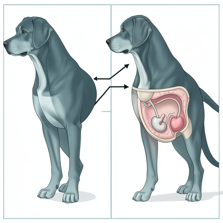
Normal vs. Abnormal Testicular Descent
AI-generated conceptual illustration (mongrel breed dog). It explains the idea and does not show study data.
View source evidence ↗Figures & tables
From the original paper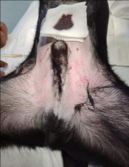 Figure 1
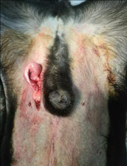 Figure 2
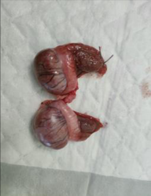 Figure 3
Introduction
Undescended testicles, known as cryptorchidism, are a urological condition present at birth and are characterized by the incomplete or absent descent of one or both testicles. In canines, the testicles typically descend through the inguinal canal 3-4 days after birth and settle in the scrotum by day 35 (Johnston SD et al., 2001) [3] . The absence or incomplete development of the testicles may be due to abnormalities in the growth of the gubernaculum and the presence of cranial gonadal suspensory ligaments, which can hinder the migration of the testis through the inguinal canal (Johnston SD et al., 2001, Kersten W et al., 1996) [3,4] . Cryptorchidism presents in different forms based on the involvement of one or both testicles and their location (Johnston SD et al., 2001, Griffin B et al. 2020) [3,1] , which can be bilateral or unilateral depending on the side (Bhuvaneshwari V. et al., 2018) [10] . The unilateral form is the most common, with the right testicle being retained twice as often (Yates D et al., 2013, Tobias KM et al., 2013) [11,9] Apart from this report, no case reports on bilateral cryptorchid condition with congenital agenesis of scrotum in a dog have been published in India. In the case report, both testicles were removed via orchiectomy, and the procedure is mentioned in details.
Case presentation
One nine months old male mongrel breed dog was presented the clinics of West Bengal University of Animal and Fishery Sciences (WBUAFS) for castration. By physical examination, it was revealed that the dog is having no scrotal sac. By digital manipulation, both the testicles are not found under the abdominal skin. Assuming the presences of testicles inside the abdominal cavity and after taking concern from the owner of the animal orchiectomy was performed under general anesthesia. During general anesthesia injection Atropine sulphate [(containing Atropine -0.6mg/ ml) (company name-Samarth Life Sciences Pvt Ltd)] was administered @0.04 mg/ kg body weight subcutaneously. After 10 minutes of 1 st injection, as induction of anesthesia Injection Xylaxin [(containing Xylazine -23.32 mg/ ml) (company name-Indian Immunologicals Ltd)] was administered intramuscularly @ 1.5 mg/ kg body weight. During operation injection Ketmin [(containing Ketamine -50 mg/ ml) (company name-Themis Medicare Ltd)] @5 mg/kg body weight and injection Calmpose [(containing Diazepam -10 mg/ ml) (company name-Sun Pharmaceutical Industries Ltd)] @ 0.50 mg/kg body weight was administered intravenously as a maintenance. At first with the help of Bp blade a 4-centimeter-long incision was given parallel to the preputial sheath of the animal at right side. Then muscles and peritoneum were incised with mayo scissors. After that the right testicle was located inside the abdomen with the thumb and index finger and exteriorized. A stump was given with Vicryl no 1 in the spermatic cord and right testis was removed. Same procedure was performed on the left side testicle. After removal of both testicles, muscles and peritoneum were sutured with Vicryl no 1 by simple interrupted suture technique. [11,6] . It is a common condition seen in small animal veterinary clinics, with incidence rates of 1.2-12.9% in dogs (Yates D et al., 2013, Tobias KM et al., 2013) [11, 9] and 1.7-3.8% in cats (Yates D et al., 2013, Tobias KM et al., 2013) [11,9] . In dogs, cryptorchidism is an inherited autosomal recessive trait, and it is more prevalent in small breeds compared to large breeds (Tobias KM et al., 2013) [9] . Breeds at high risk of cryptorchidism are Chihuahuas, Boxers, and German Shepherds, with incidences of 30.4%, 20.6%, and 14.0% respectively (Moon et al., 2014) [7] . Purebred dogs have a higher incidence of cryptorchidism compared to crossbred dogs (Khan FA et al., 2018) [5] . Unlike unilateral cryptorchid dogs, because of high-temperature sperm production bilateral cryptorchid dogs are always sterile ((Zhao et al., 2009) [12] . The treatment of cryptorchidism in dogs with orchiopexy is a debated topic due to its hereditary nature and the continued risk of testicular neoplasia in these animals (Higgins M et al., 2020) [2] . For this reason, orchiopexy surgery is not entertained in veterinary clinics. Therefore, orchiectomy is the best choice instead of orchiopexy, like in our present case.
Conclusion
This rare case report details the surgical care of a nine-monthold male mongrel dog with bilateral cryptorchidism and congenital agenesis of the scrotum, including an orchiectomy under general anaesthesia. It highlights the rareness of this condition in dogs.
Acknowledgement
AcknowledgementI would like to sincerely thank my со аuthоrs fоr their suрроrt and kind gesture tо соmрlete this mаnusсriрt in time.
Figures and tables from the paper
References
9 in the original paper- Higgins M, Smith DE, Gao D, Wilcox D, Cost NG, Saltzman AF. The impact of age at orchiopexy on testicular cancer outcomes. World J Urol. 2020;38:25312536.
- Kersten W, Molenaar GJ, Emmen JM, van der Schoot P. Bilateral cryptorchidism in a dog with persistent cranial testis suspensory ligaments and inverted gubernacula: report of a case with implications for understanding normal and aberrant testis descent. J Anat. 1996 Aug;189(Pt 1):171-176.
- Khan FA, Gartley CJ, Khanam A. Canine cryptorchidism: an update. Reprod Domest Anim. 2018;53:1263-1270.
- Mattos MRF, Simões-Mattos L, Domingues SFS. Cryptorchidism in dog. Ciência Animal. 2000;10:61-70.
- Moon JH, Yoo DY, Jo YK, Kim GA, Jung HY, Choi JH, et al. Unilateral cryptorchidism induces morphological changes of testes and hyperplasia of Sertoli cells in a dog. Lab Anim Res. 2014;30(4):185-189.
- Spangenberg C. Canine cryptorchidism: a concise review of its origin, diagnosis, and treatment. Biol Eng Med Sci Rep. 2021;7:1-3.
- Bhuvaneshwari V, Madeena Begum M. Surgical management of cryptorchidism in Beagle dogs -a report of 7 cases. Indian Vet J. 2018 Aug;95(8):82-83.
- Yates D, Hayes G, Heffernan M, Beynon R. Incidence of cryptorchidism in dogs and cats. Vet Rec. 2003;152(16):502-504.
- Zhao X, Zhiqiang D, Kim G, Max RF. Candidate gene discovery for retained testicles in dogs. Anim Ind Rep. 2009;655(1):49.