
Research article · 2024 · Full text
International Journal of Veterinary Sciences and Animal HusbandryFetotomic delivery of a ‘monocephalic thoracopagus tetrabrachius tetrapus dicaudatus’ monster in a Murrah buffalo
Volume 9(5) · Pages 78–80 · DOI: 10.22271/veterinary.2024.v9.i5b.1647 ↗
Research areasComparative Animal Anatomy StudiesGenetic and phenotypic traits in livestockVeterinary Medicine and Surgery
Abstract
Author-writtenA seven-year-old, pluriparous (3rd parity) Murrah buffalo with a history of full-term gestation and straining from the last 6 hrs. was reported to the ‘Referral Veterinary Polyclinic’ ICAR-Indian Veterinary Research Institute, Izatnagar. A Fetotomic delivery of monocephalic thoracopagus tetrabrachius tetrapus dicaudatus monster through percutaneous fetotomy in Murrah buffalo has been accomplished in this case report.
Summary
Source-checkedPlain-language reading of the full paper, prepared with AI and checked against the text. Each point links to the authors' own words.
Why it matters
- Dystocia occurs when a dam has difficulty delivering a fetus and requires human intervention. View evidence ↗
- Fetal monstrosities can cause structural deformations that lead to dystocia in bovine animals. View evidence ↗
- The incidence of fetal monstrosities in Murrah buffaloes is reported to range between 7.9% and 12.8%. View evidence ↗
The question
- The study aimed to resolve dystocia in a Murrah buffalo caused by a 'monocephalic thoracopagus tetrabrachius tetrapus dicaudatus' fetal monster using fetotomy. View evidence ↗
What they did
- The subject was a seven-year-old Murrah buffalo in her third parity who had been straining for six hours after a full-term gestation. View evidence ↗
- Examination showed a fully dilated cervix and a dead fetus in a lumbosacral posterior presentation with four hind limbs and two forelimbs visible at the vulva. View evidence ↗
- The procedure involved lateral recumbency, 7 ml of 2% lignocaine hydrochloride for low epidural anesthesia, and a 40mg intramuscular dose of dexamethasone. View evidence ↗
- The fetus was amputated using Thygeson’s fetotome and a BP knife, then extracted in five pieces using disarticular hooks and obstetrical chains. View evidence ↗
- Post-procedure, six Nitrofurazone and urea boluses were administered intra-uterinely to prevent infection and clear debris. View evidence ↗
What they found
- Gross examination identified the fetus as a dead female conjoined monocephalic twin fused at the thorax, possessing four forelimbs, four hindlimbs, and two pelvises. View evidence ↗
- Dissection of the fetal monster revealed the presence of four kidneys and a duplicated spleen and reproductive tract. View evidence ↗
What it means
- The authors conclude that percutaneous fetotomy is a safer, more time-efficient, and practical alternative to caesarean sections for managing non-viable fetal monstrosities in buffaloes. View evidence ↗
Visual summary
1 visualsIllustrations explain the paper; they do not show study data.
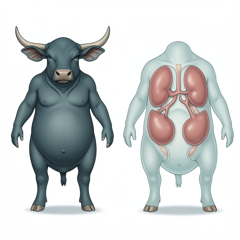
Anatomical Layout of the Fetal Monster
AI-generated conceptual illustration (Murrah buffalo fetus). It explains the idea and does not show study data.
View source evidence ↗Figures & tables
From the original paper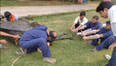 Figure 1
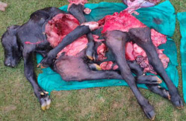 Figure 2
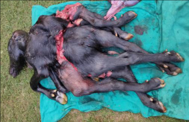 Figure 3
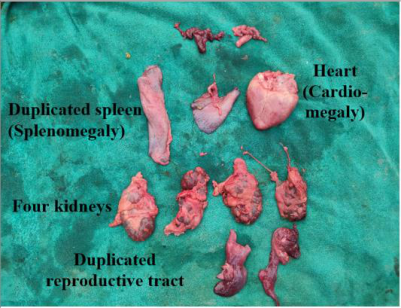 Figure 4
Introduction
Dystocia is defined as the dam's difficulty to deliver its fetus and requires human assistance. Any fetal problem, such as fetal monster, can cause deformation of body structure and causes dystocia in bovines (Kumar et al., 2014) [3] . It occurs in roughly one out of every 100,000 births in cows (Arthur, 1956) [1] . Disruption of inner cell mass in the embryo results in partial duplication of body parts, leading to congenital fetal abnormalities. Replication of the anterior portion is more common than caudal and dystocia is the common sequelae of fetal monstrosities. In Murrah buffalo, incidence of monstrosities ranged from 7.9 to 12.8%. (Phogat et al., 1992) [7] . The majority of the monstrosities are seen in water buffaloes, with little data available for swamp buffaloes (Kumar et al., 2018) [2] . Fetotomy is a good alternative for relieving the fetal monster that causes dystocia (Vermunt, 2009) [10] . There are poor survival rates in buffaloes following delivery of calf by caesarean section compared to pervaginally delivery by fetotomy. A 'monocephalic thoracopagus tetrabrachius tetrapus dicaudatus' fetal monster was relieved by fetotomy in Murrah buffalo presented with dystocia in OPD of institute referral veterinary polyclinic.
Case History and Clinical Observations
A seven-year-old Murrah-graded buffalo in 3 rd parity was presented to clinics with a history of straining and completed a normal gestation period. The animal was straining for the last 6 hours having an abnormal number of limbs in the vagina. The water bags had already ruptured as the case was manipulated by local quacks without success. The animal was dull, restless, and on sternal recumbency with a rectal temperature of 100.8ºF. vulva was edematous and the Vaginal mucus membrane was congested and moist. Per vaginal examination revealed a fully dilated cervix with a dead fetus in posterior presentation with a lumbosacral position having four hind limbs, two forelimbs, and some intestinal loops presented on the vulva. Since the fetus was dead and it was a monster hence percutaneous fetotomy was performed.
Treatment and Discussion
The animal was restrained in lateral recumbency following low epidural anesthesia with 7 ml of inj. 2% of lignocaine hydrochloride. A preoperative dose of inj. dexamethasone (40mg intramuscular) was administered. following the lubrication of the birth canal sufficiently with liquid paraffin. The extended limbs of the monster were amputated by Thygeson's fetotome. Amputated limbs and the remaining fetus were extracted by applying disarticular hooks and obstetrical chains (Fig. 1). BP knife was also used to achieve the sectioning of the fetus in a total of five pieces (Fig. 2). Following the procedure, six Nitrofurazone and urea bolus were administered intra-uterine to clean the uterine debris and prevent uterine infection. The animal was treated with an Antibiotic Ceftiofur sodium (@1.1mg/kg body weight I/M), Antihistaminic (inj. Chlorpheniramine maleate 0.5mg/kg body weight I/M) antiinflammatory (inj. Flunixin meglumine @1.1mg/kg body weight I/M), and multivitamins (inj. Tribivet @10 ml I/M). calcium supplement (inj. Mifex @450 ml slow I/V) along with fluid therapy. The gross examination of the monster revealed a female dead conjoined monocephalic twin fused at thoracic region (thoracopagus) with four forelimbs (Tetrabrachius), four hindlimbs (Tetrapus) and two pelvises (Dicaudatus) (Fig. 3). On dissecting the fetus four kidneys and duplicated spleen and the reproductive tract was recorded (Fig. 4). Dystocia due to conjoint twin had already reported in buffalo (Patil et al., 2009: Prasad et al., 2015) [6,8] and in cow. The research suggests that caesarean sections have mostly been performed to treat such cases of dystocia (Monfared et al., 2013) [5] . But, in this case, percutaneous fetotomy was done to relieve dystocia in buffaloes. The approach is safer and takes less time; it may be used effectively as an alternative to a caesarean operation.
Conclusion
This case report highlights a successful fetotomic delivery of a conjoined twin monster, specifically a monocephalic thoracopagus tetrabrachius tetrapus dicaudatus fetus, in a seven-year-old Murrah buffalo. The condition, characterized by severe fetal malformation, resulted in dystocia that was resolved through percutaneous fetotomy. The approach proved to be a practical and safer alternative to caesarean section, particularly when the fetus is non-viable. The procedure facilitated the effective removal of the fetus with minimal complications and may be preferred for managing similar cases in buffaloes, given its time efficiency and reduced post-operative risks. This case underscores the importance of timely intervention and appropriate clinical management in cases of fetal monstrosity-induced dystocia.
Acknowledgement
AcknowledgementThe authors thank the Director of the Indian Veterinary Research Institute (IVRI), Izatnagar, the head of the Division of Animal Reproduction, and In-charge of the Referral Veterinary Polyclinic for providing the facilities required at the polyclinic for the treatment of animals owned by lowincome farmers.
References
7 in the original paper- Arthur GH. Conjoined twins-the veterinary aspect. Vet Rec. 1956;68:389-393.
- Kumar P, Basir S, Singh M, Sharma A. Conjoined sternophagus twin monster: a cause of dystocia in Murrah graded buffalo. Reprod. 2018;13:69-70.
- Kumar P, Sharma A, Singh M, Sood P, Barman P. Dystocia due to a dicephalus monster fetus in a buffalo. Buffalo Bull. 2014;33(1):13-15.
- Lombard JE, Garry FB, Tomlinson SM, Garber LP. Impacts of dystocia on health and survival of dairy calves. J Dairy Sci. 2007;90(4):1751-1760.
- Patil AD, Raghuwanshi DS, Kumbhar UB, Choudhari KS, Thorat MG. Thoracopagus sternopagus conjoined twin in a Marathwadi buffalo calf. Vet Pract. 2009;10(2):155-156.
- Prasad JK, Das GK, Krishnappa B, Kumar B, Ramteke S, Rakesh HB, et al. Monocephalic tetrabrachius tetrapus International Journal of Veterinary Sciences and Animal Husbandry https://www.veterinarypaper.com monster in a Murrah buffalo. Indian J Anim Reprod, 2015, 33(1).
- Shukla SP, Mudasir Q, Nema SP. Dystocia due to a conjoined twin monster foetus in a female buffalo. Buffalo Bull. 2011;30(1):12-13.