
Research article · 2024 · Full text
International Journal of Veterinary Sciences and Animal HusbandrySurgical management of cystolith in a shih TZU bitch: A case report
Volume 9(4) · Pages 419–420 · DOI: 10.22271/veterinary.2024.v9.i4g.1633 ↗
Research areasVeterinary Medicine and SurgeryUrological Disorders and TreatmentsPediatric Urology and Nephrology Studies
Abstract
Author-writtenA four years old Shih Tzu bitch was presented to Veterinary clinic with history of urinary inconsistency, inappetence and haematuria, since last six months. Radiography revealed the presence of urolith in the urinary bladder. Cystolithotomy was performed and the dog had a complete recovery.
Summary
Source-checkedPlain-language reading of the full paper, prepared with AI and checked against the text. Each point links to the authors' own words.
Why it matters
- Uroliths are deposits of inorganic and organic solutes that form when urine becomes oversaturated. View evidence ↗
- When these uroliths develop specifically within the urinary bladder, they are referred to as cystoliths. View evidence ↗
- The formation of uroliths in dogs and cats is caused by multiple factors, including breed, sex, diet, water intake, and infections. View evidence ↗
The question
- The study aimed to resolve a case of cystolith in a dog using a surgical procedure called cystolithotomy. View evidence ↗
What they did
- The subject was a four-year-old intact female Shih Tzu presenting with a six-month history of lethargy, vomiting, inappetence, dysuria, and occasional blood in the urine. View evidence ↗
- A lateral abdominal radiograph was used to diagnose the condition, revealing a single large radiopaque mass in the bladder. View evidence ↗
- The dog was prepared for surgery with Ringer lactate (10 ml/kg IV), Ceftriaxone (20mg/kg IV), and meloxicam (0.3 mg/kg IM), and anesthetized using xylazine (1 mg/kg) and ketamine (5 mg/kg). View evidence ↗
- The surgical approach involved a ventral midline incision, after which the bladder was accessed via a dorsal incision. View evidence ↗
What they found
- The surgery successfully removed a single large urolith measuring approximately 1.2 x 2.4 cm. View evidence ↗
- Post-removal catheterization confirmed that the urethral opening was patent and no obstruction existed. View evidence ↗
- Blood analysis showed neutrophilia, indicating infection, and a marked increase in creatinine, suggesting potential urine obstruction. View evidence ↗
- Based on physical characteristics, the retrieved stone was tentatively identified as a struvite urolith. View evidence ↗
- Following the cystolithotomy, the dog achieved a complete recovery. View evidence ↗
What it means
- The authors conclude that confirmatory diagnosis and surgical intervention combined with post-operative care lead to satisfactory outcomes for urolithiasis. View evidence ↗
- The specific case of the female dog suffering for six months was successfully treated without any post-operative complications. View evidence ↗
Visual summary
1 visualsIllustrations explain the paper; they do not show study data.
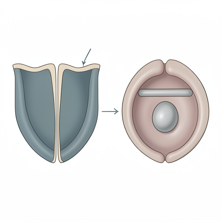
Surgical Approach Concept
AI-generated conceptual illustration (Shih Tzu dog bladder). It explains the idea and does not show study data.
View source evidence ↗Figures & tables
From the original paper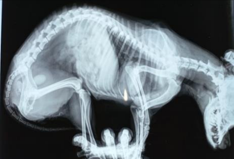 Figure 1
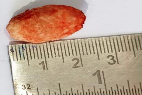 Figure 2
Introduction
Uroliths are organized inorganic and organic solutes that precipitate around the nidus because of oversaturated urine (Devi et al. 2020) [3] . Uroliths that develop in the bladder are cystoliths. Cystolithotomy refers to the surgical opening made in the urinary bladder to remove the formed stones from the bladder (Sharun et al. 2021) [7] . The occurrence of uroliths in cats and dogs have multifactorial etiologies which include breed, sex, water intake, dietary origin, abnormal urine pH, urinary tract infections, drug-induced formation, genetic disorder and metabolic or endocrine causes (Appel et al. 2008, Lekcharoensuk et al. 2002) [1,6] . There are mainly four types of mineral deposits in uroliths: calcium, struvite (magnesium ammonium phosphate), urate and cystine. The most common mineral deposits are calcium-containing urolith and struvite (Tion et al. 2015) [8] . The formation of struvite urolith is primarily linked to urinary tract infections (Tiruneh, 2017) [9] . In this study, the case of cystolith was relieved by cystolithotomy.
Materials and Methods
An intact Shih Tzu bitch aged around four years was brought to Veterinary Clinic with a complaint of inappetence, dysuria, lethargy, occasional haematuria and vomition for last six months. Previously the treatment was done for urinary tract infection before it was referred to the clinic. On clinical examination, the dog was emaciated, anemic, distended abdomen, dehydrated. After general clinical examination, venous blood sampling was carried out for complete blood count (CBC) and the serum biochemical analysis for blood urea nitrogen and creatinine. Radiographic examination of the lateral abdomen revealed the presence of one big radiopaque mass in the urinary bladder which was diagnosed as Cystolith (Figure .1). Following abdominal radiography, it was prepared for aseptic surgery. Bitch was premedicated with Ringer lactate @ 10 ml/kg BW IV, Ceftriaxone @ 20mg/kg IV and meloxicam @ 0.3 mg/kg BW IM. The caudal ventral abdomen was prepared for aseptic laparotomy. The bitch was anesthetized with xylazine @ 1 mg/kg and ketamine @ 5 mg/kg, a ventral midline approached was performed, the bladder was grasped and an incision was made from the dorsal approach (Yadav et al, 2011) [11] . One large sized urolith measuring around 1.2 x 2.4 cm was retrieved and a catheter was passed through the bladder to the urethral opening to check the patency, however, no obstruction was evident. Bladder was closed with double row of Lambert's suture using vicryl. Abdominal incision was closed in a routine manner.
Results and Discussion
Clinical and imaging findings were compatible with the diagnosis of urolithiasis. The blood picture revealed neutrophilia which showed the presence of infection and also marked increase in creatinine, which may indicate an obstruction of urine due to urolith. Elevated creatinine level could be due to renal damage caused by hydrostatic pressure on the kidney and increased reabsorption of creatinine from bladder because of prolonged stasis of urine in the urinary bladder (Kim and Oh, 2023) [4] . Struvite cystoliths are the most common stones in bitches with urinary tract infection. Uroliths in bitches are usually located in the urinary bladder (Kokila et al. 2023) [5] . Study shown that small breeds of dogs produce less urine, urinate less frequently and have more concentrated urine, which also suggested as a predisposing factor to urolithiasis. Shih Tzu is one of the most commonly affected breed (Arun et al. 2015) [2] . In the present case, the urolith was large, single and ellipsoidal in shape with surface porosity. The bladder is the most common site of phosphate urolith, and phosphate has been reported as the main constituent in canine urolithiasis especially uroliths of female dogs (Uma et al. 2018) [10] . The major component of phosphate uroliths is magnesium ammonium phosphate (Struvite). A similar physical characteristics and findings have been described for struvite urolith in earlier reports in bitches (Figure 2). The urolith retrieved from the urinary bladder was tentatively diagnosed as struvite.
Conclusion
Confirmatory diagnosis and treatment improve the outcome of urolithiasis. Surgical intervention along with post-operative care will give satisfactory results in the affected animals. The case of urolithiasis in bitch suffering from last 6 months was successfully treated with no post operative complication.
Figures and tables from the paper
References
11 in the original paper- Appel SL, Lefebvre SL, Houston DM. Evaluation of risk factors associated with suture-nidus cystoliths in dogs and cats: 176 cases (1999-2006). J Am Vet Med Assoc. 2008;231:1889-1895.
- Arun CS, Shridhar NB, Kumar CNS. Surgical management of pyometra and urocystoliths in a bitch. Intas Polivet. 2015;16(I):161-162.
- Devi TR, Das D, Konwar B, Bayan H, Saikia B, Sarma K, et al. Diagnosis and management of ammonium urate cystoliths in a bitch. J Entomol Zool Stud. 2020;8(4):1141-1144.
- Kim HJ, Oh SH. Comprehensive prediction of urolithiasis based on clinical factors, blood chemistry and urinalysis: urolithiasis score. Sci Rep. 2023;13:14885.
- Kokila S, Manjuriya R, Aruneshwaran EL, Kandasamy S, Sankar P, Vijayakumar M, et al. Successful surgical management of cystic and urethral calculi in a dog: a case report. Pharma Innov J. 2023;12(8):1069-1071.
- Lekcharoensuk C, Osborne CA, Lulich JP. Associations between dietary factors in canned food and formation of calcium oxalate uroliths in dogs. Am J Vet Res. 2002;63:163-169.
- Sharun K, Manjusha KM, Kumar R. Prevalence of obstructive urolithiasis in domestic animals: an interplay between seasonal predisposition and dietary imbalance. Iraqi J Vet Sci. 2021;35(2):227-232.
- Tion MT, Dvorska J, Saganuwan SA. A review on urolithiasis in dogs and cats. Bulg J Vet Med. 2015;18(1):1-18.
- Tiruneh D, Abdisa T. Review on canine urolithiasis. Am J Vet Res. 2017;1(1):1-7.
- Uma S, Kumar R, Lakkawar AW, Nair MG. Cystolith in a dog: a case report. J Entomol Zool Stud. 2018;6(1):924927.
- Yadav S, Shrestha R, Joshi N. Bladder stones in a female dog and its surgical removal. Intas Polivet. 2011;12(II):372-374.