
Research article · 2024 · Full text
International Journal of Veterinary Sciences and Animal HusbandrySuccessful surgical management of intestinal foreign body in a golden retriever dog
Volume 9(4S) · Pages 198–200 · DOI: 10.22271/veterinary.2024.v9.i4sd.1627 ↗
Research areasVeterinary Oncology ResearchForeign Body Medical CasesVeterinary Orthopedics and Neurology
Abstract
Author-writtenDogs have a tendency to ingest various non-edible objects due to their indiscriminate feeding habits. A golden retriever dog was presented with a history of anorexia, frequent vomiting, dilated abdomen for last 7 days. Physical examination revealed a palpable hard mass in the abdomen. Palpation and ultrasonography, confirmed the presence of an intestinal foreign body. The foreign body was successfully removed via enterotomy. The enterotomy incision was closed in a single layer interrupted suture pattern followed by a lambert suture pattern. Postoperatively, the dog was treated with fluid therapy, antibiotics, analgesics, prokinetics and a restricted diet. The animal received standard supportive care and had an uneventful recovery.
Summary
Source-checkedPlain-language reading of the full paper, prepared with AI and checked against the text. Each point links to the authors' own words.
Why it matters
- Intestinal obstruction is a frequent medical issue in cats and dogs, typically resulting from the ingestion of foreign objects. View evidence ↗
- Dogs are prone to ingesting items like rubber, plastic, and stones that may be too large to pass through the intestines, even if they fit through the esophagus. View evidence ↗
- Non-linear foreign bodies often cause luminal distention that cuts off blood supply, which can lead to tissue death (necrosis), swelling (edema), and bacterial growth. View evidence ↗
The question
- The study describes the successful surgical treatment of a golden retriever dog suffering from intestinal obstruction caused by a piece of cloth. View evidence ↗
- The authors aim to outline the diagnostic and surgical protocols used to manage foreign body obstructions in dogs. View evidence ↗
What they did
- The subject of the case was an 8.5-year-old golden retriever treated at the Teaching Veterinary Clinical Complex in Nagpur. View evidence ↗
- Intestinal obstruction was confirmed using a Sonosite M-Turbo ultrasonography machine, which showed distended intestinal loops. View evidence ↗
- Anesthesia was induced with xylazine hydrochloride (1 mg/kg) and maintained with a combination of Ketamine (6 mg/kg), Diazepam (0.5 mg/kg), and Isoflurane (1.5 mg/kg). View evidence ↗
- The surgical procedure involved washing the intestine with normal saline and metronidazole, followed by suturing with Catgut 2-0 (Lambert pattern), Vicryl-0 (simple interrupted), and Prolene-0 (horizontal mattress). View evidence ↗
What they found
- The dog presented with a 7-day history of a dilated abdomen, frequent vomiting, and anorexia. View evidence ↗
- Blood tests showed an increase in white blood cells (WBCs) and neutrophils, alongside a decrease in lymphocyte count and red blood cell distribution. View evidence ↗
- Surgery revealed a foreign body that was completely obstructing the intestine, which was removed via an incision at the caudal end. View evidence ↗
- The foreign body retrieved from the dog's intestine was identified as a sock. View evidence ↗
- Following standard supportive care, the dog experienced an uneventful recovery. View evidence ↗
What it means
- The authors conclude that immediate surgical intervention, based on timely diagnosis from history and clinical findings, is necessary for favorable outcomes in intestinal obstructions. View evidence ↗
- To prevent such incidents, the authors recommend keeping dangerous objects away from dogs and providing only durable toys that cannot be swallowed. View evidence ↗
Visual summary
1 visualsIllustrations explain the paper; they do not show study data.
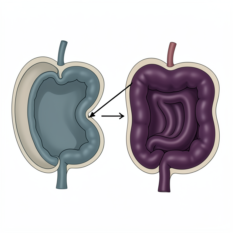
Mechanism of Intestinal Obstruction
AI-generated conceptual illustration (golden retriever dog intestine). It explains the idea and does not show study data.
View source evidence ↗Figures & tables
From the original paper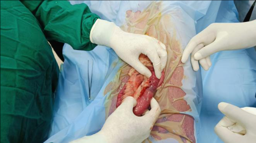 Figure 1
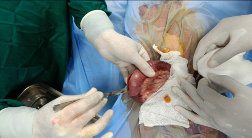 Figure 2
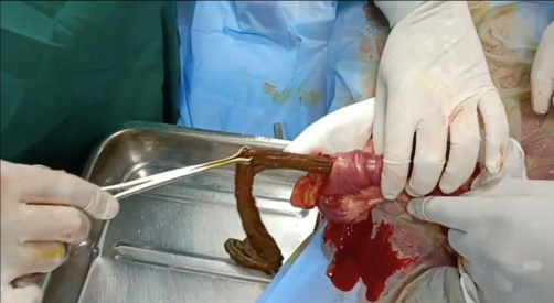 Figure 3
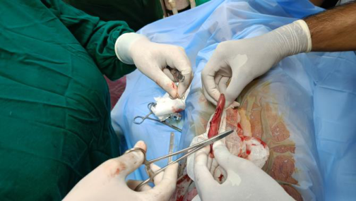 Figure 4
Introduction
Intestinal obstruction is a common condition in dogs and cats, often caused by the ingestion of foreign bodies. Intestinal foreign bodies can lead to partial or complete obstruction depending on their size (Papazoglou et al., 2003) [1] . Dogs, particularly those with large esophagi, can safely ingest objects much larger than what can be safely passed through their intestines, commonly consuming stones, plastic, and rubber as foreign bodies. Gastrointestinal foreign bodies may cause complete or partial obstruction, with complete obstruction typically associated with more dramatic clinical signs and rapid deterioration, while partial obstruction may present with more chronic signs of maldigestion and malabsorption (Papazoglou et al., 2003) [1] . Clinical signs such as anorexia, vomiting, diarrhea, abdominal discomfort, and lethargy are commonly associated with gastrointestinal foreign bodies, although they are nonspecific (Hayes, 2009) [3] . Foreign body obstruction can be classified based on the degree of obstruction (complete or partial), location along the intestinal tract (proximal, middle, distal, or colonic), and pathophysiologic alterations (simple, mechanical, or strangulating) (Papazoglou et al., 2003) [1] . In complete simple intestinal obstruction, accumulation of gas and fluid contributes to luminal distention proximal to the obstruction. Partial intestinal obstruction is associated with maldigestion and malabsorption of nutrients, leading to stagnant loop syndrome (Papazoglou et al., 2003) [1] . The majority of obstructive non-linear intestinal foreign bodies compromise the blood supply to the intestinal segment by luminal distention, leading to intestinal wall edema, progressive necrosis, ileus, and an increase in pathogenic intraluminal bacteria. These factors contribute to the breakdown of the mucosal barrier and systemic endotoxemia (Ellison, 1993) [4] . This paper presents a case of intestinal obstruction in a golden retriever dog caused by a foreign body consisting of a piece of cloth, and its successful surgical management. The study aims to provide an overview of the diagnostic protocol and surgical management of intestinal foreign body obstruction in canine patients.
Case History and Observation
A 8.5 year old golden retriever dog was presented at Teaching Veterinary Clinical Complex, Nagpur with symptoms suggestive of intestinal obstruction. It exhibited anorexia, vomiting, and reduced defecation, appearing dull, emaciated, and weak on examination, with normal physiological parameters. Clinical palpation revealed hard mass on palpation. Ultrasonography with the Sonosite M-Turbo machine revealed a distended intestinal loops cranial to the hyperechoic shadowing area indicating intestinal obstruction. Biochemical parameters were normal and Hematology indicated increased WBC's, Neutrophil"s and decreases Lymphocyte count and Red blood Cell Distribution.
Treatment and Discussion
The surgical site was shaved and prepared aseptically, and pre-operative fluid therapy with cold normal saline intravenously was initiated. The dog was pre-anaesthsized with atropine @ 0.045 mg/kg body weight, intamox @ 500 mg body weight intravenously, Ranitidine @ 2mg/kg body weight, Butorphenol @ 0.02 mg/kg body weight, Vetalgin @ 1 ml & Perinorm @ 0.1 mg/kg body weight The anesthesia was induced with xylazine hydrochloride @ 1 mg/kg body weight Maintenance of anesthesia was done using Ketamine@6 mg/kg body weight, Diazepam @ 0.5mg/kg body weight & Isoflurane @1.5mg/kg body weight. The dog was positioned in dorsal recumbency. The intestinal loops were explored for the foreign body. Obstruction was identified and the incision was given at caudal end to relieve the foreign body that was completely obstructing the intestine. The foreign body was retrieved using forceps. The intestine was washed with normal saline and metronidazole to remove the contaminants and was sutured using using Catgut 2-0 in Lambert pattern and the muscles were surtured with Vicryl-0 in simple interrupted pattern. The skin was sutured using Prolene -0 in horizontal mattress pattern Post-operatively, the animal was treated using intravenous fluids (DNS & RL), and the owner was advised to give liquid diets for a period of 3-5 days. Antibiotic therapy was initiated using Monocef @ 20 mg/kg body weight intravenously, Metronidazole 60 ml, Butorphenol @ 0.02 mg/kg body weight intravenously, Perinorm @ 0.1 mg/kg, Vitamin C @ 30 mg/kg body weight along with Pantoprazole @ 1 mg/kg body weight. The suture site was dressed using povidone-iodine ointment. Foreign bodies lodged in the gastrointestinal tract can cause ulceration, hemorrhage, anorexia, dehydration, perforation, peritonitis, and can result in death if not treated in time (Anoop et al., 2010) [5] . Intestinal obstruction should be considered a surgical emergency, as delayed management can result in necrosis and irreparable damage to the intestine (Das et al., 2015) [6] . The jejunum is the most common site for foreign body obstruction in small animals, and enterotomy is found to be the most effective treatment (Capak et al., 2001) [7] . Dogs commonly ingest materials like stones, plastic, and rubber objects, and treatment is more successful in dogs below two years of age (Capak et al., 2001) [7] . Complications like intra-operative spillage, wound dehiscence, or perforations in the lower small intestine or colon can lead to higher mortality rates (Ellison et al., 2011) [4] . Puppies and kittens are most susceptible to foreign body obstruction due to indiscriminate feeding habits, and the condition may also occur in adults (Kavirajan et al.) [6] . Balls and other smooth, round foreign bodies have the potential to completely clog the intestine and result in pressure necrosis of the intestinal wall, while sharp foreign bodies can pierce the intestinal wall and result in adynamic ileus and septic peritonitis (Papazoglou et al., 2003) [1] . Diagnostic imaging modalities, such as contrast radiography, are important to rule out intestinal obstruction from other conditions (Papazoglou et al., 2003) [1] . [10,11] .
Figures and tables from the paper
References
10 in the original paper- Papazoglou LG, Patsikas MN, Rallis T. Intestinal foreign bodies in dogs and cats. Compend Contin Educ Pract Vet. 2003;25(11):830-845.
- Hoffmann KL. Sonographic signs of gastro-duodenal linear foreign body in 3 dogs. Vet Radiol Ultrasound. 2003;44(4):466-469.
- Hayes G. Gastrointestinal foreign bodies in dogs and cats: a retrospective study of 208 cases. J Small Anim Pract. 2009;50:576-583.
- Anoop S, Kattor JJ, Mohsin A, Dilved RS, Jyaprabha C, Ltheef SK, et al. Foreign body obstruction of the small intestine and its surgical management in a dog. J Indian Vet Assoc. 2010;8:42-43.
- Das J, Behera SS, Sahoo AK, Jena B. Intestinal obstruction in a dog and its surgical management. Indian J Can Pract. 2015;7(2):157-159.
- Capak D, Simpraga M, Maticić D, Bali R, Janoska B. Incidence of foreign-body-induced ileus in dogs. Berl Munch Tierarztl Wochenschr. 2001;114(7-8):290-296.
- Kavirajan S, Simon MS, Kannan TA. Intestinal obstruction in dogs: A review of five cases. Indian J Vet Sci Biotechnol. 2016;11(3):45-46.
- Hobday MM, Pachtinger GE, Drobatz KJ, Syring RS. Linear versus non-linear gastrointestinal foreign bodies in 499 dogs: clinical presentation, management and shortterm outcome. J Small Anim Pract. 2014;55:560-565. DOI: 10.1111/jsap.12271.
- Kamalakar G, Thara Singh D, Lamani LS, Suresh K, Saigunaranjan, Nagaraja BN. Surgical management of intestinal obstruction due to pebbles in a Labrador dog. J Livest. Sci. 2018;10:19-21. DOI: 10.33259/JLivestSci.2019.19-21.
- Mahesh V, Jyothi Shree S, Lathamani VS, Nagaraja BN. Surgical retrieval of foreign body in a dog: A case report. Int J Sci. 2019;8(5):1044-1048.