
Research article · 2024 · Full text
International Journal of Veterinary Sciences and Animal HusbandryRespiratory disease caused by Citrobacter fruendii in Ball Python
Volume 9(4) · Pages 568–570 · DOI: 10.22271/veterinary.2024.v9.i4i.1606 ↗
Research areasMicrobial infections and disease researchAnimal Virus Infections StudiesAquaculture disease management and microbiota
Abstract
Author-writtenA six-month-old ball python with a severe respiratory illness was the subject of a rare case at Birdinex Avian & Exotic Pet Speciality Hospital in Palarivattom, Ernakulam. However, the Department of Veterinary Microbiology was performed the identification of the casative agent and antibiogram analysis. Citrobacter freundii was isolated by the culture and biochemical methods. According to this study, every category of antibiotic showed signs of resistance. In the groups Aminoglycoside, Polymyxins, Quinolones, Tetracyclines, Cephalosporins, Sulfonamides, and Penicillin, this strain exhibits multiple antibiotic resistance. Gene mutation and high variant resistance can result from the improper use of antibiotics and the transfer of resistant genes from one person to another.
Summary
Source-checkedPlain-language reading of the full paper, prepared with AI and checked against the text. Each point links to the authors' own words.
Why it matters
- There is increasing concern regarding how the global trade of wildlife, both legal and illegal, contributes to the spread of diseases. View evidence ↗
- Reptiles kept in captivity often act as reservoirs for bacteria that can cause disease. View evidence ↗
The question
- The study aimed to isolate, identify, and determine the antibiotic susceptibility (antibiogram) of a pathogen causing a respiratory tract infection in a ball python. View evidence ↗
What they did
- The subject was a six-month-old ball python presenting with respiratory distress in August 2024. View evidence ↗
- An oral sample was collected and cultured on MacConkey’s agar and brain-heart Infusion agar, with incubation at 37 °C for 24 to 48 hours. View evidence ↗
- Identification of the colonies was performed using staining, morphology, and a battery of biochemical tests, including catalase, oxidase, and various sugar fermentations. View evidence ↗
- The antibiotic susceptibility was tested using the disc diffusion method against 22 different antibiotics. View evidence ↗
What they found
- Based on biochemical tests and colony morphology, the causative agent was identified as Citrobacter freundii. View evidence ↗
- The isolated strain of Citrobacter freundii exhibited a multi-drug resistant (MDR) profile, showing resistance to all tested antibiotics. View evidence ↗
- The strain showed resistance across multiple antibiotic groups, including Penicillin, Cephalosporins, Tetracyclines, Quinolones, Polymyxins, Aminoglycosides, and Sulfonamides. View evidence ↗
- The isolate demonstrated maximal resistance to carbapenems, which are typically reserved for critical human medical use. View evidence ↗
What it means
- The authors state it is crucial to recognize exotic pets as potential sources of zoonotic bacterial pathogens to protect human health. View evidence ↗
Visual summary
1 visualsIllustrations explain the paper; they do not show study data.
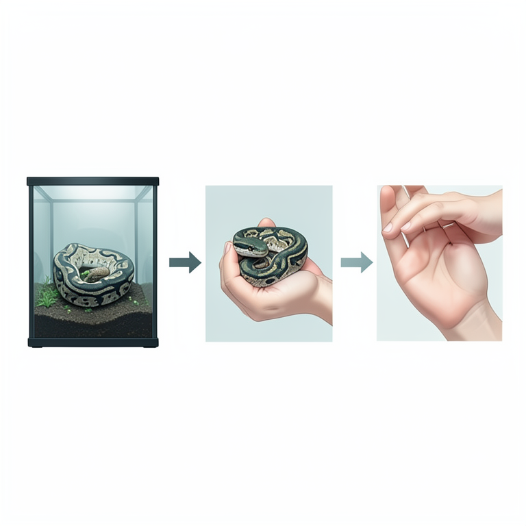
Zoonotic Transmission Cycle
AI-generated conceptual illustration (Ball python and a human hand). It explains the idea and does not show study data.
View source evidence ↗Figures & tables
From the original paper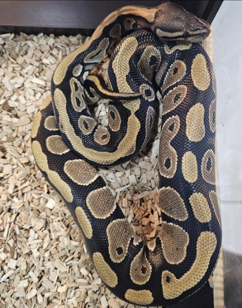 Figure 1
Introduction
According to Mitchell (2009) [10] , the demand for exotic pets such as reptiles around the world is relatively new. But their popularity has grown to such an extent that, in the global exotic pet trade, they are now believed to be the second most species-rich vertebrate class (after birds) (Bush et al. 2014) [3] . In recent decades, there has been growing concern about the role that the global trade in wildlife, whether legal or illegal, plays in the transmission of diseases (Smith et al. 2009; Can et al. 2019) [14,4] . These worries are specifically about the manner in which diseases spread when people take wild animals out of their natural environments, transport them by land, sea, and air, and then trade them-dead or alive-for goods in other countries or regions of the world (e.g., Morens et al. 2004; Karesh et al. 2005) [11,7] . Reports of infections related to reptiles, like salmonellosis and other bacterial infections are widely available for certain species (Arena et al. 2012; Bosnjak et al. 2016; Green et al. 2020) [1, 2, 6] . Captive reptiles are frequently found to be reservoirs of pathogenic bacteria. The isolation, identification, and antibiogram of the isolate from the respiratory tract infection on ball python are the main objectives of this study.
Materials and Methods
A six month old ball python (Fig. 1) with respiratory distress was brought to Birdinex Avian & Exotic Pet Speciality Hospital in Palarivattom, Ernakulam in August 2024. An oral sample was obtained for the examination. The sample was cultured on brain-heart Infusion agar (BHIA), MacConkey's agar (MAC), then incubated at 37 °C for 24-48 hrs. The colonies were identified by morphological, staining and biochemical techniques like indol, MR, VP, citrate, urease, arabinose, xylose, rhamnose, raffinose trehalose, lactose, catalase, oxidase and triple sugar iron (TSI) (Koneman et al., 1983; Quinn et al., 1994) [8,12] . Antibiogram was performed with 22 antibiotics by disc diffusion method as per The following antibiotics were used: Vancomycin (VA) 5 mcg, Piperacillin (PI) 100 mcg, Moxifloxacin (MO) 5 mcg, Cefoperazone (CPZ) 5 mcg , Levofloxacin (LE) 5 mcg, Doxycyclin Hydrocholoride (DO) 30 mcg, Amikacin (AK) 30 mcg, Tetracyclin (TE) 30 mcg, Piperacillin Tazobactum (PIT) 100/10 mcg, Gentamicin (GEN) 30 mcg, Chloramphenicol (C) 30 mcg, Imipenem (IPM) 10 mcg, Extapenem (ETP) 10 mcg, Clindamycin (CD) 2mcg, Azithromycin (AZM) 15 mcg, Ceftriaxone Sulbactum (CIS) 30 mcg, Cefixime, (CFM) 10 mcg, Neomycin (N) 30 mcg, Streptomycin (S) 10 mcg, Pencillin-G (P) 10 mcg, Cefipime (CPM) 30 mcg, Metranidaxole (MT) 5 mcg. The zones of inhibition were measured in mm and compared with CLSI.
Results and Discussion
In BHIA the organism showed large, mucoid, greyish colonies and pinkish colonies with lactose fermentation on MAC is observed. On Gram staining the organism appearance as Gram-negative coccobaccilli. In the biochemical test, the organism gave a positive result in the catalase, lactose, arabinose, xylose, rhamnose, raffinose, trehalose, citrate utilization and methyl red tests, sugar fermentation occurs and had production. The organism showed results in negative on Voges proskauer, indole, urease and oxidase. The bacteria was identified as Citrobacter freundii according to the morphology of the colonies and biochemical tests. However, in this present study, the antibiotics all antibiotics were exhibits resistant pattern. Due to their multi-drug resistance MDR profiles. It has occasionally been linked to septic arthritis, particularly in older patients with long-term illnesses (Liu et al., 2016) [9] However, Citrobacter fruendii demonstrated a potential for maximal resistance to carbapenems in this study, which are only allowed for absolutely necessary applications in human medicine. With roughly half of the isolates exhibiting resistance to aminoglycosides and chloramphenicol, these antimicrobials turned out to be the most successful options when considering other antimicrobials authorised for use in veterinary medicine. Globally, doctors are starting to worry more about K. pneumoniae, an MDR infection that is considered to be a serious zoonotic agent with consequences for both human and animal health (Schmiedel et al., 2014) [13] .
Conclusion
Isolation, identification and antimicrobial resistance in exotic pet Ball python have not been widely studied. Intimate interactions between people and exotic species like this increase the risk of bacterial infections that are incurable and dangerous to both human and animal health. To safeguard human health, it is crucial to be aware of pet birds as potential sources of zoonotic bacterial pathogens.
Acknowledgment
The authors thank the dean, College of Veterinary and Animal Sciences, Mannuthy for providing necessary facilities to carry out this work.
Conflict of Interest
Conflict of Interest
Not available
Not available
Financial Support
Financial Support
References
10 in the original paper- Bošnjak I, Zdravković N, Čolović S, Ranđelović S, Galić N, Radojičić M, et al. Neglected zoonosis -The prevalence of Salmonella spp. in pet reptiles in Serbia. Vojnosanit Pregl. 2016;73:980-982. https://doi.org/10.2298/VSP160809222B
- Bush ER, Baker SE, Macdonald DW. Global trade in exotic pets 2006-2012. Conserv Biol. 2014;28(3):663676. https://doi.org/10.1111/cobi.12240
- Can OE, D'Cruze N, Macdonald DW. Dealing in deadly pathogens: taking stock of the legal trade in live wildlife and potential risks to human health. Glob Ecol. Conserv., 2019, 17. https://doi.org/10.1016/j.gecco.2018.e00515
- Green J, Coulthard E, Megson D, Norrey J, Norrey L, Rowntree JK, et al. Blind trading: A literature review of research addressing the welfare of ball pythons in the exotic pet trade. Animals (Basel). 2020;10(2):193. https://doi.org/10.3390/ani10020193
- Karesh WB, Cook RA, Bennett EL, Newcomb J. Wildlife trade and global disease emergence. Emerg Infect Dis. 2005;11(7):1000-1002. https://doi.org/10.3201/eid1107.050194
- Liu H, Zhu J, Hu Q, Rao X. Morganella morganii, a nonnegligent opportunistic pathogen. Int. J Infect Dis. 2016;50:10-17.
- Morens DM, Folkers GK, Fauci A. The challenge of emerging and re-emerging infectious diseases. Nature. 2004;430(6996):242-249. https://doi.org/10.1038/nature02759
- Quinn PJ, Carter ME, Markey BK, Carter GR. Clinical veterinary microbiology. London: Wolfe Mosby; 1994. p. 95-101.
- Schmiedel J, Falgenhauer L, Domann E, Bauerfeind R, Prenger-Berninghoff E, Imirzalioglu C, Chakraborty T, et al. Multiresistant extended-spectrum β-lactamaseproducing Enterobacteriaceae from humans, companion animals, and horses in central Hesse, Germany. BMC Microbiol. 2014;14:187.
- Smith KF, Behrens M, Schloegel LM, Marano N, Burgiel S, Daszak P. Reducing the risks of the wildlife trade. Science. 2009;324(5927):594-595. https://doi.org/10.1126/science.1174460