
Research article · 2024 · Full text
International Journal of Veterinary Sciences and Animal HusbandryUnilateral uterine horn rupture inducing prolapse in a Chippiparai bitch
Volume 9(4) · Pages 562–565 · DOI: 10.22271/veterinary.2024.v9.i4i.1604 ↗
Research areasVeterinary Medicine and SurgeryReproductive Physiology in LivestockVeterinary Equine Medical Research
Abstract
Author-writtenDescribed below is a rare obstetrical case in veterinary medicine in a 1.5 -year-old Chippiparai bitch diagnosed with a unique combination of uterine prolapse and uterine horn rupture. This occurrence is exceptionally uncommon in canines. The female had recently given birth without incident to five healthy puppies, and this complication arose after delivery of the last puppy. In lieu of a smooth pregnancy and parturition, no specific cause for the prolapse could be pinpointed. The protruding mass appeared swollen and reddish, suggesting a recent development. Notably, neither foetal remnants nor signs of significant bleeding or shock were observed upon examination. Prompt surgical intervention was undertaken, involving the repositioning of the uterus and subsequent ovary-hysterectomy to address the issue effectively. This case underscores the importance of vigilance and prompt veterinary care in managing such intricate reproductive complications in canines.
Summary
Source-checkedPlain-language reading of the full paper, prepared with AI and checked against the text. Each point links to the authors' own words.
Why it matters
- Uterine prolapse is a condition that occurs after giving birth; while it happens in several animal species, it is exceptionally rare in dogs, with an incidence rate of less than 0.03%. View evidence ↗
- Uterine rupture is a rare veterinary emergency often linked to difficult births (dystocia) or weak uterine walls. View evidence ↗
- The outcome for a dog with a uterine rupture depends on the size of the tear and how much material leaks into the abdominal cavity. View evidence ↗
The question
- The authors aim to describe a rare case of a 1.5-year-old Chippiparai bitch presenting with both uterine prolapse and uterine horn rupture. View evidence ↗
What they did
- The subject was a 21 kg, 1.5-year-old Chippiparai bitch who presented with a reddish mass at the vulva one day after delivering five healthy puppies. View evidence ↗
- Veterinarians used abdominal palpation and ultrasonography to confirm that no puppies remained in the uterus or abdomen. View evidence ↗
- Blood tests showed a slight decrease in hemoglobin (9.8 g/dl), an elevated white blood cell count (19,900/cmm), and a decreased albumin level (2 g/dl). View evidence ↗
- The animal was anesthetized using a combination of Atropine, Xylazine, Butorphanol, Ketamine, and Diazepam, with isoflurane used for maintenance. View evidence ↗
- The surgical procedure was performed via a midventral celiotomy to access the abdominal cavity. View evidence ↗
What they found
- Surgery revealed that the right uterine horn had completely separated from the uterine body and prolapsed through the vagina. View evidence ↗
- The separated portion of the horn and the ovarian bursa were found in the abdomen, though there was no evidence of peritonitis or internal bleeding. View evidence ↗
- The dog recovered without complications after receiving Cefpodoxime proxetil (20 mg/kg) for seven days post-surgery. View evidence ↗
- A full recovery was noted during a re-evaluation two days after surgery, and sutures were removed after ten days. View evidence ↗
What it means
- The authors conclude that prompt surgical intervention, specifically an ovariohysterectomy, was essential for the animal's recovery from the rupture and prolapse. View evidence ↗
- The authors emphasize that early recognition and rapid response to canine obstetrical emergencies are critical to prevent death, shock, or peritonitis. View evidence ↗
- The authors note that there are currently too few reported cases of this condition to determine if specific breeds or ages are more predisposed to it. View evidence ↗
Visual summary
1 visualsIllustrations explain the paper; they do not show study data.
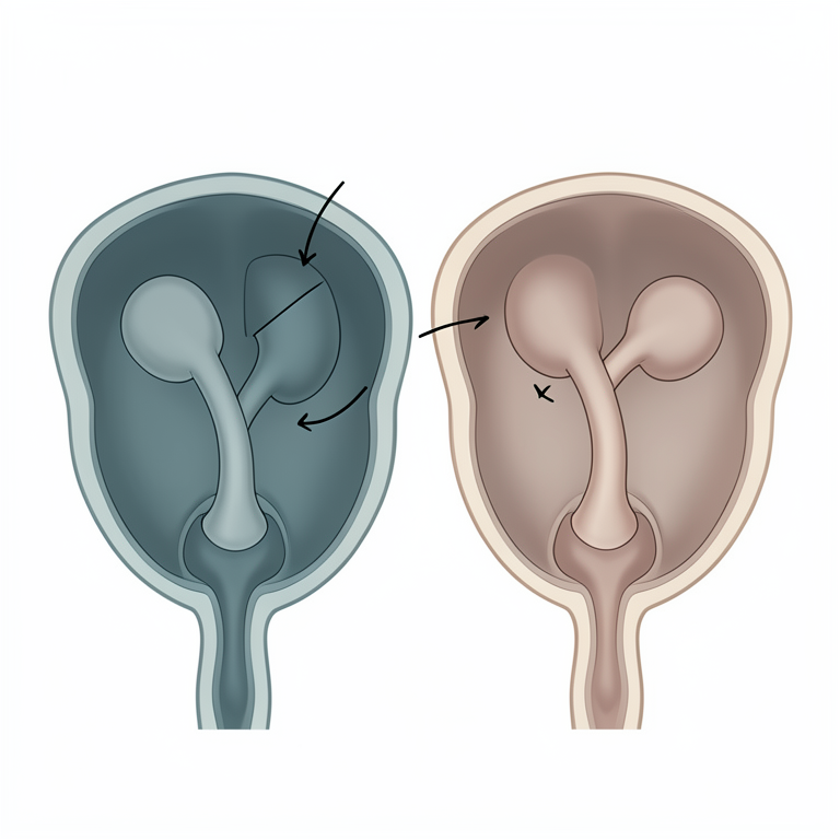
Anatomical Concept of Uterine Horn Rupture and Prolapse
AI-generated conceptual illustration (female Chippiparai dog reproductive system). It explains the idea and does not show study data.
View source evidence ↗Figures & tables
From the original paper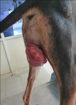 Figure 1
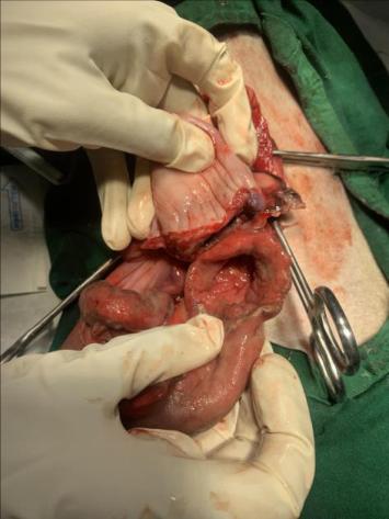 Figure 2
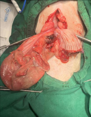 Figure 3
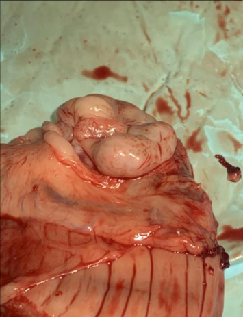 Figure 4
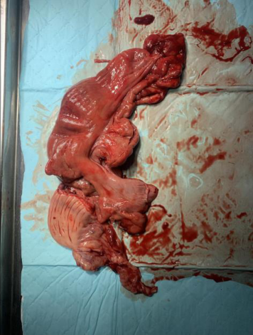 Figure 5
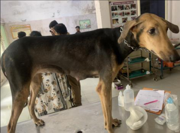 Figure 6
 Figure 6
Introduction
Uterine prolapse is a post-partum condition occurring sporadically in ewes, cattle, mares, queens and sows and is extremely rare in bitches with an incidence of <0.03%. In bitches, it may be partial or complete and usually resembles oestrus-induced vaginal prolapse manifesting within 48 hours after whelping, presenting as the protrusion of one or both uterine horns with the bifurcation distinctly through the vulva, often signalling an obstetric emergency. Prolonged whelping, dystocia, and other factors such as oversized foetuses, inappropriate obstetric manoeuvres or uterine ligament laxity contribute to its development. In this condition, a uterine segment passes through the cervix and vagina, protruding externally at the vulva. Clinical signs and prognosis depend on the duration of the prolapse and the potential occurrence of haemorrhage due to uterine vessel rupture. The presence of a protruding mass at the vulva, along with signs of dehydration, hypothermia, or shock, are indications for uterine prolapse. Uterine rupture, although rare, is also considered an emergency situation, often associated with dystocia or fragility of the uterine walls. Possible causes include trauma, uterine torsion, oversized foetuses, or faulty oxytocin administration. Spontaneous uterine rupture may occur due to unknown causes or in association with very large litters, resulting in marked stretching and thinning of the uterine wall. The severity and prognosis of uterine rupture vary depending on factors such as the size and nature of the rupture and the amount of uterine material that escapes into the abdominal cavity. This condition may also be accompanied by other complications, such as bladder or intestine prolapse through the genital tract fissure. Depending on the extent of tissue involvement, affected bitches may exhibit partial invagination without protrusion or complete inversion with prolapse of the inverted uterus. Despite these challenges, affected bitches typically do not show impairment of general health, although shock symptoms may occur depending on the severity and duration of the illness. Treatment decisions are influenced by the condition of the everted uterus and future breeding plans. The most suitable therapeutic approach for uterine prolapse is often surgery, particularly when one uterine horn is fully everted. In cases where the bitch will not be bred, ovariohysterectomy may be necessary to ensure long-term health and well-being.
Case description A 1.5 year old Chippiparai bitch weighing about 21 kgs was brought to Small Animal Gynaecology and Obstetrical Unit, Madras Veterinary College Teaching Hospital, Chennai-600 007 with the history of a large reddish mass hanging from the vulva since the previous day following the delivery of five healthy puppies. This was the animal's first whelping and it was hand mated 4 times on alternate days with the last mating done before 56 days. The pregnancy and parturition was uneventful with minimal straining by the bitch and no external manipulation. On observation, the bitch was in normal body condition with rectal temperature, heart rate and respiratory rate within normal parameters and the mucous membranes were pink and moist. The owner reported that the bitch had a satisfactory appetite, had urinated normally but had not voided faeces since the incident. On examination of the external genetalia, a pinkish tubular mass was noticed along with a serosanguineous discharge. The exposed mass included one uterine horn but the rest of the structures were ambiguous. There was no evidence of external haemorrhage or necrosis. Abdominal palpation and ultrasonography ruled out the presence of retained foetuses in the uterus and abdomen. A tentative diagnosis of a postpartum uterine prolapse was made. The mass was edematous and was not suitable for manual reduction. A complete blood count revealed slight decrease in haemoglobin values (Hb -9.8 g/dl) and elevated WBC count (19900/cmm). The biochemical values indicated decreased albumin value (2 g/dl). Hence considering the situation the bitch was prepared for ovariohysterectomy.
Treatment
The everted tissue was first cleansed with Normal saline and antiseptic solution (povidone iodine). Pre medication was done with Atropine (@ 0.04 mg/kg b.wt), Xylazine (@ 1 mg/kg b.wt) and Butorphenol (@ 0.2 mg/kg b.wt) following induction with Ketamine and Diazepam cocktail at a 4:1 ratio (@ 1 ml/ 10 kg b.wt). Maintenance was done with isoflurane. The ventral abdominal wall was prepared aseptically followed by a prompt abdominal approach via midventral celiotomy. During manipulation, it was found that the right uterine horn was separated from the uterine body and had prolapsed through the vaginal tract. The separated part of the horn along with the ovarian bursa were present in the abdomen. There were no signs of internal haemorrhage or peritonitis. The abdominal cavity was flushed with normal saline and metronidazole solutions to prevent infections due to leakage of uterine fluids into the cavity. Routine three-layer closure of abdominal muscles (PGA 2-0), subcutaneous tissue (PGA 2-0) and skin (Polyamide 2-0) was done. At the end of the surgery, Tramadol (@3 mg/kg b.wt) was administered intravenously. The bitch made an uneventful recovery from the surgery. During the postoperative period, the animal was medicated with Cefpodoxime proxetil (@20 mg/kg b.wt) for 7 days. Owner was advised to maintain the puppies with their mother for nursing. The animal was scheduled for reevaluation after 2 days wherein it had made a full recovery. The removal of the sutures was done after 10 days of surgery.
Discussion
The above described case report is a rare obstetrical case especially in canines involving uterine rupture and subsequent prolapse right after parturition. In some reported unique cases prolapse has occurred before completion of delivery of all the foetuses and evolves into a maternal cause of dystocia due to obstruction of the vaginal passage. According to various literatures, it usually occurs only after induction by oxytocin and/or calcium administration, vigorous digital manipulation of the uterus, prolonged dystocia involving continuous straining, oversized foetuses and a change in progesterone to oestrogen dominance. In this case however, the female whelped normally within 2 hours to five average sized puppies and no external manipulation was carried out by the owners. The uterus prolapsed right after delivery of the last puppy suggesting that the rupture would have also occurred at that point. This may be attributed to a narrow birth canal in the primiparous animal. There have been too few reports of this condition to arrive at an age or breed predisposition factor in these cases. In clinical presentations like this case, it is very simple to arrive at a diagnosis suggesting a uterine prolapse but the subsequent methodical assessment to trace the exact prolapsed structure of the uterus, quick decision of the veterinarian for surgical approach of the case leading to the concurrent diagnosis of the uterine rupture which is an emergency condition with peritonitis, shock and possible death as its sequelae is most crucial. When the prolapsed uterine tissue is lacerated or necrotic, surgical removal seems to be the only treatment of choice. But in this case despite the delay the mass was considerably fresh though covered with debris. Surgical management was considered due to two major factors, one due to the uncertainty pertaining to the diagnosis of the exact portion of the reproductive tract that had prolapsed and the second being the hardened and edematous nature of the prolapsed mass which did not facilitate manual reduction. Even in cases where manual reduction is a viable treatment option, surgical intervention is superior as there is no guarantee of the bitch carrying forthcoming pregnancies to term due to the severe stretching of the broad ligament which may provide reduced suspensory support to the uterus. The prognosis largely depends on the extent and nature of the rupture and concurrent internal haemorrhage as well as contamination of the abdominal cavity with retained foetuses, foetal membranes and/or uterine and foetal fluids. These complications manifest as dehydration, hypovolaemic or septic shock and hyperthermia followed by hypothermia in the affected animal. This condition should be differentiated from pre or postpartum vaginal prolapse, vaginal fibromas and vaginal hyperplasia or neoplasia by history and careful clinical examination. Thus, a quick and correct diagnosis followed by prompt medical or surgical intervention is necessary for achieving maximum success in treatment of these emergency cases as even a slight delay could end up in grave consequences.
Conclusion
This case highlights the rare and complex combination of uterine prolapse and uterine horn rupture in a Chippiparai bitch following parturition, a condition that is extremely uncommon in canines. Despite the absence of typical risk factors such as prolonged dystocia, oversized fetuses, or external manipulation, the complication emerged suddenly after the normal delivery of five healthy puppies. The prompt surgical intervention, including an ovary-hysterectomy, was crucial in addressing the prolapsed and ruptured uterine tissues, ensuring the animal's recovery. The case underscores the importance of early recognition and swift veterinary response to obstetrical emergencies in canines, as delays in treatment could lead to severe outcomes, including peritonitis, shock, and potentially death. This report also serves as a reminder that even in cases of uncomplicated pregnancies, vigilance is necessary post-partum to manage unexpected complications effectively.
Figures and tables from the paper
References
9 in the original paper- Payan-Carreira R, Albuquerque C, Abreu H, Maltez L. Uterine prolapse with associated rupture in a Podengo bitch. Reprod Domest Anim, 2012, 47(4).
- Jadhao A, Ingole RS, Surjagade SR, Bansod A, Ingawle MV. Uterine prolapse in bitch: A case report. J Entomol Zool Stud. 2020;8(3):1282-1284.
- Sathiamoorthy T, Cecilia J, Sridevi P, Kulasekar K. Manual replacement of bilateral uterine horn prolapse coupled with retained fetus in a Great Dane bitch. Int. J Vet. Sci. 2013;2(4):125-127.
- Ağaoğlu AR, Kocamüftüoğlu M, Çetin Y, Çelik MT. Uterine prolapse in a Pointer bitch. Eurasian J Vet. Sci. 2012;28(3):182-184.
- Latha C, Rao JR, Mohan KM. Management of uterine prolapse in a spitz bitch. Intas. Polivet. 2014;15(2):452453.
- Greiling MS, Packeiser EM, Gerhauser I, Goericke-Pesch S. Uterine prolapse in a non-pregnant bitch. Reprod Domest Anim. 2023;58(12):1773-1776.
- Prassinos N, Adamama-Moraitou K, Ververidis H, Anagnostou T, Kladakis S. Vaginal rupture and evisceration in a dog. Acta Vet Hung. 2010;58(3):309315.
- Ekstrand C, Linde-Forsberg C. Dystocia in the cat: A retrospective study of 155 cases. J Small Anim Pract. 1994;35(9):459-464.
- Darvelid AW, Linde-Forsberg C. Dystocia in the bitch: A retrospective study of 182 cases. J Small Anim Pract. 1994;35(8):402-407.