
Research article · 2024 · Full text
International Journal of Veterinary Sciences and Animal HusbandryUse of tilapia fish (Oreochromis niloticus) skin graft for chronic wound management in six dogs
Volume 9(4) · Pages 522–526 · DOI: 10.22271/veterinary.2024.v9.i4h.1601 ↗
Research areasElectrospun Nanofibers in Biomedical ApplicationsPeriodontal Regeneration and TreatmentsAquatic life and conservation
Abstract
Author-writtenSecond-intention healing can lead to joint dysfunction and loss of mobility. The extracellular matrix maintains the tissue structure and regulates cell growth and migration. The use of fish collagen in pigs and rats has shown early healing. TADM is also of good use in acute wound healing. Six clinical dog cases with different wound patterns were selected for the study. After descaling and myotomy of the under-skin muscle, various treatments of normal saline and glycerol were given. Two cooperative dogs were applied without any sedation or anaesthesia. For the other dog, xylazine with propofol was given as anaesthesia. Two cases required reapplication of graft. The young dog shows early wound healing after the graft.
Summary
Source-checkedPlain-language reading of the full paper, prepared with AI and checked against the text. Each point links to the authors' own words.
Why it matters
- Healing by second intention in skin wounds can lead to complications such as joint dysfunction, skin friability, and the formation of unwanted scar tissue. View evidence ↗
- Skin transplantation is considered an effective repair method for skin defects that are larger than 4 cm. View evidence ↗
- Tilapia fish skin grafts contain 27% collagen protein and provide moisture, which helps promote epithelialization in chronic nonhealing wounds. View evidence ↗
The question
- The study aimed to evaluate the use of tilapia fish (Oreochromis niloticus) skin grafts for managing chronic wounds in six dogs. View evidence ↗
What they did
- Out of 10 cases presented at the Veterinary Clinical Complex, six dogs with various wound patterns (including dog bites, accidents, and post-surgical dehiscence) were selected for the study. View evidence ↗
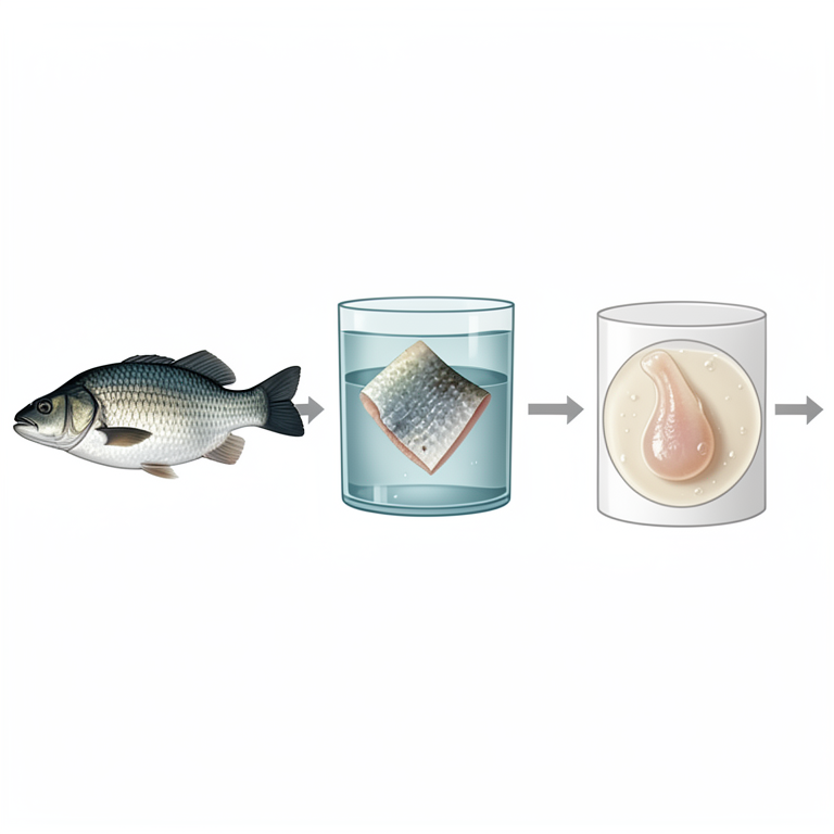
- The tilapia skin was prepared by descaling, rinsing with 0.9% saline, soaking in 2% chlorhexidine gluconate for 60 minutes, soaking in a 75% glycerol/25% saline mixture for one hour, and finally soaking in 100% glycerol for 3 hours after a 5-minute massage. View evidence ↗
- Depending on the dog's cooperation, grafts were applied either without sedation or under anesthesia using xylazine and propofol. View evidence ↗
What they found
- Case 4, which had the largest wound (17 cm x 7 cm), required 12 days for granulation and 27 days for healing. View evidence ↗
- Two of the six cases (Case 1 and Case 3) required the graft to be reapplied. View evidence ↗
- The authors found that younger animals and those with smaller wound sizes experienced faster healing. View evidence ↗
- No hemodynamic complications were reported following the application of the fish skin grafts. View evidence ↗
What it means
- The authors conclude that tilapia skin grafts are a cost-effective method that increases the rate of wound healing and decreases pain in animals. View evidence ↗
- The authors recommend this xenograft as a potential substitute for treating partial-thickness and superficial wounds. View evidence ↗
Visual summary
1 visualsIllustrations explain the paper; they do not show study data.
Tilapia Skin Processing Workflow
AI-generated conceptual illustration (Tilapia fish (Oreochromis niloticus) skin). It explains the idea and does not show study data.
View source evidence ↗Figures & tables
From the original paper Table 1
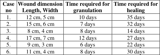 Table 2
Introduction
The simplest technique to achieve reliable healing should be used for every skin wound. One should realize that Second-intention healing can result in joint dysfunction due to skin contractures and might result in unwanted scar tissue and skin friability. Skin grafting is a novel technique that is used whenever trauma or oncologic surgery has resulted in significant skin loss. Wounds with large surfaces and wounds at the distal limb should be considered for the graft. Full-thickness skin lost due to trauma and the lack of dermis will lead to difficulty in the epithelialization of keratinocytes in the natural epidermis. Skin defects exceeding 4 cm can be effectively repaired by skin transplantation With the removal of the cellular structure and collagen extraction from animal skin, the remaining graft has relatively weak immunogenicity. Extra Cellular Matrix (ECM) maintains the tissue structure and regulates cell growth and migration. It also plays a crucial role in the specific process of skin repair. Additionally, fish collagen has also been widely studied. Acellular fish skin provides better healing on deep partial thickness burn wounds in porcine. Collagen extracted from Tilapia quickly and effectively promotes full-thickness wound healing in rats also, the extracted marine collagen peptides from tilapia skin promote wound healing such as deep Ⅱ degree scald in rabbits. Different studies have shown that collagen-rich fish skin materials have high biodegradability, so we judge that the obtained Tilapia Skin acellular dermal matrix (TADM) has a good potential for application in acute wound repair and regenerative medicine. Tilapia skin is rich in type I collagen, which helps form supra-molecular assembly in the extracellular matrix. It forms a perfect matrix with rigidity and resistance to stretching; collagen, by its chemotactic nature, attracts fibroblast, supports new blood vessels, and helps in the formation of granulation tissue. Tilapia fish skin graft promotes epithelialization by a large amount of moisture and collagen protein (27%) for chronic nonhealing wounds and wounds at medical management requiring fast healing for better prognosis.
Materials and Methods Evaluation of Patients
A total of 10 cases were presented to the Veterinary Clinical Complex, KNP College of Veterinary Science, Shirwal Dist. Satara with history (Table 1). All cases were examined for physio-biochemical and cardiopulmonary evaluation.
| Case no | Breede | Sex | Age | Area | History |
|---|---|---|---|---|---|
| 1. | Spitz | Female | 2 year | Left Shoulder | Post-injury wound dehiscence |
| 2. | German cross | Male | 7 month | Right hindlimb | Dog bite |
| 3. | Non-descript | Female | 2.5 month | Left forelimb | Accident |
| 4. | Doberman | Female | 3year | Ventral abdomen | Post-tumour excision suture dehiscence |
| 5. | Non-descript | Female | 5 month | Right hindlimb | Accident |
| 6. | Pitbull | Male | 3.5 year | Thoracic region | Boar attack |
Table text extracted from the original PDF; superscript letters are shown inline. The original table image is below.
View original table image
Preparation of Tilapia Fish Graft
Freshly caught tilapia fish from the local fishermen was procured. The fish was examined and checked for any skin discontinuations. Scales were brushed and obliterated from the skin. With the help of a sterile surgical blade, incisions were made across the three borders to avoid tearing of the skin while debriding. The skin was separated from the underlying muscles, and debridement of underlying tissues was done. The fish skin is rinsed with 0.9% saline and soaked in 2% chlorhexidine gluconate in a sealed container for 60 min. It was rinsed with 0.9% saline and soaked in a mixture of 75% glycerol and 25% saline solution in a sterile sealed container for an hour. The fish skin is rinsed again with sterile 0.9% saline and moved to another sealed sterile container filled with 100% glycerol, in which it is massaged for 5 min and kept soaked for 3 h. The skin is packed in sealed plastic envelopes and stored at 4 degrees Celsius.
Pic 1: Descaling of tilapia Fish
Pic 2: Tilapia Fish Skin Graft
Application of Tilapia Fish Graft
Two cooperative dogs with injury at the right forelimb and right hindlimb were treated by putting a tilapia graft and applying vet wrap around it. Other animals, after pretreatment with atropine @ 0.04 mg/kg S/C, were sedated with inj. Xylazine @1.0 mg/kg. Induction of anaesthesia was done with Inj. Propofol @4 mg/kg.
Preparation of wound for graft
Wound scrubbing was done with betadine 5% solution, and surgical debridement was done with BP blade no 21. Foreign debris is removed with thumb forceps, and necrotic tissue is removed by sharp dissection until healthy bleeding is encountered. Wound edges were also debrided. An appropriate-size graft was cut and applied to the wound. Stay sutures were taken with Nylone no 3-0 at the edges to hold the graft.
Results and Discussion
Case 1 and Case 2, with 12cm × 5 cm and 15cm × 6 cm wounds, showed granulation after 10 and 7 days, respectively. In case no 3 ND, a 2.5-month-old puppy showed granulation in three days and healing in 14 days. Case no 4, with 17cm ×7cm showed granulation in 12 days, but healing was seen after 37 days. Case 5, with 9 cm ×3 cm, showed granulation after six days, and healing was seen after 22 days. Case 6, with a wound 11 cm ×4 cm, showed granulation in 8 days; healing was seen after 30 days. The time required for wound healing is less, with minimal complications. Lima et al. 2019, reported similar findings with tilapia grafts in human cases after gunpowder explosion wounds. Luze et al. (2022) [8] reported similar findings after applying tilapia grafts to burn cases. No other hemodynamic complications were seen after fish grafting. A similar finding was reported by Alam and Jeffery (2019) [9] . Cases 1 and 3 required reapplications of graft. Young animal shows early wound healing. Wound with small wound sizes also shows early healing. Technique choice also influences the success of skin grafts. Grada et al. (2018) [1] also observed that the age of animals affects wound healing. A similar finding with Nile tilapia graft. He mentioned that the Nile tilapia graft is an excellent candidate among xenografts in treating wounds. This displays an excellent wound management method.
Figures and tables from the paper
References
9 in the original paper- Grada A, Mervis J, Falanga V. Research techniques made simple: animal models of wound healing. J Investig Dermatol. 2018;138(10):2095-105.
- Edmar ML, Manoel OMF, Bruno AC, Francisco VF, Maria EAM, Francisco RS, et al. Innovative treatment using tilapia skin as a xenograft for partial thickness burns after a gunpowder explosion. J Surg Case Rep. 2019;(6):1-4.
- Ghosh B, Sánchez VD, Parvati P, Anam SMA, Dominique O, Shahtaj AS, et al. Use of Nile tilapia fish skin in treatment for burn victims. Int J Surg Glob Health. 2023;6(5):1-5.
- Khurshid A, Steven LAJ. Acellular fish skin grafts for management of split thickness donor sites and partial thickness burns: A case series. Mil Med. 2019;184(Supplement_1):16-20. Available from: https://doi.org/10.1093/milmed/usy280
- Lv K, Wang L, He X, Li W, Han L, Qin S. Application of tilapia skin acellular dermal matrix to induce acute skin wound repair in rats. Front Bioeng Biotechnol. 2022;9:110.
- Xuewei Z, Chen X, Hua H, Rubei H, Jiashang L, Changsheng L, et al. Decellularized extracellular matrix scaffolds: recent trends and emerging strategies in tissue engineering. Bioact Mater. 2022;10:15-31. Available from: https://doi.org/10.1016/j.bioactmat.2021.09.014
- Yang CK, Polanco TO, Lantis JC. A prospective, postmarket, compassionate clinical evaluation of a novel acellular fish-skin graft which contains omega-3 fatty acids for the closure of hard-to-heal lower extremity chronic ulcers. Wounds. 2026;28(4):112-118.
- Luze H, Nischwitz SP, Smolle C, Zrim R, Kamolz LP. The use of acellular fish skin grafts in burn wound management -A systematic review. Medicina. 2022 Jul 9;58(7):912.
- Alam K, Jeffery SL. Acellular fish skin grafts for management of split thickness donor sites and partial thickness burns: A case series. Military medicine. 2019 Mar 1;184(Supplement_1):16-20.
Cited by 1
From OpenAlex, OpenCitations and reference lists read by Scinovex
- Marine collagen xenografts accelerate tissue repair in open canine skin wounds: clinical and histopathological evidence ↗
Fabbini Andrés Loor Moreira, Mauren Nayeli Lascano–Pionce, Fernando Rincón–Acosta, María–Karolina López–Rauschemberg · Revista Científica de la Facultad de Ciencias Veterinarias · 2025
The use of marine collagen xenografts represents an innovative therapeutic alternative in regenerative veterinary medicine due to their ability to accelerate tissue repair and improve the structural quality of cutaneous wound healing. This biomaterial of ichthyologic origin exhibits high biological compatibility, low immunogenic risk, and strong similarity to mammalian dermal collagen. The present study evaluated its efficacy in the regeneration…
❝ 0PDF