
Research article · 2024 · Full text
International Journal of Veterinary Sciences and Animal HusbandryPyometra in a jungle cat: Necropsy and histopathology findings
Volume 9(4) · Pages 509–513 · DOI: 10.22271/veterinary.2024.v9.i4h.1591 ↗
Research areasVeterinary Medicine and SurgeryReproductive Biology and FertilityNeuroendocrine regulation and behavior
Abstract
Author-writtenPyometra is a lethal disease characterized by the accumulation of pus within the uterus. The condition is commonly reported in dogs and has been reported captive large felids, but it has not been documented in the jungle cat (Felis chaus). This article presents an analysis of necropsy and histopathology findings from a geriatric female jungle cat afflicted with pyometra at the Zoological Garden, Trivandrum. The cat, which had been undergoing treatment for poor health, collapsed in the zoo hospital, prompting further investigation. Postmortem examination revealed a distended uterus filled with greenish thick cheesy material, alongside morphological changes in the kidney, epicardial blood vessels and the lungs exhibiting areas of emphysema and atelectasis. Histopathology confirmed pyometra. The death of the jungle cat was attributed to cardiopulmonary failure and renal dysfunction, presumably due to toxaemia associated with pyometra. This case report emphasizes the importance of considering pyometra as a potential differential diagnosis in felids, particularly in cases involving reproductive or systemic health issues. To the best of our knowledge, this study represents the first documentation of closed cervix pyometra in jungle cats, supported by histopathological evidence.
Summary
Source-checkedPlain-language reading of the full paper, prepared with AI and checked against the text. Each point links to the authors' own words.
Why it matters
- Pyometra is a potentially fatal condition where pus accumulates inside the uterus. View evidence ↗
- While pyometra is common in dogs and some captive large felids, it had not been previously documented in jungle cats. View evidence ↗
- The disease is driven by prolonged progesterone exposure, which causes changes in the uterine lining and leads to fluid buildup. View evidence ↗
The question
- The study aims to analyze the necropsy and histopathology of a geriatric female jungle cat suspected of having pyometra. View evidence ↗
What they did
- The subject was a 17-year-old female jungle cat weighing 3.4 kg. View evidence ↗
- Prior to death, the cat received Amoxicillin Sulbactum (12.5 mg/kg), Enrofloxacin (5 mg/kg), intravenous normal saline (10 mL/kg), and B complex vitamins. View evidence ↗
- Tissues from the lungs, liver, kidney, and uterus were preserved in 10% formalin and analyzed using Haematoxylin and eosin (H&E) staining. View evidence ↗
What they found
- Necropsy revealed distended uterine horns filled with thick, greenish, purulent material. View evidence ↗
- Histopathology of the uterus showed moderate thickening of the endometrium caused by an infiltration of lymphocytes, macrophages, and neutrophils. View evidence ↗
- Kidney analysis showed interstitial fibrosis, extensive tubular necrosis, and glomerular atrophy. View evidence ↗
- The heart tissue exhibited focal mixed inflammatory cell infiltration, hemorrhages, vascular congestion, and extensive myocardial necrosis. View evidence ↗
- Liver findings included vascular congestion, biliary hyperplasia, MNC infiltration, and mild portal fibrosis, which indicated systemic toxemia. View evidence ↗
- The respiratory system showed atelectasis and diffuse areas of emphysema in collapsed lungs. View evidence ↗
What it means
- The authors concluded that the cat died from renal dysfunction and cardiopulmonary failure caused by toxemia related to pyometra. View evidence ↗
- The authors recommend that pyometra be considered as a differential diagnosis for felids presenting with systemic or reproductive health problems. View evidence ↗
Visual summary
1 visualsIllustrations explain the paper; they do not show study data.
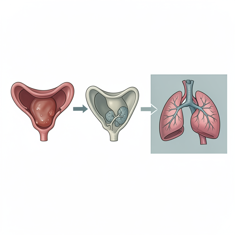
Pathological Cascade of Pyometra
AI-generated conceptual illustration (jungle cat (Felis chaus)). It explains the idea and does not show study data.
View source evidence ↗Figures & tables
From the original paper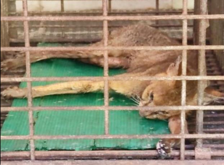 Figure 1
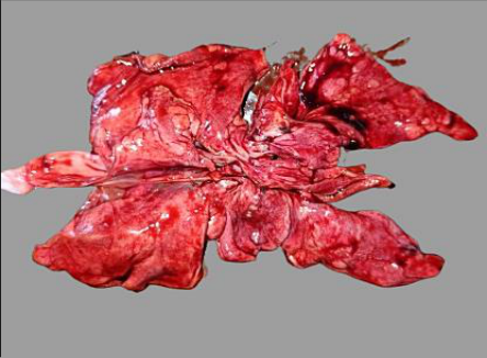 Figure 2
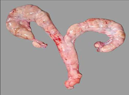 Figure 3
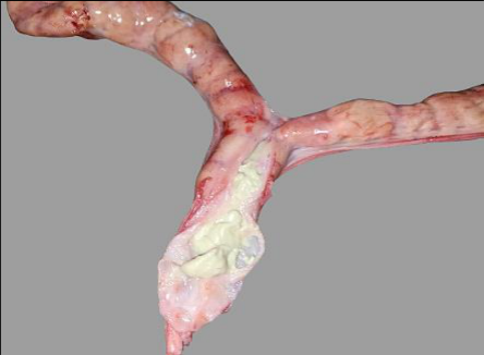 Figure 4
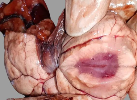 Figure 5
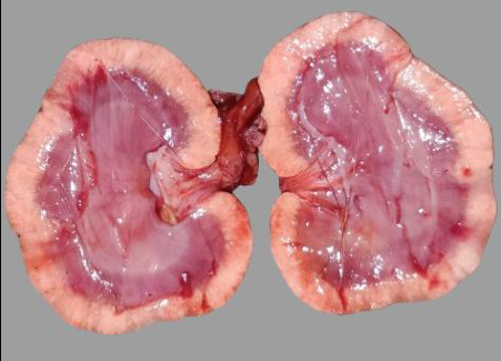 Figure 6
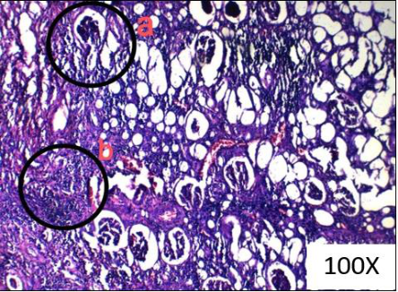 Figure 7
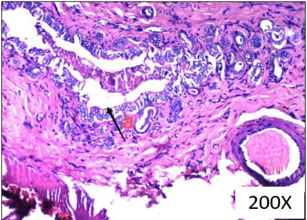 Figure 8
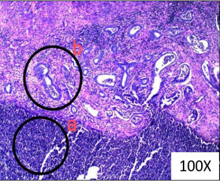 Figure 9
Introduction
The jungle cat (Felis chaus), classified as of least concern by the International Union for Conservation of Nature (IUCN), presents an intriguing subject for studying reproductive biology within the felid family. These medium-sized felids exhibit a varied diet, primarily comprising small mammals, birds, and amphibians, displaying partial omnivorous behaviour. Pyometra, characterized by the accumulation of purulent material within the uterine cavity, is a well-documented ailment in domestic felines. The condition is also reported in geriatric lioness and igress (Carlson et al., 2022) [1] . Despite its relatively lower prevalence, pyometra can affect cats across a wide age range, from one year to as old as 20 years. This disorder stems from inflammation of the uterine wall, often compounded by ascending infections with vaginal microbes (Johnston et al., 2001) [2] . Sustained progesterone exposure induces cystic hyperplastic changes within the endometrium, leading to fluid accumulation in the uterus and endometrial glands. Additionally, immune suppression and reduced myometrial contraction create an environment conducive to bacterial colonization and infection establishment (Greene, 2012) [3] . While pyometra is more commonly associated with dogs, its prevalence in cats remains notably lower (McCain et al., 2009) [4] , attributed to differences in reproductive physiology, mainly the lack of prolonged progesterone exposure in cats due to their status as induced ovulators (Greene, 2012) [3] . Earlier theories linking pyometra with immune-complex glomerulonephritis have been revisited, expanding our understanding of the disorder's pathogenesis. The occurrence of pyometra in jungle cats remains considerably rare and understudied. In this study, we present a comprehensive analysis of necropsy and histopathological findings from a female jungle cat (Felis chaus) afflicted with pyometra.
Materials and Methods
Materials and Methods
Signalment
Signalment
Anamnesis
The subject of investigation was a geriatric female jungle cat residing in the zoological garden of Trivandrum, India.
Having exhibited signs of declining health, the feline was undergoing treatment before succumbing to death within the confines of the zoo hospital. The animal had a history of illness characterized by weakness, lethargy, poor appetite, and severe cachexia. Upon admission to the zoo hospital, the cat appeared weak, dull, and emaciated.
Treatment was initiated with parenteral administration of Amoxicillin Sulbactum (12.5 mg/kg body weight), Enrofloxacin (5 mg/ kg body weight) and supportive therapy of intravenous normal saline (10 mL/ kg body weight) and B complex vitamin. Despite medical intervention of two weeks, the animal succumbed. Following the death of the jungle cat, its necropsy was done.
Results
Results
Necropsy Findings
Postmortem examination revealed significant pathological findings including poor body condition, generalized hair loss, and dental attrition consistent with advanced age (Fig. 1). Lower incisors were worn out till the base, lower canines were worn out exposing the root canal and few molars were also absent. From the dentition, the age of cat was estimated to be nearly 17 years. Examination of the respiratory system showed collapsed lungs with diffuse areas of emphysema and atelectasis, suggesting respiratory compromise (Fig. 2). Distended uterine horns containing thick, viscous purulent material with a mild greenish tinge were evident, indicating possible uterine pathology (Fig. 3 and Fig. 4). Gross architectural loss of the kidneys was evident. Renal cortex had the appearance of gyri of the brain and was pale compared to the medulla (Fig. 5 and Fig. 6). Examination of heart revealed absence of epicardial fat, moderately congested epicardial blood vessels, and no evidence of blood clots in the ventricles and auricles suggesting that rigor have passed.
Histopathology Findings
Tissue samples from the uterus, kidney, liver, and lungs were collected and preserved in 10 per cent formalin for histopathological examination. Haematoxylin and eosin (H&E) staining was performed to visualize cellular morphology and tissue architecture. Histopathology of collected samples substantiated the diagnosis of pyometra, shedding light on the underlying pathological processes. Histopathology of the uterine tissue revealed moderate endometrial thickening primarily attributed to mixed inflammatory cell infiltration, comprising neutrophils, macrophages, and lymphocytes (Fig. 9). Severe vascular congestion was evident across all layers of the uterus, indicative of disrupted blood flow. Dilated endometrial glands containing desquamated epithelial cells and inflammatory cells were observed, along with vacuolated endometrial glandular epithelium, suggesting cellular degeneration. Furthermore, some endometrial glands were entirely replaced by inflammatory cells, indicative of extensive inflammatory activity. Histopathological examination of the kidney revealed glomerular atrophy with extensive tubular necrosis and interstitial fibrosis (Fig. 7). Severe vascular congestion and haemorrhages in the interstitium with pronounced mononuclear cell infiltration in focal areas were noted. Histopathology of heart tissue revealed extensive myocardial necrosis along with vascular congestion, haemorrhages, and focal mixed inflammatory cell infiltration and that of liver showed biliary hyperplasia and vascular congestion along with mild portal fibrosis and MNC infiltration supporting the condition of systemic toxaemia (Fig. 8).
Discussion
Feline pyometra, characterised by inflammation of the uterine wall and bacterial involvement, is an important reproductive disorder in female cats. The condition results from progesterone stimulation of the uterus and ascending infection with vaginal microbes through the relaxed cervix during oestrus (Wadas et al., 1996) [5] . Most commonly isolated bacteria from pyometra affected uteri is E. coli (Johnston et al., 2001) [2] . Domestic cats being induced ovulators, with low progesterone dominance due to seasonality, the occurance of pyometra in domestic cats are rare when compared to canines (Greene, 2012 [3] ; Hagman et al., 2014) [6] . In sexually intact female cats, the prevalence of pyometra increases with age (Tanjila et al., 2021) [7] . Common clinical signs in pyometra affected queens include purulent vulvar discharge (if cervix is open), anorexia, dehydration, lethargy, pyrexia, abdominal distension, polydipsia, and polyuria. In cats, polyuria and polydipsia is less common than in dogs (Johnston et al., 2001) [2] . Extragenital lesions include degenerative changes in the kidney, spleen, liver, and congestion of the lungs (Johnston et al., 2001) [2] . The disease is more severe in the case of closed cervix pyometra, leading to systemic illness and potentially fatal complications such as endotoxemia and bacteraemia (Hollinshead and Krekeler, 2016) [8] . Pathological changes similar to glomerulonephritis in human have been documented (Fransson, 2023) [9] . Feline closed cervix pyometra should be addressed as emergency cases requiring immediate surgery. Antimicrobial therapy should be initiated immediately and adjusted as necessary based on culture and sensitivity tests. quinolones can reach higher concentration in uterine fluid. Aminoglycoside groups are not recommended due to it potent nephrotoxic effect (Greene, 2012) [3] . Higher concentration of endotoxins can lead to disseminated intravascular coagulation, fatal shock and multiple organ failure (Okano et al., 1998) [10] . Azotemia has been reported in a significant proportion of dogs affected with pyometra (Fransson, 2003) [9] . Histological changes associated with pyometra include endometritis, endometrial atrophy, cystic endometrial hyperplasia, and polyps in the uterus. Glandular hyperplasia and suppurative endometritis with neutrophil infiltration of endometrial glands and inter-glandular space has been reported. (Gayakawad et al., 1998) [11] . Histological changes associated with pyometra include endometritis, endometrial atrophy, cystic endometrial hyperplasia, and polyps in the uterus. Glandular hyperplasia and suppurative endometritis with neutrophil infiltration of endometrial glands and inter-glandular space has been reported. (Gayakawad et al., 1998) [11] .
Diagnosis can be made based on signalment, previous oestrus, clinical signs, physical examination, ultrasonography, and hematobiochemical analysis. The gold standard for diagnosing pyometra is histopathology. The recommended treatment for feline pyometra is ovariohysterectomy together with fluid therapy and antibiotics. The decision to continue with medical therapy or surgical intervention is according to the physical status and breeding potential of animal (Tanjila et al., 2021) [7] . Pyometra should be treated as a medical emergency in cats and dogs due to the potential for rapid deterioration resulting from sepsis. Immediate treatment should focus on uterine evacuation, fluid administration, and antimicrobial therapy. Fluid therapy should be continued throughout medical management. Antimicrobial therapy should be promptly initiated and adjusted based on the culture and sensitivity results of uterine contents. Aminoglycosides are contraindicated in pyometra due to their significant renal toxicity. Enrofloxacin and other quinolones are preferred as they achieve high concentrations in uterine fluid (Greene, 2012) [3] .
In cases of open cervix pyometra, uterine evacuation can be achieved with PGF 2 α at 0.05-0.5 mg/kg body weight subcutaneously once or twice daily for two to five days. Synthetic prostaglandins have not been used in cats. Natural PGF 2 α is not recommended in cats with closed cervix pyometra due to the risk of uterine rupture. PGF 2 α is generally effective in treating open cervix pyometra in dogs and cats, but successful medical management of closed cervix pyometra in queens has not been reported. Aglepristone, an anti-progestin, at 15 mg/kg body weight subcutaneously on days one, two, and seven can also be administered (Greene, 2012) [3] . In this case, the Jungle cat was suffering from illness for two weeks. There were no signs of discharge from the vulva. Despite medical treatment with antibiotics, fluids and supportive therapy, the Jungle cat succumbed. The death can beattributed due to the systemic toxaemia causing multiple organ failure, as evident from gross and histopathology finding.
Summary
The Jungle cat was having severe weakness and cachexia.
During the entire period of treatment, cat had shown no signs of vulval discharge. After the death of Jungle cat, a postmortem examination was conducted, and tissue samples were sent for histopathological analysis. Gross lesions observed included pus-filled uterine horns, structural loss in the kidneys, and in lung, conditions such as atelectasis and emphysema. Prominent histopathological findings revealed glomerular atrophy, extensive tubular necrosis, and interstitial fibrosis in the kidneys, severe congestion throughout the layers of the uterus, dilated endometrial glands, endometrial thickening from mixed inflammatory cell infiltration, and extensive myocardial necrosis. Biliary hyperplasia and portal fibrosis were indicative of severe systemic toxaemia. These histopathological findings confirmed the diagnosis of pyometra, elucidating the underlying pathological mechanisms. This study provides valuable insights into the veterinary care and management of captive wild felids, underscoring the importance of vigilance in addressing reproductive health issues in such populations.
Acknowledgements
The authors sincerely thank Dr. Anand.J.R, BVSc & AH, for his sincere efforts and indispensable technical support in completing the draft.
Conflict of Interest Not available
Conflict of Interest Not available
Financial Support
Financial Support
References
8 in the original paper- Carlson AK, Ramsay EC, Sun X, Chaffins D, Sula MJ. Endometrial hyperplasia and pyometra in captive lions (Panthera leo) and tigers (Panthera tigris). Vet Pathol. 2022;59:1003-1011.
- McCain S, Ramsay E, Allender MC, Souza C, Schumacher J. Pyometra in captive large felids: a review of eleven cases. J Zoo Wildl Med. 2009;40(1):147-151.
- Wadas B, Kuhn I, Lagerstedt AS, Jonsson P. Biochemical phenotypes of Escherichia coli in dogs: comparison of isolates from bitches suffering from pyometra and urinary tract infection with isolates from feces of healthy dogs. Vet Microbiol. 1996;52:293-300.
- Hagman R, Holst BS, Moller L, Egenvall A. Incidence of pyometra in Swedish insured cats. Theriogenology. 2014;82:114-20.
- Tanjila H, Monir H, Nazifa T, Afzal H, Musleh U. Pyometra in a cat: a clinical case report. Biomed J Sci Tech Res. 2021;37(5):29851-29856.
- Hollinshead F, Krekeler N. Pyometra in the queen: to spay or not to spay? J Feline Med Surg. 2016;18:21-33.
- Okano S, Tagawa M, Takas K. Relationship of the blood endotoxin concentration and prognosis in dogs with pyometra. J Vet Med Sci. 1998;60:1265-1267.
- Gayakawad SG, Ranganathi BN, Jayadevappa SM, Srinivas CL, Llazani M, Qoku A, et al. Laboratory findings, vaginal cytology, and histopathology in bitches with cystic endometrial hyperplasia-pyometra complex. Eur J Biol Biotechnol. 2021;2(3):61-63.