
Research article · 2024 · Full text
International Journal of Research in AgronomyAssessment and management of diverse isolates of Colletotrichum falcatum associated with red rot disease in sugarcane
Volume 7(7) · Pages 417–425 · DOI: 10.33545/2618060x.2024.v7.i7e.1062 ↗
Research areasPlant Pathogens and Fungal DiseasesSugarcane Cultivation and ProcessingBanana Cultivation and Research
Abstract
Author-writtenRed rot, a devastating disease affecting sugarcane worldwide, is caused by the pathogen Colletotrichum falcatum. This menace poses a serious threat to sugarcane production, affecting all parts of the sugarcane plant, including leaves, midribs, and stalks. In our comprehensive study, we investigated the morphological, cultural, and pathogenic variability among isolates from midribs and stalks infected with the red rot pathogen. Our research revealed significant variations in mycelium growth among the three isolates from midribs and stalks. These isolates displayed distinct characteristics such as raised fluffy or flat white mycelium growth with light orange pigmentation and a medium to high level of sporulation. The conidia exhibited a falcate shape with an average size of 28.3 µm x 6.75 µm. Pathogenicity was assessed using both the pinprick and plug methods, both of which effectively evaluated disease severity in the CoJ 85 sugarcane variety. An epidemiological investigation highlighted the significant influence of relative humidity on disease development. To combat red rot, we evaluated seven different treatments. Among these, Bavistin, Trichoderma harzianum, and T. viride emerged as the most effective approaches. In addition to fungicides, biocontrol agents have shown promise in managing red rot by targeting C. falcatum and inducing systemic resistance in treated plants.
Summary
Source-checkedPlain-language reading of the full paper, prepared with AI and checked against the text. Each point links to the authors' own words.
Why it matters
- Red rot is a destructive global disease of sugarcane caused by the fungus Colletotrichum falcatum, which can infect the plant's stalks, midribs, and leaves. View evidence ↗
- The disease is critical to manage because it reduces both the overall crop yield and the quality of the resulting sugarcane juice and commercial sugar. View evidence ↗
- Because red rot is complex, single-method approaches are ineffective, making integrated disease management (IDM) essential for the industry. View evidence ↗
The question
- The study aimed to assess the morphological, cultural, and pathogenic variability of C. falcatum isolates and evaluate their management using IDM protocols. View evidence ↗
What they did
- The researchers examined approximately three isolates each from the midribs and stalks of infected sugarcane. View evidence ↗
- Pathogenicity was tested on the CoJ 85 variety, with midrib isolates tested in the field and stalk isolates tested in a laboratory setting. View evidence ↗
- A field experiment during the 2022-2023 season used a randomized block design with three replications to test seven different IDM treatments on CoJ 85 variety setts. View evidence ↗
What they found
- Midrib isolates showed rapid growth, with colony diameters increasing from 27.5–35.5 mm at 3 days to 79.5–84.5 mm by day 7. View evidence ↗
- Stalk isolates also grew rapidly, though they were generally smaller than midrib isolates, ranging from 24.5–29.5 mm at 3 days to 70.5–79.5 mm by day 7. View evidence ↗
- Stalk isolates had larger average conidia (30.5 µm x 7.4 µm) compared to midrib isolates (26.1 µm x 6.1 µm). View evidence ↗
- Stalk isolate KS-1 was highly virulent, causing 100% spread across two internodes, while JS-2 showed intermediate virulence with over 50% spread across nearly one internode. View evidence ↗
- Bavistin (0.25% sett application and foliar spray) was the most effective treatment, resulting in 9.32% disease intensity before harvest, compared to 29.89% in the C. falcatum spore treatment. View evidence ↗
What it means
- The authors conclude that Bavistin, T. harzianum, and T. viride are the most effective agents for managing red rot in sugarcane. View evidence ↗
- The authors state that biocontrol agents are beneficial because they directly attack the pathogen and induce systemic resistance in the plants. View evidence ↗
Visual summary
3 visualsIllustrations explain the paper; charts are replotted from the paper's own table values.
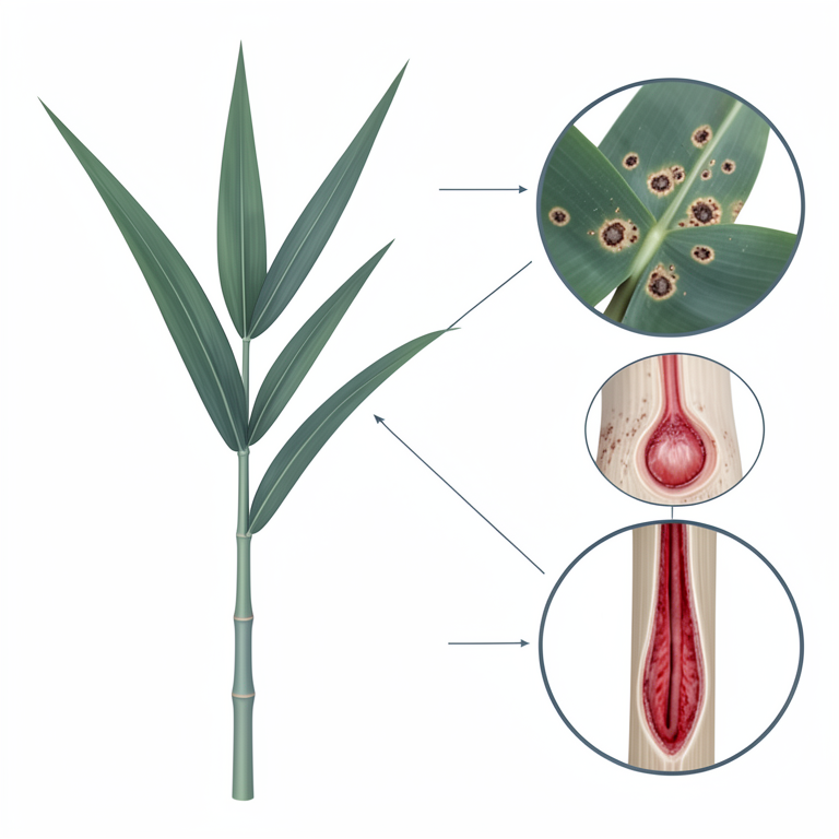
Pathogen Distribution in Sugarcane
AI-generated conceptual illustration (Sugarcane plants (Saccharum officinarum) and Colletotrichum falcatum fungus). It explains the idea and does not show study data.
View source evidence ↗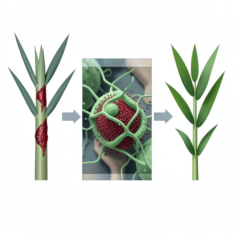
Integrated Management Strategy
AI-generated conceptual illustration (Sugarcane plants and Colletotrichum falcatum fungi). It explains the idea and does not show study data.
View source evidence ↗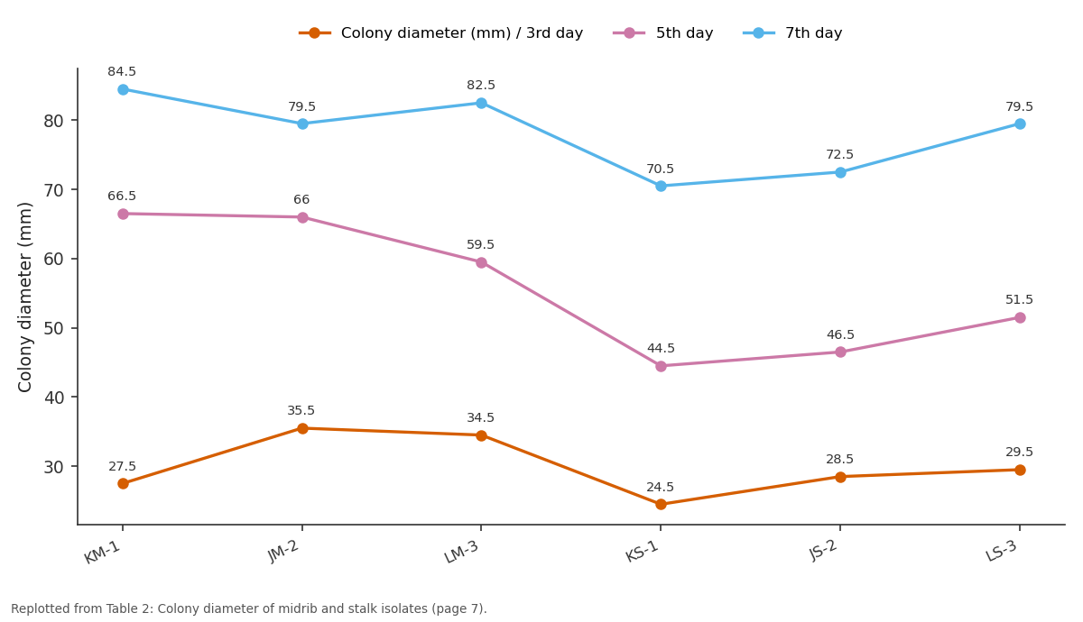
Colony diameter (mm) / 3rd day and 5th day and more: KM-1 vs JM-2 vs LM-3 vs KS-1 vs JS-2
Replotted from Table 2 (page 7) of the original paper. Every bar is the paper's own table value.
View source evidence ↗Figures & tables
From the original paper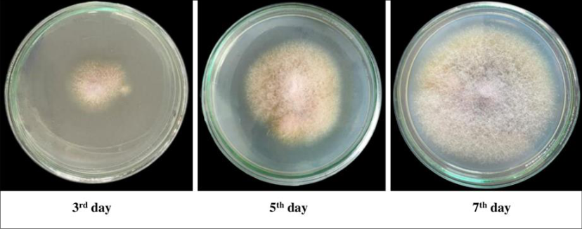 Figure 3
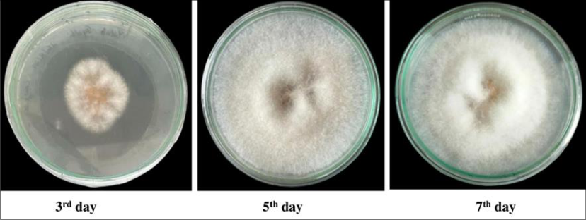 Figure 2
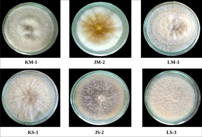 Figure 1
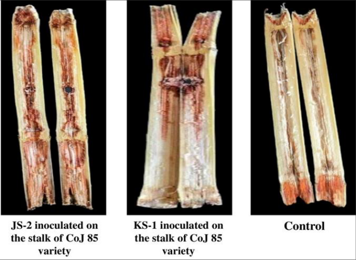 Figure 6
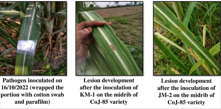 Figure 5
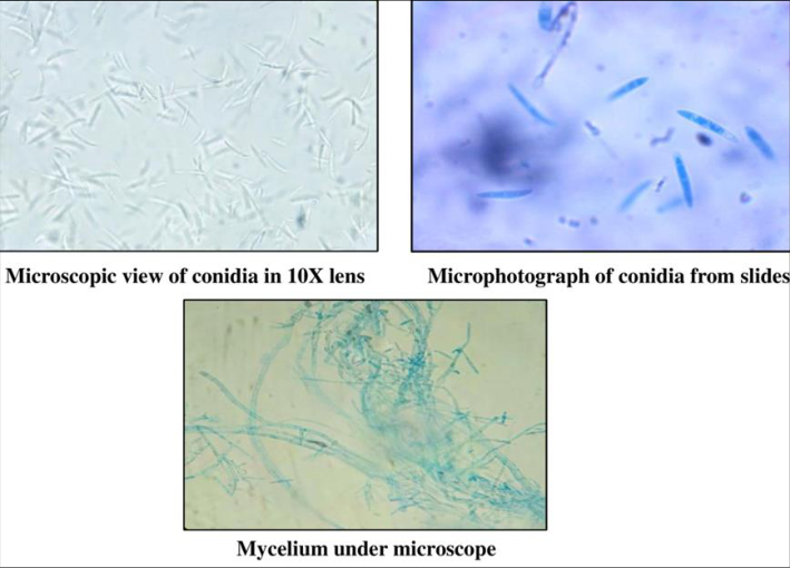 Figure 4
 Table 1
 Table 2
 Table 3
 Table 4
 Table 5
 Table 6
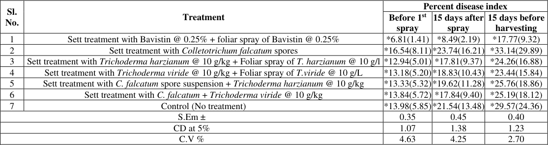 Table 7
Introduction
Sugarcane (Saccharum officinarum L.) is a perennial grass of the Poaceae family, extensively grown for its juice, which serves as the primary raw material for sugar production. This adaptable crop flourishes in tropical and subtropical regions of India and holds significant economic importance. Besides its pivotal role in sugar production, sugarcane plays a crucial part in the biofuel sector, where it is utilized for ethanol production. According to the FAO's 2021 [10] report, the leading sugarcane-producing countries include Brazil, India, Thailand, Pakistan, Mexico, and Australia. India emerges as a significant global player in sugarcane cultivation, covering an expansive area of 48.7 million hectares. The impressive productivity of sugarcane was notable during the 2021-2022 periods, with the country's production surpassing 500 million metric tonnes (Anon., 2022a) [3] . A crucial contributor to this achievement is Punjab, where sugarcane holds pivotal importance as a cash crop. In 2022, Punjab achieved substantial yields, producing a total of 6.6 million metric tonnes on 0.92 million acres of land (Anon., 2022b) [4] . Sugarcane growers face a myriad of challenges in production, resulting in significant losses due to both living organisms and environmental factors. Environmental challenges such as droughts, floods, typhoons, extreme heat, frost, and poor soil fertility greatly affect crop yields (Talukder et al., 2000) [23] . Additionally, sugarcane is susceptible to various pathogenic microorganisms including fungi, bacteria, viruses, mycoplasma, and nematodes. Among these, red rot caused by Colletotrichum falcatum Went (also known as Glomerella tucumanensis (Speg.) V. Arx and E. Muller in its perfect stage) consistently threatens sugarcane cultivation across different regions in the country (Viswanathan et al., 2002) [26] . Severe outbreaks of this disease can lead to extensive damage in the fields and complete loss of crops, often referred to as the 'Cancer' of sugarcane (Khan et al. 2011) [14] . This ailment not only reduces yield but also diminishes the quality of commercial cane sugar and sugarcane juice, highlighting the urgent need for effective management strategies. In 1893 [28] , Went documented the emergence of red rot disease in Java (now part of Indonesia). He attributed the pathogen to Colletotrichum falcatum Went. The devastating impact of red rot was particularly notable in India between 1895 and 1899, affecting the Red Mauritius cultivar in the Godavari delta of the Madras Presidency (Barber, 1901) [5] . This disease subsequently spread throughout tropical India, causing significant crop damage and resulting in the rejection of several commercial varieties such as CoJ 64, CoJ 82, CoJ 84, and CoJ 1148 within the region (Viswanathan 2010) [24] . Identification of Colletotrichum falcatum involves detailed examination of its distinct morphological and cultural characteristics, including acervuli with setae, presence or absence of a teleomorph, colony color, sporulation patterns, and mycelium growth (Viswanathan et al., 2003) [25] . Managing red rot in sugarcane has long been recognized as a challenging task. Pathogen virulence is influenced by environmental factors, plant genotypes, and the presence of aggressive pathogens. Current management strategies primarily focus on agricultural methods aimed at reducing pathogen prevalence in fields. Given the complexity of the issue, relying on a single approach is ineffective. Therefore, integrated disease management (IDM) is essential in the sugarcane industry (Agnihotri 1996; Ghazanfar et al. 2017) [2,11] . The primary objective of IDM techniques is to mitigate the risks of red rot during replanting while simultaneously enhancing sugarcane yields. IDM includes a variety of practices such as cultural methods, the use of disease-free setts, resistant varieties, physical interventions, biological control, and chemical measures. These practices have been shown to significantly improve growth parameters and enhance sugarcane quality by reducing the prevalence of red rot compared to non-IDM techniques. Implementing integrated disease management techniques has become indispensable for successful sugarcane cultivation in India and other countries. This research aims to achieve two objectives: i) assess various isolates of Colletotrichum falcatum for morphological, cultural, and pathogenic variability, and ii) evaluate the management of these isolates through in vitro testing using integrated disease management protocols.
Materials and Methods
Red rot-affected canes and leaves from various sugarcane fields in Punjab were collected, encompassing different varieties. Approximately three isolates each from midribs and stalks were examined for morphological, cultural, and pathogenic variability. Pathogenic variability was assessed both in vivo and in vitro using differentials.
Collection, Isolation and Purification
The diseased midrib and stalk samples were gathered from farms in Jalandhar, Kapurthala, and Lovely Professional University (LPU) fields and transported to the Laboratory of Plant Pathology at LPU in Phagwara. After thorough washing with tap water, the samples were air-dried. Isolation procedures were conducted under sterile laminar airflow conditions. The midrib and stalk samples were finely chopped and surface sterilized with 1% sodium hypochlorite (NaOCl) for 1 minute, followed by multiple rinses with sterile water. The sterilized samples were then dried on sterile tissue paper. Isolation was carried out on water-agar medium (20 g agar in 1000 ml distilled water), and Petri plates were incubated at a controlled temperature of 28±1 °C in a BOD incubator or at room temperature for 7-10 days. Subsequently, the pathogen was sub-cultured using the single hyphal tip culture method on potato dextrose agar (PDA) medium (200 g peeled potato, 20 g dextrose, and 20 g agar agar in 1000 ml distilled water) and incubated at 28±1 °C in a BOD incubator for 15 days to promote proper sporulation.
Identification based on morphological and cultural characters of pathogen
Identification of the causative organism's genus relied on analyzing its morphological and cultural characteristics. These included observing conidial shape and size, mycelial coloration, colony growth patterns, pigmentation, and other growth traits. Additionally, pertinent scientific literature was consulted to aid in precise identification. Inoculated samples were incubated in Petri plates at a controlled temperature of 28±1 °C for 15 days to facilitate active sporulation of the pathogen. Post-incubation, morphological and cultural features such as sporulation, pigmentation, mycelial growth, colony diameter (measured in millimetres), and overall colony appearance were meticulously examined and recorded. Conidial sizes were measured using an ocular micrometer, and spore counts were determined by preparing a mycelial suspension (1×10³ CFU/ml) from a 10-day-old culture in sterile distilled water, followed by quantification using a haemocytometer. Furthermore, microphotographs of slides were taken to document conidial shape and size.
Pathogenic variation in stalk and midrib isolates on differential
To evaluate the pathogenicity of Colletotrichum falcatum, pure cultures of freshly prepared isolates obtained from stalks and midribs were utilized. These isolates, aged between 7 to 10 days, were employed in our experiments. For midrib isolates, strain CoJ 85 was selected, and pathogenicity tests were conducted in the field using healthy sugarcane plants. Specifically, we targeted the upper, middle, and lower leaves of each plant. A spore suspension with a concentration of 1×10 3 CFU/ml was meticulously prepared in distilled water using a homogenizer. Inoculation of midrib isolates was performed using both the pin prick and cotton swab methods. Disease progression was monitored at 7-day intervals over a period of 21 days. For stalk isolates, testing was carried out in a controlled laboratory environment using the same sugarcane variety. Stalk isolates were introduced via the plug method and allowed to incubate at room temperature for 20 days.
Symptomology of disease
Disease data were meticulously recorded concerning infections and symptoms appearing on the leaf midrib and stalk. The observations concentrated on measuring lesion lengths along the midrib. Furthermore, canes were meticulously split open to examine infections, evaluating factors such as lesion width, the number of internodes breached by the pathogen, and the presence of white spots on the inner tissues.
Integrated disease management of Colletotrichum falcatum on sugarcane
Sugarcane is of considerable economic importance, but it faces a significant threat from red rot, a fungal disease that severely impacts its yield. To combat Colletotrichum falcatum effectively, one of the most damaging fungal pathogens in sugarcane, an integrated management study was conducted at the agricultural field of LPU campus in Punjab. The experiment used the CoJ 85 variety, planted as two-budded setts, and tested seven different treatments. Each treatment was replicated three times following a randomized block design during the 2022-2023 growing season (Table 1). Throughout the study, the sugarcane crop was meticulously monitored to evaluate disease intensity. Initial observations were recorded promptly upon the disease's detection in the field, using a predefined formula. Subsequent assessments were conducted at 15-day intervals to track the disease's progression and assess the effectiveness of the treatments.
| Sr. No. | Treatments |
|---|---|
| T1 | Sett treatment with Bavistin @ 0.25% + foliar spray of Bavistin @ 0.25% |
| T2 | Sett treatment with Colletotrichum falcatum spores |
| T3 | Sett treatment with Trichoderma harzianum @ 10 g/kg + Foliar spray of T. harzianum @ 10 g/l |
| T4 | Sett treatment with Trichoderma viride @ 10 g/kg + Foliar spray of T. harzianum @ 10 g/L |
| T5 | Sett treatment with C. falcatum spore suspension + Trichoderma harzianum @ 10 g/kg |
| T6 | Sett treatment with C. falcatum + Trichoderma viride @ 10 g/ kg |
| T7 | Control (no treatment) |
Table text extracted from the original PDF; superscript letters are shown inline. The original table image is below.
View original table image
Results and Discussion
Red rot, caused by the fungus C. falcatum Went, poses a major threat to the cultivation of sugarcane varieties in states where sugarcane is grown, especially under the specific conditions found in Punjab. This study explores various scientific aspects, integrating pertinent weather variables and management strategies to tackle this urgent problem.
Isolation, Purification, and Identification
Various isolates obtained from infected midrib and stalk samples of red rot were extracted using the method outlined in the Materials and Methods section (2.1 Collection, Isolation, and Purification), depicted in Figure 1. These cultures were subsequently transferred to test tube slants and stored in a refrigerator for further investigation.
Identification
Red rot samples were collected from different regions in Punjab, including Kapurthala, Jalandhar, and the LPU field. From these samples, we isolated distinct midrib and stalk strains: Kapurthala midrib isolates (KM-1), Kapurthala stalk isolates (KS-1), Jalandhar midrib isolates (JM-2), Jalandhar stalk isolates (JS-2), LPU field midrib isolate (LM-3), and LPU field stalk isolate (LS-3) of C. falcatum. These isolates were cultured on Potato Dextrose Agar (PDA). Identification involved analyzing the pathogen's morphological and cultural characteristics, comparing the isolates with references in scientific literature. According to descriptions in previous studies, a creamy-grey fungus was isolated from both midrib and stalk samples (Sarkar, 1960; Prakasan and Venkatareddy, 1961; Sharma, 1970) [19,17,20] (Fig. 2).
Morphological and Cultural characters of pathogen
Morphological and cultural traits of C. falcatum were analyzed through direct observation and stereobinocular microscopic examination. Characteristics such as sporulation, pigmentation, mycelium growth, colony diameter (in mm), and overall colony development were evaluated. Conidia size for each isolate was measured using an ocular micrometer, and spore counts were determined with a hemocytometer. Microphotographs of slides were taken to record conidia shape and size.
Colony diameter of midrib and stalk isolates
The three midrib and stalk isolates were cultured on potato dextrose agar (PDA) medium in Petri plates, and the colony diameters were measured at 3, 5, and 7 days post-incubation at 28±1 °C in a BOD incubator. The data are presented in Table 2.
| Sr. No. | Isolates | Colony diameter (mm) | — | — |
|---|---|---|---|---|
| 3rd day | 5th day | 7th day | ||
| Midrib isolates | KM-1 | 27.5 | 66.5 | 84.5 |
| JM-2 | 35.5 | 66.0 | 79.5 | |
| LM-3 | 34.5 | 59.5 | 82.5 | |
| Stalk isolates | KS-1 | 24.5 | 44.5 | 70.5 |
| JS-2 | 28.5 | 46.5 | 72.5 | |
| LS-3 | 29.5 | 51.5 | 79.5 |
Table text extracted from the original PDF; superscript letters are shown inline. The original table image is below.
View original table image
Midrib Isolates
Among the midrib isolates (KM-1, JM-2, and LM-3), rapid growth was observed with colony diameters ranging from 27.5 to 35.5 mm after 3 days of incubation. KM-1 exhibited the smallest colony diameter at 27.5 mm. By the 5th day, the midrib isolates showed significant growth; with diameters ranging from 55.5 to 59.5 mm. LM-3 displayed the smallest diameter at 59.5 mm. By the 7th day, the midrib isolates had nearly covered the Petri dishes; with colony diameters ranging from 79.5 to 84.5 mm. JM-2 had the smallest diameter at 79.5 mm (Fig. 3).
Stalk Isolates
On the 3rd day, the stalk isolates (KS-1, JS-2, and LS-3) also showed rapid growth, with colony diameters ranging from 24.5 to 29.5 mm. By the 5th day, their diameters had increased to 44.5-51.5 mm. LS-3 exhibited the largest diameter at 51.5 mm. By the 7th day, the stalk isolates had grown further, with diameters ranging from 70.5 to 79.5 mm (Fig. 4). As indicated in Table 1, there was significant variation in mycelial growth among the three midrib and stalk isolates of Colletotrichum falcatum. Previous studies by and Viswanathan et al. (2003) [26] also reported significant differences in colony growth patterns when investigating C. falcatum pathotypes.
Mycelium characters of midrib and stalk isolates
The growth pattern, sporulation, and pigmentation of the colonies isolated from midribs and stalks were observed after 7 days of incubation on PDA medium. Detailed data can be found in Table 3 and Fig. 4.
| Isolates | Colony Characters | — | — |
|---|---|---|---|
| Mycelium growth | Pigmentation | Sporulation | |
| KM-1 | Raised fluffy | White cottony mass with light orange pigment | +++ |
| JM-2 | Raised fluffy | Whitish mycelium with greyish orange pigment | ++ |
| LM-3 | Raised fluffy | White mycelium with light orange colour | +++ |
| KS-1 | Flat | Whitish-grey mycelium | ++ |
| JS-2 | Flat | White mycelium with dark violet pigment | ++ |
| LS-3 | Less fluffy | Whitish with pale orange pigmentation | +++ |
| +++ - High sporulation, ++ - Medium sporulation, + - Less sporulation |
Table text extracted from the original PDF; superscript letters are shown inline. The original table image is below.
View original table image
Midrib Isolates 3.2.2.1.1. Mycelium growth pattern and Sporulation
All three midrib isolates of Colletotrichum falcatum (KM-1, JM-2, and LM-3) exhibited a raised, fluffy mycelial growth pattern with abundant sporulation. However, JM-2 showed moderate sporulation compared to the other midrib isolates.
Colony colour
The midrib isolates exhibited raised fluffy mycelial growth. The KM-1 isolate displayed a whitish cottony mass when viewed from the front, with a light orange pigmentation on the reverse side of the culture. In contrast, the JM-2 isolate featured white mycelium with light orange pigmentation on the front view and orange with grey pigmentation on the reverse of the culture plate. The LM-3 isolate resembled the KM-1 isolate; showing white mycelial masses with light orange pigmentation (see Fig 2).
Stalk Isolates 3.2.2.2.1. Mycelium growth pattern and Sporulation
Two out of the three stalk isolates exhibit a flat colony growth pattern: KS-1 and JS-2. Meanwhile, the LS-3 isolate demonstrates a less fluffy mycelium growth pattern. All three isolates are expected to show medium to high levels of sporulation.
Colony colour
In stalk isolates, KS-1 exhibited a whitish-grey pigmentation. In contrast, isolate JS-2 displayed white mycelium accompanied by dark violet pigmentation in culture. The LS-3 isolate featured a less fluffy, whitish mycelium with a pale orange hue on the culture (Fig 3).
Conidial characters of midrib and stalk isolates
Conidial size for each isolate was measured using an ocular micrometer. Spore count was determined by preparing a mycelial suspension (1×10³ CFU/ml) from 10-day-old cultures in distilled sterilized water using a homogenizer, and counting with a hemocytometer. Microphotographs of slides were taken to record the shape and size of conidia, and detailed data are presented in Table 4 and Fig. 4.
| Midrib isolates | Spore shape | Spore size (µm) (L x B) | Spore count (spores/ml) | Stalk isolates | Spore shape | Spore size (µm) (L x B) | Spore count (spores/ml) |
|---|---|---|---|---|---|---|---|
| KM-1 | Falcate | 26.7 x 6.2 | 104 x 10² | KS-1 | Falcate | 34.5 x 7.9 | 125 x 10² |
| JM-2 | Falcate | 27.0 x 6.9 | 116 x 10² | JS-2 | Falcate | 27.8 x 7.0 | 113 x 10² |
| LM-3 | Falcate | 24.7 x 5.3 | 107 x 10² | LS-3 | Falcate | 29.3 x 7.4 | 100 x 10² |
| Mean size | 26.1 µm x 6.1 µm | Mean size | 30.5 µm x 7.4 µm |
Table text extracted from the original PDF; superscript letters are shown inline. The original table image is below.
View original table image
Midrib Isolates
The average dimensions of conidia from midrib isolates ranged around 26.1 µm x 6.1 µm. Among these isolates, JM-2 exhibited the largest conidia measuring 26.7 µm x 6.2 µm, while LM-3 had the smallest, measuring 24.7 µm x 5.3 µm. All conidia observed were falcate in shape, with sporulation counts ranging from 104 x 10² to 116 x 10² per isolate.
Stalk Isolates
Stalk isolates displayed an average spore size of approximately 30.5 µm x 7.4 µm. These spores exhibited a falcate shape, with sporulation rates ranging from 100 x 10² to 125 x 10². Among these isolates, KS-1 had the largest spores, measuring 34.5 µm x 7.9 µm, while JS-2 exhibited the smallest spores at 27.8 µm x 7.0 µm. Our observations regarding the morphological and cultural characteristics of various midrib and stalk isolates align closely with earlier studies by Chona and Srivastava (1960) [9] . They noted that conidia from different isolates varied in length from 10 µm to 36 µm. Similarly, other studies have reported colony characteristics and conidial dimensions, with sizes ranging from 17.1 µm to 2.84 µm x 4.5 µm to 6.8 µm (Pandey and Sakel, 1974) [16] . Jothi (1989) [13] categorized different C. falcatum isolates based on criteria such as color, texture, and sporulation.
Pathogenicity assay
Pathogenicity tests of midrib and stalk isolates of Colletotrichum falcatum were conducted under both field and laboratory conditions using the CoJ 85 variety of sugarcane. For midrib isolates, inoculation was performed using two methods: the pin prick and cotton swab techniques on the upper, middle, and lower leaves of healthy plants. Disease progression was monitored at 7-day intervals for up to 21 days after inoculation. Stalk isolates were inoculated using the plug method with a pure culture of the pathogen and were kept at room temperature. Disease symptoms were observed and recorded after a 20-day incubation period.
Midrib Isolates
Isolates such as KM-1 and JS-2 were introduced into the CoJ 85 variety of sugarcane under field conditions. Using the pinprick method, these isolates were inoculated into the upper, middle, and lower leaves of each plant. Data was collected on the 7th, 10th, 16th, and 21st days post-inoculation. The lesions on the midribs of the inoculated leaves revealed larger sizes on the lower leaves and smaller sizes on the middle leaves of sugarcane. When comparing KM-1 and JS-2 isolates, KM-1 induced larger midrib lesions than JS-2. The details are presented in Table 5 and Figure 5. The pinprick method proved to be effective and efficient for evaluating the pathogenicity of Colletotrichum falcatum isolates in sugarcane. Previous studies by Abraham et al. (1980) [1] and Mehetre (2009) [15] have demonstrated that creating wounds using the pinprick method and inoculating with spore suspensions are reliable techniques for assessing the pathogenicity of Colletotrichum species on plants.
| Midrib isolates | Leaf | 7th day | 10th day | 16th day | 21st day |
|---|---|---|---|---|---|
| KM-1 | Upper | 1.9 cm | 2.3 cm | 2.4 cm | 2.6 cm |
| Middle | 1.3 cm | 1.3 cm | 1.4 cm | 1.5 cm | |
| Lower | 4.5 cm | 5.0 cm | 5.3 cm | 6.0 cm | |
| JM-2 | Upper | 2.1 cm | 2.4 cm | 2.5 cm | 2.5 cm |
| Middle | 1.5 cm | 1.7 cm | 1.9 cm | 2.0 cm | |
| Lower | 2.5 cm | 3.3 cm | 3.8 cm | 4.0 cm |
Table text extracted from the original PDF; superscript letters are shown inline. The original table image is below.
View original table image
Stalk Isolates
Pathogenicity testing was conducted in a laboratory setting on stalk isolates such as KS-1 and JS-2 using the plug method on the sugarcane variety CoJ 85. Each stalk, inoculated with a 5 cm diameter piece from pure cultures of KS-1 and JS-2, was left at room temperature for 20 days before observations were made. The results showed significant differences: the KS-1 inoculated cane exhibited lesions covering two internodes with 100% spread, indicating susceptibility of CoJ 85 to KS-1. Meanwhile, JS-2 resulted in lesions covering nearly one internode with more than 50% spread, indicating an intermediate susceptibility of CoJ 85 to JS-2. These findings align with previous studies; Sandhu et al. (1974) [18] noted that stalk isolates tend to be more virulent than midrib isolates. Additionally, Shukla et al. (2001) [21] suggested that while midrib isolates currently pose no immediate threat to sugarcane cultivation, they could potentially adapt to local varieties and infect stalks under natural conditions.
Integrated disease management of Colletotrichum falcatum on sugarcane
A field experiment was conducted at the agriculture field of LPU campus in Punjab to assess integrated disease management strategies against Colletotrichum falcatum in sugarcane during 2022-2023. Sugarcane of the CoJ 85 variety, planted with twobudded setts, was subjected to seven different treatments in a randomized block design with three replications per treatment. Initial disease incidence data were recorded before the first spray application, timed upon the appearance of initial disease symptoms in the field. Subsequent data collections occurred 15 days post-spray and again 15 days prior to harvesting. The disease intensity observations for each treatment are detailed in Table 7. Before the initial spray, the least disease intensity (1.41%) was noted in the treatment involving sett application of Bavistin @ 0.25% combined with foliar spray of Bavistin @ 0.25%. Conversely, the highest disease intensity (8.11%) occurred in the treatment using C. falcatum spores on setts. Other effective treatments included sett applications with Trichoderma harzianum @ 10 g/kg plus foliar spray @ 10 g/l (5.01%), Trichoderma viride @ 10 g/kg plus foliar spray @ 10g/l (5.20%), C. falcatum spore suspension with Trichoderma harzianum @ 10g/kg (5.32%), and C. falcatum with Trichoderma viride @ 10g/kg (5.72%), while the control showed 5.85% disease intensity. After 15 days post-spray, the treatment with Bavistin @ 0.25% sett application plus foliar spray maintained the lowest disease intensity (2.19%). Following closely were treatments with Trichoderma harzianum @ 10g/kg plus foliar spray @ 10g/l (9.37%), C. falcatum with Trichoderma viride @ 10g/kg (9.40%), Trichoderma viride @ 10g/kg plus foliar spray @ 10g/l (10.43%), C. falcatum spore suspension with Trichoderma harzianum @ 10g/kg (11.28%), and the control with 13.48% disease intensity. The highest disease intensity (16.21%) was found in the treatment using C. falcatum spores on setts. Prior to harvesting, the treatment with Bavistin @ 0.25% sett application plus foliar spray showed the best disease management (9.32%). The highest disease intensity (29.89%) was observed in the treatment using C. falcatum spores on setts. Other effective treatments included Trichoderma viride @ 10 g/kg plus foliar spray @ 10g/l (15.84%), Trichoderma harzianum @ 10 g/kg plus foliar spray @ 10 g/l (16.88%), C. falcatum with Trichoderma viride @ 10 g/kg (18.12%), C. falcatum spore suspension with Trichoderma harzianum @ 10 g/kg (18.86%), and the control with 24.36% disease intensity. Bharadwaj and Sahu (2014) [7] reported complete inhibition of C. falcatum mycelial growth with Bavistin, underscoring its efficacy. Hence, among the treatments evaluated, Bavistin @ 0.25% sett application plus foliar spray emerged as the most effective, followed by treatments involving Trichoderma harzianum @ 10 g/kg plus foliar spray @ 10 g/l and Trichoderma viride @ 10 g/kg plus foliar spray @ 10 g/l. The application of T. harzianum and T. viride aids in red rot control by directly impacting C. falcatum and inducing systemic plant resistance (Yadav et al. 2008) [29] .
Conclusion
Red rot, caused by Colletotrichum falcatum, poses a severe threat to global sugarcane production as it affects all parts of the plant-leaves, midribs, and stalks. A study was conducted to analyze the morphological, cultural, and pathogenic variability among midrib and stalk isolates of the red rot fungus. Significant differences were observed in mycelial growth among the three isolates, each displaying either raised fluffy or flat white mycelium with light orange pigmentation and medium to high levels of sporulation. Conidia were consistently falcate-shaped, averaging 28.3 µm x 6.75 µm in size. Pathogenicity tests using the pinprick and plug methods confirmed the effectiveness of both techniques in assessing disease severity on the sugarcane variety CoJ 85. Epidemiological studies conducted under natural conditions demonstrated a linear disease progression, highlighting the pivotal role of relative humidity in disease development. In managing red rot, a comparative evaluation of seven treatments revealed that applications containing Bavistin, T. harzianum, and T. viride were most effective. Beyond fungicides, the application of biocontrol agents has proven beneficial by directly impacting C. falcatum and inducing systemic resistance in sugarcane plants propagated from treated setts.
Figures and tables from the paper
| Stalk isolates | Lesion length | Nodal transgression |
|---|---|---|
| KS-1 | 100% | ++ |
| JS-1 | > 50% | + |
| Control | < 25% | - |
| ++ - Both internodes, + - Only one internode, - - No internodes |
Table text extracted from the original PDF; superscript letters are shown inline. The original table image is below.
View original table image
References
25 in the original paper- Abraham M, Karunakaran P, Mathew J. Leaf spot disease of Dioscorea alata Linn. Agricultural Research Journal of Kerala. 1980;18(1):132-133.
- Agnihotri VP. Current sugarcane disease scenario and management strategies. Indian Phytopathology. 1996;49:109-126.
- Anonymous. Press Information Bureau, Government of India. Ministry of Consumer Affairs, Food and Public Distribution. Posted on October 5; c2022a. Available from: https://www.pib.gov.in/PressReleseDetailm.aspx?PRID=18 65320 (Accessed July 5, 2024).
- Anonymous. Tractor Junction. Sugarcane production in India -largest production states. Posted on February 3; c2022b. Available from: https://www.tractorjunction.com/blog/sugarcaneproduction-in-india-largest-producing-states/ (Accessed July 5, 2024).
- Barber CA. Sugarcane disease in Godawari and Ganjam districts. Madras Department Land Records and Agriculture Bulletin. 1901;512:181-194.
- Barber CA. Studies on sugarcane. Memoirs of Department of Agriculture India Botany Series. 1919;10:155-179.
- Bharadwaj N, Sahu RK. Evaluation of some fungicides, botanicals and essential oils against the fungus Colletotrichum falcatum causing red rot of sugarcane. The Bioscan. 2014;9(1):175-178.
- Butler EJ, Khan AH. Some new sugarcane diseases. Part I, wilt. Memoirs of Department of Agriculture India Botany Series. 1913;6:180-190 [8] .
- Chona BL, Srivastava DN. Variations in Colletotrichum falcatum, the causal organism of red rot of sugarcane. Indian Phytopathology. 1960;13:58-65.
- FAO. OECD-FAO Agricultural Outlook 2021-2030. OECD Agriculture Statistics (Database); c2021. p. 150-162. Available from: https://www.fao.org/3/cb5332en/Sugar.pdf (Accessed July 5, 2024).
- Ghazanfar MU, Raza W, Gondal SK. Screening of sugarcane cultivars against Colletotrichum falcatum causing red rot disease and its control with different fungicides under laboratory conditions. Pakistan Journal of Phytopathology. 2017;29(1):103. https://doi.org/10.33866/phytopathol.029.01.0381
- Hughes CG. The economic importance of ratoon stunting disease. Proceedings of the International Society of Sugar Cane Technologists. 1974;15:213-217.
- Khan A, Awais M, Raza W, Zia A. Identification of sugarcane lines with resistance to red rot. Pakistan Journal of Phytopathology. 2011;23(2):98-102.
- Pandey LN, Sakel R. New pathogenic strain of Glomerella tucumanensis (Speg.) Arx and Muller in Uttar Pradesh. Indian Sugar. 1974;24:707-709.
- Prakasam R, Venkatareddy TC. Occurrence of light race of Colletotrichum falcatum Went in Andhra Pradesh. Science and Culture. 1961;27:250-251.
- Sandhu SS, Mehan VK, Singh K. Role of leaf midrib lesions in epidemiology of red rot caused by Colletotrichum falcatum Went. in the Punjab. Indian Sugar. 1974;24:391395.
- Sarkar A. Comparative study of two races of Colletotrichum falcatum Went. Science and Culture. 1960;26:83-84.
- Sharma MN. Occurrence of a new strain of Physalospora tucumanensis Speg. Nizamabad District of Andhra Pradesh. Science and Culture. 1970;36:52-54.
- Shukla RK, Pandey AK, Singh SP, Verma KP, Singh RR. Studies on comparative pathogenic behaviour of stalk and midrib isolates of red rot pathogen (Colletotrichum falcatum Went.). Indian Sugar. 2001;51(3):175-180.
- Sydow H. Notizen Uber Ushlagineen. Annales Mycologici. 1924;22:277.
- Talukder MI, Alam MS. Effect of moist hot air treatment (MHAT) on control of white leaf disease and growth of sugarcane. Pakistan Sugar Journal. 2000;15(3):16-21.
- Viswanathan R, Sundar AR, Padmanaban P, Mohanraj D. Red rot disease of sugarcane and its management. In: Upadhyay RK, Mukherji KG, Dubey OP, editors. IPM Systems in Agriculture. key pathogens and diseases. New Delhi: Aditya Books. 2002;8:277-301.
- von Arx JA, Muller E. Die Gattungen der amerosporen Pyrenomyceten. Beiträge zur Kryptogamenflora der Schweiz. 1954;11:1-434.
- Went FAFC. Het Rood Snot (Summary in English). Archief voor de Java Suikerindustrie. 1893;1:265-282.
- Yadav RL, Singh SN, Srivastava R, Lal SK, Awasthi BB. Use of Trichoderma harzianum for the control of red rot disease of sugarcane. Sugarcane International. 2008;26(4):28-33.