
Research article · 2024 · Full text
International Journal of Veterinary Sciences and Animal HusbandryEmergency cesarean sections in goats with abnormal fetal presentations: A case study
Volume 9(4) · Pages 148–150 · DOI: 10.22271/veterinary.2024.v9.i4c.1526 ↗
Research areasAnimal health and immunologyReproductive Physiology in LivestockCongenital Anomalies and Fetal Surgery
Abstract
Author-writtenTwo goats presented with anorexia, restlessness and prolonged straining due to dystocia necessitated immediate cesarean sections at Devashraya Animal Hospital. Clinical examination revealed elevated heart and respiration rates but normal body temperature. Radiographic evaluation indicated abnormal fetal presentations—twin fetuses in one case and a single fetus with atypical positioning in the other. Surgery involved intramuscular analgesics and sedatives, followed by left flank celiotomy under regional anesthesia. Cesarean sections were performed successfully, with meticulous surgical technique to prevent contamination. Postoperatively, both goats received antimicrobials, analgesics and wound care. Both cases resulted in live births with uneventful recoveries, highlighting the importance of timely intervention and surgical expertise in managing dystocia in small ruminants.
Summary
Source-checkedPlain-language reading of the full paper, prepared with AI and checked against the text. Each point links to the authors' own words.
Why it matters
- Dystocia is defined as slow or difficult labor that occurs when the delivery of fetuses is interrupted or prolonged, requiring assistance. View evidence ↗
- In small ruminants, dystocia leads to significant economic losses because of increased uterine infections, longer kidding intervals, and the death of both mothers and offspring. View evidence ↗
- Fetal factors that cause dystocia primarily include developmental abnormalities, oversized fetuses, malpositioning, or having multiple fetuses in the pelvic canal. View evidence ↗
The question
- The study aims to present the causes of dystocia and the outcomes for two goats treated with cesarean sections via left flank laparotomy while in a sedated recumbent position. View evidence ↗
What they did
- The study involved two full-term goats presented at Devashraya Animal Hospital with symptoms of anorexia, restlessness, and straining for over 6-8 hours. View evidence ↗
- The goats were cast in right lateral recumbency after receiving an intramuscular combination of xylazine (0.2mg/kg), ketamine (0.4mg/kg), and butorphanol (0.05mg/kg) for sedation and analgesia. View evidence ↗
- Regional anesthesia for the left flank was achieved using an inverted-L block with the infiltration of 2ml of 2% lignocaine hydrochloride solution. View evidence ↗
- The surgical approach involved a 10-12cm vertical skin incision in the lower flank region, located midway between the tuber coxae and the last rib. View evidence ↗
- Post-operative care included 5 days of intramuscular Flunixin (1.1mg/kg) and Amoxycillin-sulbactam (10mg/kg), and 10 days of povidone iodine antiseptic dressing. View evidence ↗
What they found
- Radiographs of the first goat revealed twin fetuses in a ventral position with posterior longitudinal presentation and both hindlimbs in the pelvic canal. View evidence ↗
- Radiographs of the second goat showed a single fetus in a dorso-sacral position with anterior longitudinal presentation, featuring a head-breast posture and a foot-nape posture. View evidence ↗
- Both surgical interventions were successful, resulting in the delivery of live fetuses (twins in one case and a single kid in the other). View evidence ↗
- Both goats recovered without major complications, and their skin sutures were removed on the 12th post-operative day. View evidence ↗
What it means
- The authors conclude that timely surgical intervention and expertise are critical for successfully managing dystocia in small ruminants. View evidence ↗
- The authors state that delaying the decision to perform surgery increases the risk of death for both the mother and the fetus. View evidence ↗
Visual summary
1 visualsIllustrations explain the paper; they do not show study data.
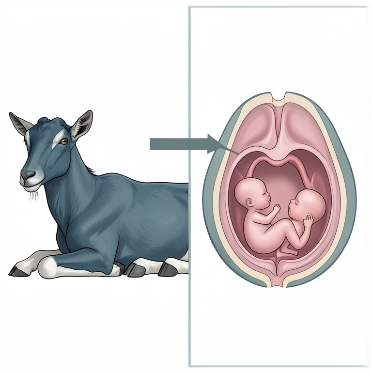
Surgical Approach Overview
AI-generated conceptual illustration (female goat). It explains the idea and does not show study data.
View source evidence ↗Figures & tables
From the original paper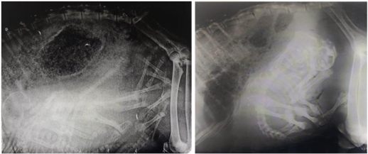 Figure 1
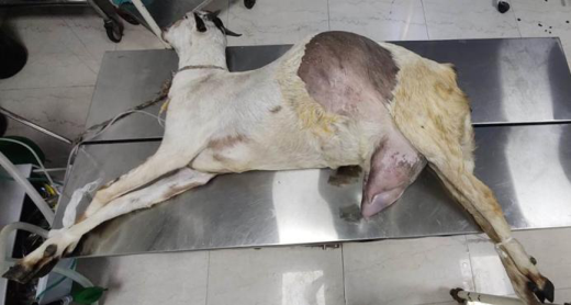 Figure 2
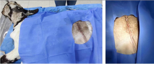 Figure 3
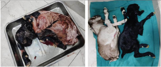 Figure 4
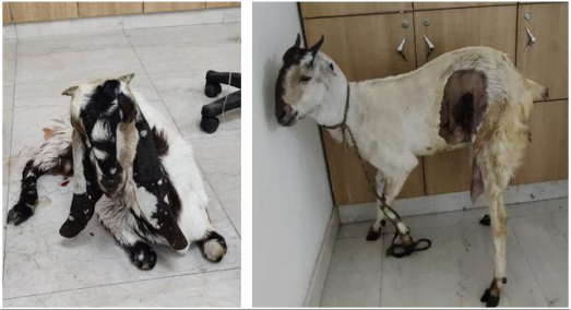 Figure 5
Introduction
Dystocia is characterized as slow or difficult labor (Venes, 2013) [12] . It occurs when labor is prolonged, interrupted, or when delivery of the fetus (es) cannot proceed in a timely manner without assistance. Dystocia in small ruminants contributes significantly to economic losses due to perinatal deaths of dams and fetuses, increased incidence of retained placentas, uterine infections and longer lambing and kidding intervals (Ghosh et al., 1992; Brounts et al., 2004; Scott, 2005) [5,1,10] . It can arise from maternal or fetal factors (Noakes et al., 2009) [9] . Maternal factors contributing to dystocia typically involve narrow pelvis, uterine inertia and incomplete cervical dilatation (e.g., ring womb) (Majeed and Taha, 1989b; Thomas, 1992; Noakes et al., 2009) [8,11,9] . Fetal causes include primarily malpositioning, oversized fetuses, presence of multiple fetuses in the pelvic canal and developmental abnormalities (Majeed and Taha, 1989a; Noakes et al., 2009) [7,9] . Cesarean section involves making an incision through the abdominal wall and uterus to deliver the kid. It is associated with high survival rates for both the dam and offspring, and is often quicker, safer and less physically demanding than fetotomy. The primary objectives of performing a cesarean section are to ensure the survival of both the dam and kid and to manage dystocia effectively. The procedure is typically performed in standing or sedated recumbent ruminants using a left oblique laparotomy or left paralumbar fossa laparotomy approach. Alternative approaches such as ventral midline, ventrolateral and paramedian under general anesthesia have also been described (Gilbert et al., 2017) [6] . This communication presents the causes of dystocia and the outcomes for two dams undergoing cesarean section in sedated recumbent position via left flank laparotomy to alleviate dystocia in small ruminants.
History and clinical observations
Two goats were presented to Devashraya Animal Hospital by Sarvodaya Foundation, Faridabad, Haryana with chief complaint of anorexia, restlessness and straining since more than 6-8 hours with full term gestation period. Clinical examination revealed moderate elevation in both heart and respiration rate in both the cases with body temperature within the normal limits. In both the case, per vaginal examination revealed dilated cervix but there was insufficient room for manipulation of the fetus inside the uterus. Radiographic evaluation of abdomen of one doe had shown presence of twin fetuses with posterior longitudinal presentation, ventral position with their hindlimbs in the pelvic canal simultaneously. The other doe had revealed presence of one fetus with anterior longitudinal presentation, dorso-sacral position with complete downward deviation of the head (head-breast posture) along with curling of one forelimb over the head (foot-nape posture) on radiographic evaluation. In both the cases there were extreme deviation from the normal presentation, position and posture of fetuses (Fig. 1). Usually for normal delivery of fetus it should be in anterior longitudinal presentation, dorso-sacral position with complete extension of head and forelimbs in the pelvic canal. On the basis of the history, clinical and radiographic evaluation it was decided to perform immediate cesarean section in both the cases, as delay in the decision to perform surgery may result in fetal or maternal death.
Surgery and perioperative procedure
For proper analgesic and sedative effect, combination of butorphanol @ 0.05mg/kg, ketamine @ 0.4mg/kg and xylazine @ 0.2mg/kg (Ketamine stun) was injected intramuscularly followed by casting in right lateral recumbency. Perioperative antimicrobial and NSAID injected intramuscularly were Amoxycillin-sulbactam @ 10mg/kg and Flunixin @ 1.1mg/kg respectively, along with adequate intravenous fluid therapy to correct any dehydration and acidbase imbalances. The left paralumbar fossa was clipped, aseptically prepared and draped for surgery in a routine fashion for performing cesarean section via left flank celiotomy approach (Fig. 2). An inverted-L block regional anesthesia was given by infiltration 2ml of 2% lignocaine hydrochloride solution for desensitization of the left flank region. An approximate 10-12cm vertical skin incision was made at the lower flank region midway between the last rib and the tuber coxae of the same side followed by blunt dissection and separation of the abdominal muscles. After incising the peritoneum carefully to avoid any damage to the underneath organs, the gravid uterus was separated and packed with sterile surgical drapes to avoid any spillage of the uterine content in the abdominal cavity which might lead to intraabdominal sepsis. An incision with size just enough to exteriorize the fetus was given on the gravid uterus followed by extraction of both the fetus and placenta. The incised uterus was repaired by applying double layer of continuous inversion suture pattern i.e. cushing and lambert sutures, with synthetic absorbable suture (Polyglactin-910) no. 0. A thorough lavage of the inner surface of uterus and peritoneal cavity with normal saline was performed to reduce chances of contamination before closure of the abdominal cavity. The incised peritoneum and the individual muscular layers were sutured by applying continuous simple interrupted sutures with Polyglactin-910, no. 0 suture. Apposition of the skin incision was achieved with application of horizontal mattress sutures with synthetic non-absorbable suture material (Polyamide, no. 0) (Fig. 3). Goats continued to receive intramuscular antimicrobials and analgesics (Amoxycillinsulbactam @ 10mg/kg and Flunixin @ 1.1mg/kg respectively) for 5 days along with antiseptic dressing of the suture site with povidone iodine solution for 10 days.
Outcome and Discussion
Both the cases were successfully managed with live fetuses delivered via left flank cesarean, having one doe with twin kids and the other with single (Fig. 4 and5). The skin sutures were removed on 12 th post-operative day without any major complication with uneventful recovery in both the doe. A successful outcome of dystocia depends on several factors, including the surgeon's skill, duration of dystocia, surgical conditions, overall health of the dam, viability of the offspring and concurrent diseases. (2023) [4] , it was found that the survival rate of infants was notably higher when cesarean sections were performed with sedation compared to those done under general anesthesia. The inverted L block, described by Edwards (2001) [3] , is a regional anesthetic technique that specifically targets the tissues adjacent to the caudal aspect of the thirteenth rib and the ventral aspect of the lumbar vertebrae transverse processes. This technique offers advantages such as simplicity, minimal disruption to ambulation, and a reduced likelihood of edema and emphysema at the surgical incision site (Edmondson, 2008) [2] . In situations of dystocia, an urgent cesarean section may become necessary if the uterus or fetus is inaccessible, or if the cervix remains closed (e.g., due to ringwomb). Forceful attempts to manipulate the fetus under conditions of inadequate space or cervical dilation can potentially cause harm to the cervix or uterus. When the dam's size precludes transvaginal palpation, a prompt cesarean section may be the preferred course of action. Delaying the decision for surgical intervention could increase the risk of mortality for both the fetus and the mother.
Acknowledgement
AcknowledgmentsThanks to the Managing Director and the team of Devashraya Animal Hospital by Sarvodaya Foundation, Faridabad, Haryana for providing support and facilities require for these surgical intervention and post-operative care.
Figures and tables from the paper
References
8 in the original paper- Brounts SH, Hawkins JF, Baird AN, Glickman LT. Outcome and subsequent fertility of sheep and goats undergoing cesarean section because of dystocia: 110 cases (1981-2001). J Am Vet Med Assoc. 2004;224:275281.
- Edmondson MA. Local and regional anesthesia in cattle. Vet Clin North Am Food Anim Pract. 2008;24(2):211226.
- Edwards B. Regional anaesthesia techniques in cattle. In Pract. 2001;23:142-149.
- Elane GL, Portela DA, Hobbs KJ, Bauck AG, Biedrzycki AH. Sedated cesarean sections are associated with increased kid survival compared to general anesthesia in goats: retrospective cohort of 45 cases (2011-2021). J Am Vet Med Assoc. 2023;261(3):391-396.
- Ghosh A, Yeasmina F, Alam MGS. Studies of ring womb in Black Bengal goats. Theriogenology. 1992;37:527532.
- Scott PR, Naqvi BS, Pandey GK, Gautam KK, Joshi A, Geetha lakshmi V, et al. The management and welfare of some common ovine obstetrical problems in the United Kingdom. Vet J. 2005;170:33-40.
- Thomas JO. Survey of the causes of dystocia in sheep. Vet Rec. 1992;127:574-575.
- Vidya SP, Veni KS, Vadde KS. Dystocia due to fetal ascites with wry neck in a graded Murrah buffalo: A case report. Buffalo Bull. 2010;29:73-74.