
Research article · 2024 · Full text
International Journal of Veterinary Sciences and Animal HusbandryMolecular detection of Trichomonas foetus in women and cow in Babylon province, Iraq
Volume 9(4) · Pages 114–118 · DOI: 10.22271/veterinary.2024.v9.i4b.1523 ↗
Research areasReproductive tract infections research
Abstract
Author-writtenThe present study was conducted to detect the presence of Trichomonas foetus from reproductive tract of women and cows in Babylon Province, Iraq. The study included the collection of 200 vaginal swabs (100 women and 100 cows). These samples were tested using microscopy, measuring trophozoites, and PCR that targeted the 18S rRNA gene of the protozoan. The results revealed the presence of the parasite in 26 (26%) of woman swabs and 95 (95%) of cow samples as detected by the PCR. For the geographical correlation, the findings demonstrated significant (p
Summary
Source-checkedPlain-language reading of the full paper, prepared with AI and checked against the text. Each point links to the authors' own words.
Why it matters
- Trichomonas foetus is a protozoan that can cause temporary sterility and inflammation of the uterus, cervix, and vagina. View evidence ↗
- In humans, T. foetus causes trichomoniasis, a sexually transmitted disease affecting millions of people globally through various tract diseases. View evidence ↗
- The parasite is also well-documented to reduce reproductive efficiency in various animals, including bulls, sheep, goats, camels, cats, and dogs. View evidence ↗
The question
- The study aimed to detect the presence of Trichomonas foetus in the reproductive tracts of cows and women within the Babylon Province of Iraq. View evidence ↗
What they did
- The researchers collected a total of 200 vaginal swabs, consisting of 100 samples from women and 100 from cows. View evidence ↗
- Detection was performed using microscopy to measure trophozoites and PCR targeting the protozoan's 18S rRNA gene. View evidence ↗
- The research was conducted over a period from August 2023 to the end of January 2024. View evidence ↗
What they found
- In cows, a 100% prevalence rate (25/25) was found in Hilla City, the province capital's center. View evidence ↗
- Cows aged 2-4 and 5-6 years showed a significantly higher prevalence (100% for both groups) compared to older cows. View evidence ↗
- Among women, those from rural areas had a significantly higher prevalence rate of 30% (21/70) compared to urban areas. View evidence ↗
- Women in the 26-35 age group exhibited a significantly higher prevalence rate of 45% (18/40) than other age groups. View evidence ↗
What it means
- The authors conclude that the high prevalence of the parasite in cows suggests they may be a significant source of zoonotic disease. View evidence ↗
Visual summary
2 visualsIllustrations explain the paper; charts are replotted from the paper's own table values.
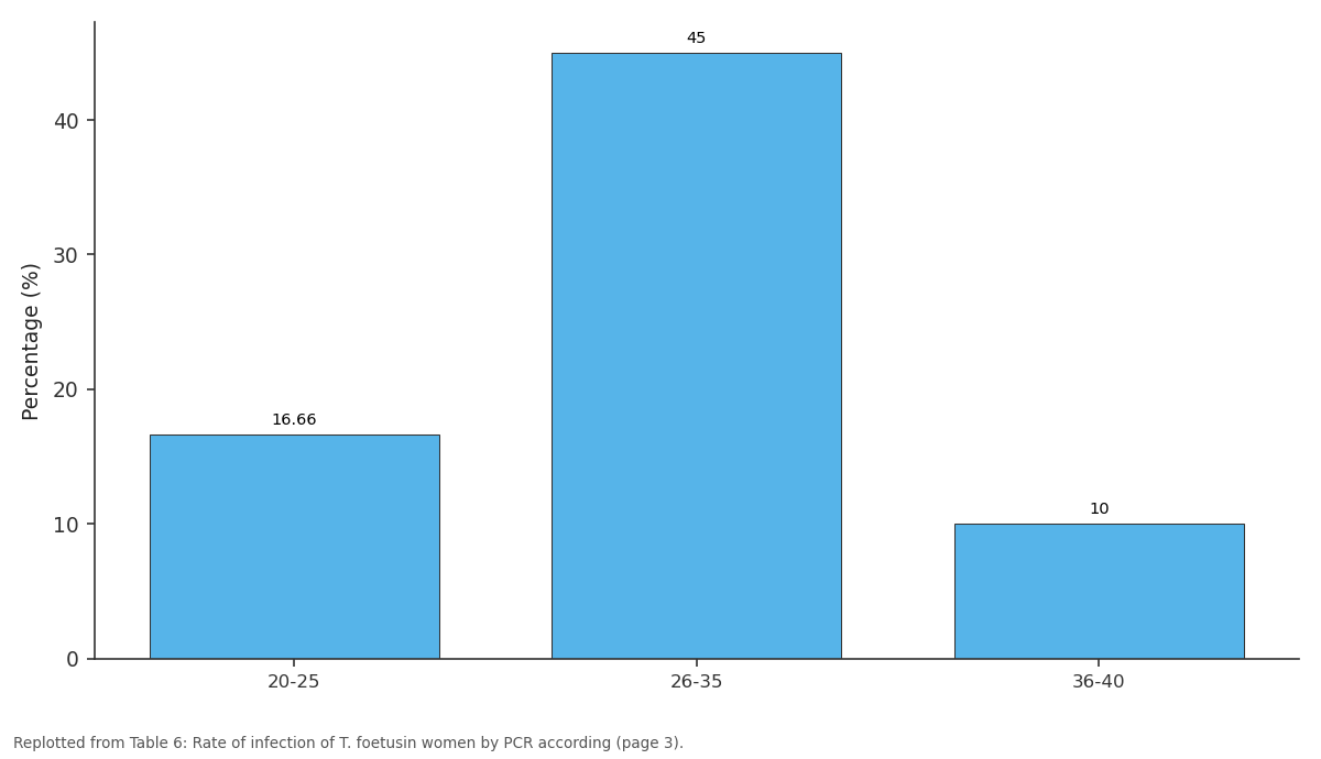
Percentage (%): 20-25 vs 26-35 vs 36-40
Replotted from Table 6 (page 3) of the original paper. Every bar is the paper's own table value.
View source evidence ↗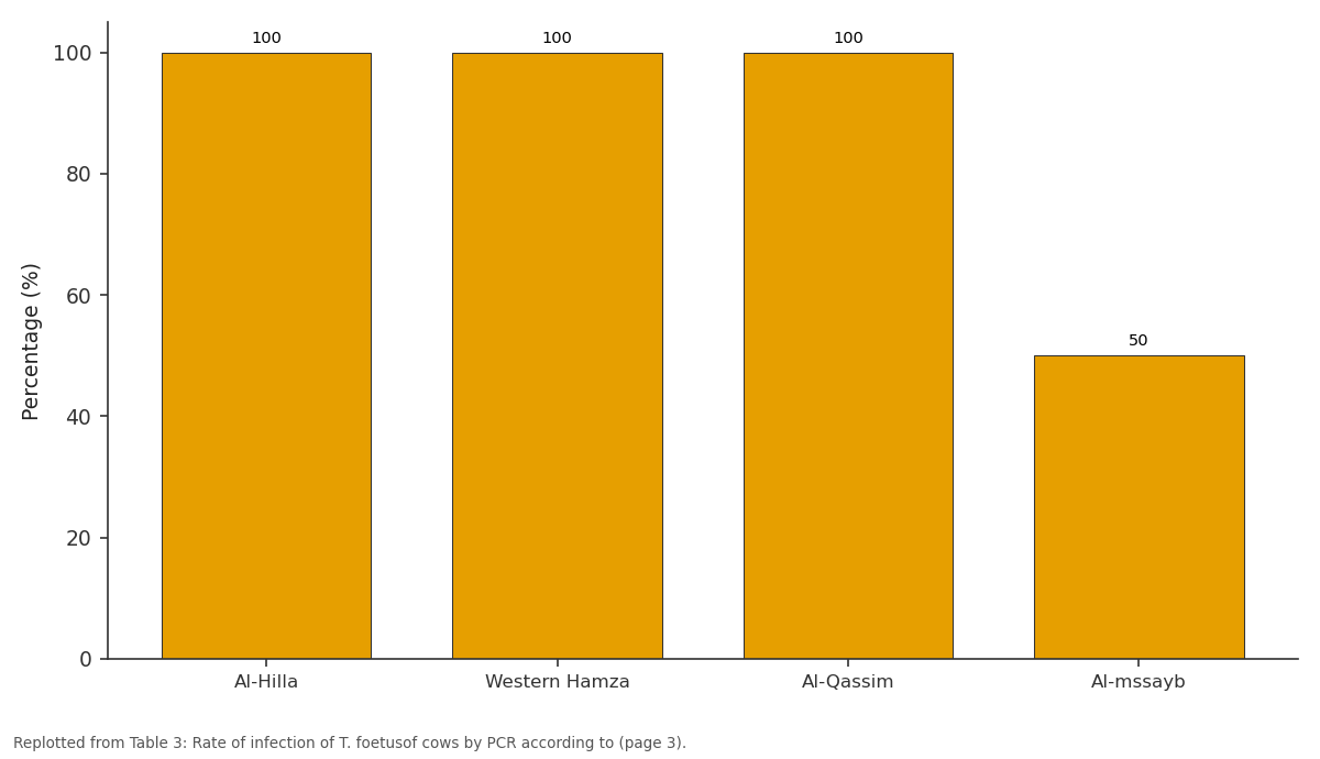
Percentage (%): Al-Hilla vs Western Hamza vs Al-Qassim vs Al-mssayb
Replotted from Table 3 (page 3) of the original paper. Every bar is the paper's own table value.
View source evidence ↗Figures & tables
From the original paper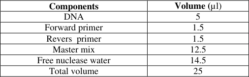 Table 1
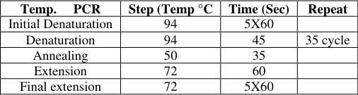 Table 2
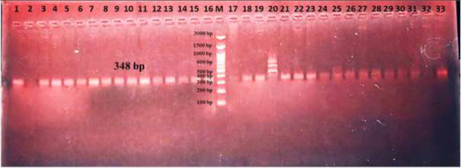 Figure 1
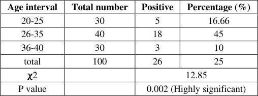 Table 6
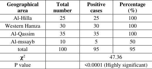 Table 3
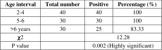 Table 4
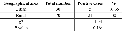 Table 5
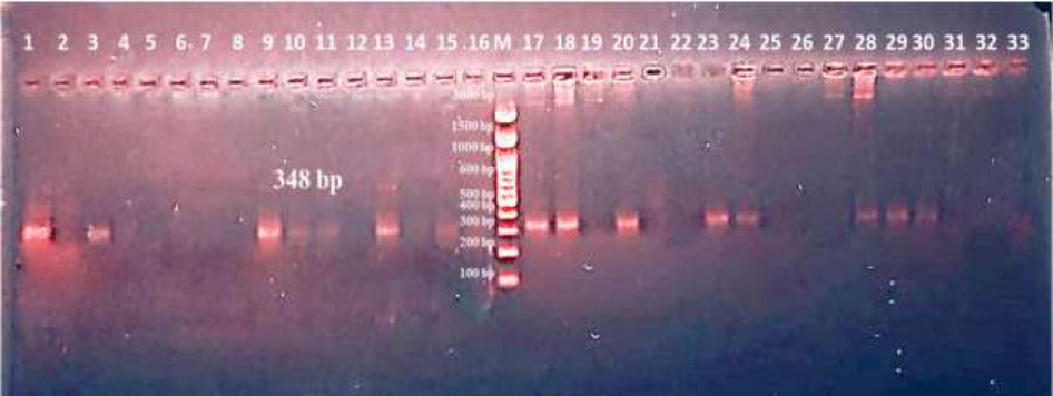 Figure 2
Introduction
The protozoan Trichomonas foetus causes sterility only temporarily, with inflammation of the vagina, cervix and uterus. It might result in fetal mortality in early pregnancy but abortion is rarely its cause. There are usually no gross lesions. Microscopically, there is fetal placental oedema together with a mild lymphocytic and histiocytic chorionitis and focal areas of trophoblast necrosis. Fetal pneumonia can be seen in about 50% of cases, with neutrophils, macrophages and sometimes multinucleate giant cells in the bronchioles [1] . Trichomonads are flagellated protozoa which have a worldwide distribution. Some species are nonpathogenic, commensal in the digestive systems of animals and humans, while other species are pathogenic parasites. T. foetus are found in both animals and humans. T. foetus of animals (sheep, goats, bulls, camels, cats, and dogs) is well-documented to affect their reproductive efficiency. T. foetus is a human pathogen that affects millions of individuals worldwide by a variety of reproductive and nonreproductive tract diseases. Foetus causes human trichomoniasis, which is considered a sexually transmitted disease (STD) [2,3] . The clinical pathogenicity of T. foetus in humans includes vaginitis, urethritis, and cervicitis of the upper reproductive tract. Symptoms may include vulvovaginal discharge with a purulent or bloody or yellowish sample, male infertility, early preterm birth, ectopic pregnancy, and serious consequences or symptoms during pregnancy. The pathogenicity of T. foetus in women is considered to be "trichomoniasis", a disease that often goes unreported [4] . There is no research to identify T. foetus by molecular and microscopic techniques in the women's reproductive tract with genital importance [5,6] . T. foetus. are simple and particle balanoposthitis. They are living parasitic flagellated protozoa, belonging to the genus T. from the class of flagellates and the subclass of Trichomonadida. They are found in the colon, reproductive, and upper respiratory tracts of the host vertebrate.
The multiple host species of T. foetus include humans, ruminants, birds, racing pigeons, pet birds, cattle, animal exhibition, and wild animals. T. foetus also infect the urinary tract of humans, cattle, pigs, and horses, causing the known disease trichomoniasis. The path of transmission and the risk factors for trichomoniasis in humans include contaminated food and water, poor hygiene, hand-to-hand contact, animal bites, contact with infected animals, and sexual contact. It is reported through histories of patients diagnosed with vaginitis or balanoposthitis that they have had contact with animals with a purulent or bloody discharge [7][8][9] . The morphology of trichomonads is mainly divided into three generally accepted structural features that are present in all known species within this group: the presence and morphology of the characteristic axostyle, the body shape, and the presence and type of the undulating membrane. The classification of trichomonads is still subject to constant divergence, and only DNA-related studies are a more accurate tool for species identification. This tool is quickly becoming available in regions where this ability was not offered before [10][11][12] . The phylum Euglenozoa is closely related to this phylum and is represented by species belonging to trypanosomatids. Trypanosomatids are responsible for important human diseases such as Chagas disease, Leishmaniasis, and African sleeping sickness. Both phyla share several ultrastructural cell characteristics. Trichomonads, which are a type of protist, have five to six free flagella (from axostyle, pelta, costa, undulating membrane, and haemastricum, if present) and a prominent parabasal filament is visible in the cytoplasm. Trichomonads are cells that lack mitochondria and instead possess hydrogenosomes and paravirus, similar to parabasalids. The list of trichomonads from different ecosystems is not complete and what is known is only the tip of the iceberg [13][14][15] . T. foetus trophozoites have a life cycle marked by distinct cellular stages [16][17][18] . Recently, the life cycle of the organisms is thought to be relatively simple and direct. This occurs in the reproductive and urinary tracts of their hosts and has four distinct morphological stages: the trophozoite, the pseudocyst, the prepubescent cell, and the spherical cell. The trophozoite is pear-shaped, has at least three anterior flagella, and an undulating membrane. Studies aimed at elucidating the life cycle in various hosts incubated infected vaginal fluid from women-hosts of T. foetus with gerbils (Meriones unguiculatus) and demonstrated that infection was caused by the swallowed organism [19][20] . The present study was conducted to detect the presence of Trichomonas spp from reproductive tract of women and cows in Babylon Province, Iraq.
Materials and Methods
Materials and Methods
Subjects and samples
The study included the collection of 200 vaginal swabs (100 women and 100 cows). These samples were tested using microscopy, measuring trophozoites, and PCR that targeted the 18S rRNA gene of the protozoan. This study was conducted in collaboration with the gynecology clinics in three public hospitals: AL-Imam AL-Sadiq Hospital, Al-Hilla Teaching Hospital, and Babil Teaching Hospital for Maternity and Children, and several private gynecology clinics in Babil province; Al.Qassime, Al-Hashemite, Al-Hilla, and Mahaweel Districts. Different ages and different areas of the Babylon province were investigated. The study was lasted from August, 2023 until end of January, 2024.
Molecular study Extraction of DNA
The samples were subjected to Genaid DNA purification kit and instructions, in which 0.5 ml vaginal swab was placed in 1.5 ml centrifuge tube which were followed by the procedure of the kit. The extracted DNA was NanoDrop measured.
PCR
The complete PCR procedures are mentioned in Tables 1,2. A 1.5%-agarose gel electrophoresis was utilized at 80 volts for 60 mins. The products were visualized by a UV-machine.
Results
The results revealed the presence of the parasite in 26 (26%) of woman swabs and 95 (95%) of cow samples as detected by the PCR (Figure 1). For the geographical correlation, the findings demonstrated significant (p<0.05) connection between the prevalence rate and the sample collection regions, in which high rates 25/25 (100%) was detected in cows from Hilla City, The city center of the province capital. For the age of cows, 2-4 and 5-6 years old showed significant (p<0.05) high prevalence 40/40 (100%) and 30/30 (100%), respectively, when compared with this from older ages. For women, the findings recorded significant (p<0.05) higher prevalence rates according to the collection regions, in which higher rates (30%) 21 out of 70 was detected in women from rural. In urban rate detected (16.66%) 5 out of 30. For the age of women, 26-35 years old showed significant (p<0.05) high prevalence 18/40 (45%) in a comparison with this from different ages (Tables 6).
Discussion
Trichomonas foetus can be infected to women due to sexual transmission, which may cause vaginitis and cervicitis and subsequently infertility and abortion. The molecular detection of T. foetus in Iranian women with Bacterial vaginosis (BV) showed a high prevalence of 27.5% compared to wet mounts and culture, in which only 6.9% were negative for T. foetus.
In addition, their special attention to the wet preparations with respect to identifying the active mobility of the organisms and their quantitative abundance in this study. Their study represents that probably the majority of women examined for BV in their laboratory were over the phase of parasite growth and multiplication in vaginal secretions, and most of the samples were part of the late phase, beginning phase of the detection with the quickly art method amongst wet mount method. Nenoff et al. without reporting the prevalence, and another study in their results showed 1.69%, although the molecular technique used in this study was a real-time PCR method [21][22][23][24] . Little is known about the prevalence in the general population of symptomatic and asymptomatic individuals, about the prevalence in women and men looking for general medical consultation, and about the treatment response. The discrepancy in the prevalence rates of T. foetus is noticed, which may be due in part to the different methods used for detection. It is confirmed that the wet preparation and cultivation are not very sensitive. The highest rates of T. foetus are reported on cultures performed in microaerophilic and macronutrient-rich medium [25,26] . In Iraq, until today and to the best of our knowledge, there is no or little previous studies using molecular techniques to detect T. foetus from women or cows. In general, these methods are relatively few and have been used for the detection of T. foetus Some other studies have detected T. vaginalis with a different rate of infection [27,29] . Alkan et al.
detected Trichomonas in 32% of women with BV using the culture method on Diamond's media. On the other hand, Smayevsky et al. detected 8% T. vaginalis with BV-negative cultures by wet mount microscopy and the culture method on the modified Diamond's media. Based on the molecular method, our results showed that the prevalence of T. foetus among women was high. This variation in the rate of infection is due to being affected by several factors. Some are personal hygiene, social and demographic factors, behavioral lifestyle, and immunity [21,30] .
Conclusion
The present study shows important data about the presence of high rates in cows, which might be an important source for a zoonosis disease.
Figures and tables from the paper
| Components | Volume (µl) |
|---|---|
| DNA | 5 |
| Forward primer | 1.5 |
| Revers primer | 1.5 |
| Master mix | 12.5 |
| Free nuclease water | 14.5 |
| Total volume | 25 |
Table text extracted from the original PDF; superscript letters are shown inline. The original table image is below.
View original table image
| Temp. PCR | Step (Temp °C | Time (Sec) | Repeat |
|---|---|---|---|
| Initial Denaturation | 94 | 5X60 | |
| Denaturation | 94 | 45 | 35 cycle |
| Annealing | 50 | 35 | |
| Extension | 72 | 60 | |
| Final extension | 72 | 5X60 |
Table text extracted from the original PDF; superscript letters are shown inline. The original table image is below.
View original table image
| Age interval | Total number | Positive | Percentage (%) |
|---|---|---|---|
| 20-25 | 30 | 5 | 16.66 |
| 26-35 | 40 | 18 | 45 |
| 36-40 | 30 | 3 | 10 |
| total | 100 | 26 | 25 |
| 𝛘2 | 12.85 | ||
| P value | 0.002 (Highly significant) |
Table text extracted from the original PDF; superscript letters are shown inline. The original table image is below.
View original table image
| Geographical area | Total number | Positive cases | Percentage (%) |
|---|---|---|---|
| Al-Hilla | 25 | 25 | 100 |
| Western Hamza | 30 | 30 | 100 |
| Al-Qassim | 35 | 35 | 100 |
| Al-mssayb | 10 | 5 | 50 |
| total | 100 | 95 | 95 |
| 𝛘2 | 47.36 | ||
| P value | <0.0001 (Highly significant) |
Table text extracted from the original PDF; superscript letters are shown inline. The original table image is below.
View original table image
| Age interval | Total number | Positive | Percentage (%) |
|---|---|---|---|
| 2-4 | 40 | 40 | 100 |
| 5-6 | 30 | 30 | 100 |
| >6 years | 30 | 25 | 83.33 |
| 𝛘2 | 12.28 | ||
| P value | 0.002 (Highly significant) |
Table text extracted from the original PDF; superscript letters are shown inline. The original table image is below.
View original table image
| Geographical area | Total number | Positive cases | % |
|---|---|---|---|
| Urban | 30 | 5 | 16.66 |
| Rural | 70 | 21 | 30 |
| 𝛘2 | 1.94 | ||
| P value | 0.164 |
Table text extracted from the original PDF; superscript letters are shown inline. The original table image is below.
View original table image
References
21 in the original paper- Henriquez FL, Mooney R, Bandel T, Giammarini E, Zeroual M, Fiori PL, et al. Paradigms of protist/bacteria symbioses affecting human health: Acanthamoeba species and Trichomonas vaginalis. Front Microbiol. 2021 Jan 7;11:616213.
- Carneiro FP, Darós AC, Darós AC, de Castro TM, de Vasconcelos Carneiro M, Fidelis CR, et al. Cervical cytology of samples with Ureaplasma urealyticum, Ureaplasma parvum, Chlamydia trachomatis, Trichomonas vaginalis, Mycoplasma hominis, and Neisseria gonorrhoeae detected by multiplex PCR. Biomed Res Int. 2020 Jul 7;2020.
- Martínez CI, Iriarte LS, Salas N, Alonso AM, Pruzzo CI, dos Santos Melo T, et al. Prolonged survival of venereal Trichomonas foetus parasite in the gastrointestinal tract, bovine fecal extract, and water. Microbiol Spectrum. 2023 Dec 12;11(6).
- Ahmed A, Ijaz M, Ayyub RM, Ghaffar A, Ghauri HN, Aziz MU, et al. Balantidium coli in domestic animals: An emerging protozoan pathogen of zoonotic significance. Acta Trop. 2020 Mar 1;203:105298.
- Jones KR, Garcia GW. Teaching of Veterinary Parasitology to University students in Trinidad, West Indies; are our local wildlife species neglected?. Braz J Biol. 2021.
- Manny AR, Hetzel CA, Mizani A, Nibert ML. Discovery of a Novel Species of Trichomonasvirus in the Human Parasite Trichomonas vaginalis Using Transcriptome Mining. Viruses. 2022;14:548.
- Kučerová J, Zdrha A, Shinde A, Harant K, Hrdý I, Tachezy J. The divergent ER-mitochondria encounter structures (ERMES) are conserved in parabasalids but lost in several anaerobic lineages with hydrogenosomes. BMC Biol. 2023 Nov 15;21(1):259.
- Manny AR, Hetzel CA, Mizani A, Nibert ML. Discovery of a Novel Species of Trichomonasvirus in the Human Parasite Trichomonas vaginalis Using Transcriptome Mining. Viruses. 2022;14:548.
- Maciejowski WJ, Gile GH, Jerlström-Hultqvist J, Dacks JB. Ancient and pervasive expansion of adaptin-related vesicle coat machinery across Parabasalia. Int J Parasitol. 2023 Apr 1;53(4):233-45.
- Stairs CW, Táborský P, Salomaki ED, Kolisko M, Pánek T, Eme L, et al. Anaeramoebae are a divergent lineage of eukaryotes that shed light on the transition from anaerobic mitochondria to hydrogenosomes. Curr Biol. 2021 Dec 20;31(24):5605-12.
- Beri D, Yadav P, Devi HN, Narayana C, Gadara D, Tatu U. Demonstration and characterization of cyst-like structures in the life cycle of Trichomonas vaginalis. Front Cell Infect Microbiol. 2020 Jan 14;9:430.
- Hsu HM, Yang YY, Huang YH, Chu CH, Tu TJ, Wu YT, et al. Distinct features of the host-parasite interactions between nonadherent and adherent Trichomonas vaginalis isolates. PLoS Negl Trop Dis. 2023 Jan 3;17(1).
- Riestra AM, de Miguel N, Dessi D, Simoes-Barbosa A, Mercer FK. Trichomonas vaginalis: lifestyle, cellular biology, and molecular mechanisms of pathogenesis. In: Lifecycles of Pathogenic Protists in Humans. Cham: Springer International Publishing; 2022. p. 541-617.
- Al-Ethafa LFM. Trichomonas vaginals: A review on pathogenicity, diagnosis and treatment. Sci Arch. 2021.
- Shiratori M, Patel A, Gerhold RW, Sullivan SA, Carlton JM. Persistent Trichomonas vaginalis infections and the pseudocyst form. Trends Parasitol. 2023 Oct 6.
- Ismael S. Prevalence of Trichomoniasis and Vulvovaginal Candidiasis among Married Women in Duhok City, Kurdistan Region, Iraq. Arch Razi Inst. 2024.
- Pouryasin M, Mousavi AS, Pakravesh J, Kamel ZD, Nooriardabili S, Khodadad S, et al. The Prevalence of HPV and non-HPV STIs Among Iranian Women and Assessment of the HPV/non-HPV STIs Co-infection on Cervical Cell Changes. J Obstet Gynecol Cancer Res. 2022 Jul 7;7(5):405-13.
- Yasin J, Ayalew G, Dagnaw M, Shiferaw G, Mekonnen F. Vulvovaginitis prevalence among women in gondar, Northwest Ethiopia: Special emphasis on aerobic vaginitis causing bacterial profile, antimicrobial susceptibility pattern, and associated factors. Infect Drug Resist. 2021 Nov 2:4567-80.
- Tamrat R, Kasa T, Sahilemariam Z, Gashaw M. Prevalence and factors associated with sexually transmitted infections among Jimma University students, Southwest Ethiopia. Int J Microbiol. 2020 Nov 10;2020:1-6.
- Hassan BJ. Detection of Trichomonas spp. from penile bulls fluid in Iraq. Asian J Infect Dis. 2020.
- Mohammed MO, Ali KN, Dyary HO. Bovine Trichomoniasis. One Health Triad, Unique Scientific Publishers, Faisalabad, Pakistan. 2023;2:107-13.