
Research article · 2022 · Full text
International Journal of Advanced Chemistry ResearchApplications of raman spectroscopy in biomedical diagnostics
Volume 4(2) · Pages 411–413 · DOI: 10.33545/26646781.2022.v4.i2f.200 ↗
Research areasSpectroscopy Techniques in Biomedical and Chemical Research
Abstract
Author-writtenRaman spectroscopy has emerged as a powerful analytical tool in biomedical diagnostics due to its non-invasive nature, high sensitivity, and ability to provide detailed molecular information. This review explores the various applications of Raman spectroscopy in the biomedical field, including cancer detection, microbial identification, and the analysis of biochemical changes in tissues and cells. We discuss the principles of Raman spectroscopy, recent technological advancements, and the integration of Raman techniques with other modalities to enhance diagnostic capabilities. The potential challenges and future directions of Raman spectroscopy in clinical diagnostics are also highlighted.
Summary
Source-checkedPlain-language reading of the full paper, prepared with AI and checked against the text. Each point links to the authors' own words.
Why it matters
- Raman spectroscopy is a valuable tool for biomedical diagnostics because it is non-invasive, highly sensitive, and provides detailed molecular data. View evidence ↗
- Compared to traditional diagnostic methods, Raman spectroscopy is better suited for clinical use because it requires minimal sample preparation. View evidence ↗
The question
- The authors aim to review the applications of Raman spectroscopy in cancer detection, microbial identification, and the analysis of biochemical changes in cells and tissues. View evidence ↗
What they did
- The technique works by using a laser to scatter monochromatic light off molecules in a sample. View evidence ↗
- The process involves inelastic scattering, which creates an energy shift in photons that corresponds to the vibrational energy levels of the molecules. View evidence ↗
- Sensitivity and spatial resolution have been improved through advancements like SERS, TERS, and CARS. View evidence ↗
What they found
- Raman spectroscopy can detect breast cancer by identifying changes in the protein and lipid content of the tissues. View evidence ↗
- Near-infrared Raman spectroscopy has been shown by Huang et al. (2003) to differentiate between cancerous and normal lung tissues. View evidence ↗
- SERS was used by Gao et al. (2017) to distinguish different strains of E. coli from other bacteria. View evidence ↗
- Raman spectroscopy can be used to detect the fungal pathogen Candida albicans in clinical samples, as shown by Berus et al. (2017). View evidence ↗
- In cardiovascular research, Raman spectroscopy can differentiate between unstable and stable atherosclerotic plaques by analyzing protein and lipid content. View evidence ↗
- Raman spectroscopy has been used to track the differentiation of human mesenchymal stem cells into osteoblasts by identifying biochemical markers. View evidence ↗
- Diagnostic capabilities are improved when Raman spectroscopy is integrated with fluorescence microscopy or optical coherence tomography (OCT). View evidence ↗
- The use of artificial intelligence and machine learning allows for the identification of subtle spectral patterns in complex Raman spectra associated with diseases. View evidence ↗
What it means
- The authors conclude that Raman spectroscopy is a promising tool for personalized medicine, as it can help develop treatment plans based on a patient's specific molecular information. View evidence ↗
- A primary limitation of the technique is the weak intensity of the Raman signal, which can restrict detection limits and sensitivity. View evidence ↗
- The complexity of biological Raman spectra makes data interpretation challenging, requiring advanced computational methods. View evidence ↗
Visual summary
1 visualsIllustrations explain the paper; they do not show study data.
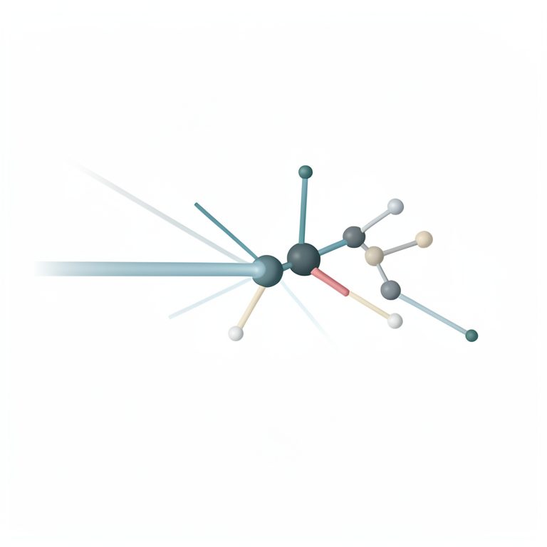
Raman Scattering Process
AI-generated conceptual illustration (a single organic molecule in a transparent biological sample). It explains the idea and does not show study data.
View source evidence ↗Introduction
Raman spectroscopy, based on the inelastic scattering of photons by molecules, has become an invaluable tool in biomedical diagnostics. Its ability to provide detailed molecular fingerprints makes it particularly useful for identifying biochemical changes in tissues and cells, detecting pathogens, and diagnosing various diseases. Unlike traditional diagnostic methods, Raman spectroscopy is non-invasive and requires minimal sample preparation, making it ideal for clinical applications. This review provides a comprehensive overview of the principles of Raman spectroscopy, its technological advancements, and its diverse applications in biomedical diagnostics.
Principles of raman spectroscopy
Raman spectroscopy relies on the scattering of monochromatic light, usually from a laser, by molecules in a sample. When photons interact with the sample, most are elastically scattered (Rayleigh scattering), but a small fraction undergoes inelastic scattering (Raman scattering). This inelastic scattering results in a shift in the energy of the scattered photons, corresponding to the vibrational energy levels of the molecules. The resulting Raman spectrum provides a unique molecular fingerprint that can be used to identify and characterize the chemical composition of the sample. Recent advancements in Raman spectroscopy, such as surface-enhanced Raman spectroscopy (SERS), tip-enhanced Raman spectroscopy (TERS), and coherent anti-Stokes Raman scattering (CARS), have significantly improved its sensitivity and spatial resolution. These techniques enhance the Raman signal, enabling the detection of low-concentration analytes and providing high-resolution imaging capabilities.
Applications in cancer detection
Raman spectroscopy has shown great promise in the early detection and diagnosis of various cancers. By analyzing the biochemical composition of tissues, Raman spectroscopy can distinguish between normal and malignant cells. For instance, studies have demonstrated the ability of Raman spectroscopy to detect breast cancer by identifying changes in lipid and protein content in breast tissues. Similarly, Raman spectroscopy has been used to diagnose cervical cancer by detecting biochemical alterations in cervical cells. One notable application is in the detection of skin cancer, where Raman spectroscopy can non-invasively identify malignant melanoma and differentiate it from benign lesions. Lieber et al. (2008) [1] demonstrated the potential of Raman spectroscopy in distinguishing between melanoma and nonmelanoma skin cancers with high sensitivity and specificity. The integration of Raman spectroscopy with confocal microscopy has further enhanced its ability to provide detailed morphological and biochemical information, aiding in accurate cancer diagnosis. In addition to skin cancer, Raman spectroscopy has been used for detecting other types of cancers such as oral, lung, and prostate cancers. For example, Huang et al. (2003) [3] demonstrated that near-infrared Raman spectroscopy could be used to differentiate between normal and cancerous lung tissues, providing a rapid and non-invasive diagnostic tool. Similarly, Raman spectroscopy has been employed to identify biochemical changes in prostate tissues, aiding in the early detection of prostate cancer.
Applications in microbial identification
Raman spectroscopy is also a powerful tool for the rapid identification of microbial pathogens. Traditional methods of microbial identification, such as culture and biochemical tests, are time-consuming and may not always provide accurate results. Raman spectroscopy, on the other hand, offers a rapid and reliable alternative by providing unique spectral fingerprints for different microbial species. SERS, in particular, has been extensively used for microbial identification due to its enhanced sensitivity. By using metal nanoparticles to amplify the Raman signal, SERS can detect low concentrations of bacteria and viruses. For example, Gao et al. (2017) [2] utilized SERS to identify different strains of Escherichia coli and distinguish them from other bacterial species. Similarly, SERS has been used to detect viral pathogens, such as influenza and herpes simplex virus, by targeting specific viral proteins and nucleic acids. The application of Raman spectroscopy in microbial identification extends beyond bacteria and viruses. It has also been used to identify fungal infections, which are often difficult to diagnose using traditional methods. For instance, Berus et al. (2017) [4] demonstrated the use of Raman spectroscopy to detect Candida albicans, a common fungal pathogen, in clinical samples. This rapid and accurate identification of pathogens can significantly improve patient outcomes by enabling timely and appropriate treatment.
Analysis of biochemical changes in tissues and cells
Raman spectroscopy provides detailed information about the biochemical composition of tissues and cells, making it a valuable tool for studying disease progression and monitoring treatment responses. By analyzing the Raman spectra of tissues, researchers can identify biochemical markers associated with various diseases. In cardiovascular research, Raman spectroscopy has been used to analyze atherosclerotic plaques and assess their composition. Huang et al. (2010) demonstrated that Raman spectroscopy could differentiate between stable and unstable plaques based on their lipid and protein content. This capability is crucial for assessing the risk of plaque rupture and subsequent cardiovascular events. In neurodegenerative diseases, Raman spectroscopy has been employed to study the aggregation of amyloid proteins, which are implicated in conditions such as Alzheimer's and Parkinson's disease. Raman spectroscopy can detect the structural changes in amyloid fibrils and provide insights into the mechanisms of protein aggregation. Additionally, Raman spectroscopy has been used to monitor the effects of therapeutic interventions on amyloid aggregation, aiding in the development of new treatments. Raman spectroscopy has also been used to study metabolic changes in cells. For example, it has been used to monitor the differentiation of stem cells by detecting changes in their biochemical composition. Noting et al. (2017) [5] used Raman spectroscopy to track the differentiation of human mesenchymal stem cells into osteoblasts, identifying specific biochemical markers associated with different stages of differentiation. This ability to monitor cellular processes in real-time provides valuable insights into cellular biology and disease mechanisms.
Technological advancements and integration with other modalities
Recent technological advancements have significantly enhanced the capabilities of Raman spectroscopy in biomedical diagnostics. The development of portable and handheld Raman devices has facilitated point-of-care diagnostics, allowing for the rapid and non-invasive assessment of patients. These portable devices are particularly useful in resource-limited settings, where access to advanced laboratory facilities may be limited. The integration of Raman spectroscopy with other imaging modalities, such as optical coherence tomography (OCT) and fluorescence microscopy, has further improved its diagnostic capabilities. Combined Raman-OCT systems provide complementary information about tissue structure and composition, enabling more accurate disease diagnosis. For instance, combined Raman-OCT has been used to assess the biochemical and structural changes in retinal tissues, aiding in the diagnosis of retinal diseases. Fluorescence-Raman dual-modal imaging systems have also been developed to provide both molecular and functional information. By combining the high sensitivity of fluorescence imaging with the molecular specificity of Raman spectroscopy, these systems offer a comprehensive approach to disease diagnosis and monitoring. For example, fluorescence-Raman imaging has been used to study the distribution of drugs in tissues, providing insights into drug delivery and efficacy. Advancements in computational techniques and data analysis have also played a significant role in enhancing the utility of Raman spectroscopy. Machine learning algorithms and artificial intelligence have been applied to analyze complex Raman spectra, enabling the identification of subtle spectral patterns associated with diseases. These advanced data analysis techniques have improved the accuracy and reliability of Raman spectroscopy in clinical diagnostics.
Challenges and future directions
Despite its numerous advantages, there are several challenges associated with the clinical implementation of Raman spectroscopy. One of the primary challenges is the weak intensity of the Raman signal, which can limit the sensitivity and detection limits of the technique. Although techniques like SERS and TERS have improved sensitivity, further advancements are needed to enhance the signal-tonoise ratio and reduce background interference. Another challenge is the complexity of data analysis and interpretation. The Raman spectra of biological samples can be highly complex, requiring advanced algorithms and computational methods for accurate analysis. The development of standardized protocols and robust data analysis tools is essential for the widespread adoption of Raman spectroscopy in clinical diagnostics. Future research should focus on addressing these challenges and exploring new applications of Raman spectroscopy in biomedical diagnostics. The development of novel Raman-active probes and nanoparticles can enhance the specificity and sensitivity of the technique. Additionally, the integration of machine learning and artificial intelligence with Raman spectroscopy can improve data analysis and enable the identification of subtle spectral patterns associated with diseases. The application of Raman spectroscopy in personalized medicine is another promising area for future research. By providing detailed molecular information about individual patients, Raman spectroscopy can aid in the development of personalized treatment plans. For example, Raman spectroscopy can be used to monitor the response of tumors to chemotherapy, enabling the optimization of treatment regimens based on individual patient profiles.
Conclusion
Raman spectroscopy has emerged as a powerful tool in biomedical diagnostics, offering non-invasive, rapid, and highly specific analysis of biological samples. Its applications in cancer detection, microbial identification, and the analysis of biochemical changes in tissues and cells demonstrate its versatility and potential in clinical diagnostics. Recent technological advancements and the integration of Raman spectroscopy with other imaging modalities have further enhanced its diagnostic capabilities. However, challenges related to sensitivity, data analysis, and clinical implementation remain. Continued research and technological innovations are needed to overcome these challenges and fully realize the potential of Raman spectroscopy in biomedical diagnostics.
References
5 in the original paper- Lieber CA, Majumder SK, Ellis DL, Billheimer DD, Mahadevan-Jansen A. In vivo nonmelanoma skin cancer diagnosis using Raman microspectroscopy. Lasers in Surgery and Medicine. 2008;40(7):461-467.
- Gao R, Cheng Z, deMello AJ. On-chip microfluidic integration of SERS detection. Journal of Micromechanics and Microengineering. 2017;27(12):123002.
- Huang Z, McWilliams A, Lui H, et al. Near-infrared Raman spectroscopy for optical diagnosis of lung cancer. International Journal of Cancer. 2003;107(6):1047-1052.
- Berus L, Matwijczuk A, Gagoś M, Szymczuk D, Cwalina B. Application of Raman spectroscopy for the identification of Candida albicans. European Journal of Clinical Microbiology & Infectious Diseases. 2017;36(6):1163-1170.
- Noting JT, Williams D, Kendall CA, et al. Raman spectroscopy as a tool for real-time monitoring of biochemical changes during the differentiation of human mesenchymal stem cells. Analytical Chemistry. 2017;89(6):3223-3231
Cited by 1
From OpenAlex, OpenCitations and reference lists read by Scinovex
- Biophotonics-Driven Advances in Biomedical Imaging: Evolving Techniques, Ongoing Challenges, and Forward-Looking Strategies ↗
M Tasdik Hasan, Himel Dey Shikder, Abir Tirtha Das, Md Kawsar Khan, Samiul Hassan, Md. Sayed Imam Mojumder et al. · 2026
Biophotonics, positioned at the intersection of photonics and life sciences, has rapidly transformed biomedical imaging by enabling minimally invasive visualization of biological structures and functions across molecular, cellular, and tissue scales. While individual imaging technologies such as Raman spectroscopy, optical coherence tomography, fluorescence-lifetime imaging, and multiphoton microscopy have been extensively studied, their collective evolution reveals a deeper set of transitions…
❝ 0