
Research article · 2024 · Full text
International Journal of Veterinary Sciences and Animal HusbandryCase report of surgical correction for persistence of congenital penile frenulum in a dog
Volume 9(4) · Pages 93–95 · DOI: 10.22271/veterinary.2024.v9.i4b.1516 ↗
Research areasUrological Disorders and TreatmentsGenital Health and DiseaseOral and Craniofacial Lesions
Abstract
Author-writtenA ten-month-old, 14.4 kg dog of unidentified breed presented to Devashraya Animal Hospital by Sarvodaya Foundation, Faridabad, Haryana, with a complaint of protruding penis and difficulty urinating for the past 8-12 hours. Clinical examination revealed a congenital penile frenulum causing ventral deviation of the penis along with paraphimosis. Surgical intervention was performed under general anesthesia, involving resection of the persistent frenulum and repair of resulting wounds with absorbable sutures. Postoperatively, the dog received antimicrobial, analgesic therapy, and topical ointment. The dog recovered uneventfully, with resolution of symptoms and normal urination by the 3rd postoperative day. Persistent penile frenulum, though often asymptomatic, can lead to urinary and reproductive issues in animals and is heritable.
Summary
Source-checkedPlain-language reading of the full paper, prepared with AI and checked against the text. Each point links to the authors' own words.
Why it matters
- External genitalia anomalies in small animals are rare but can cause significant urinary dysfunction and breeding difficulties. View evidence ↗
- The penile frenulum is a thin band of connective tissue that normally runs from the prepuce to near the tip of the penis. View evidence ↗
- In males, the penile frenulum typically ruptures spontaneously during puberty, a process influenced by androgen levels. View evidence ↗
The question
- The objective was to surgically correct a congenital penile frenulum that caused penile deviation and paraphimosis in a dog. View evidence ↗
What they did
- Anesthesia was achieved using intramuscular butorphanol (0.2 mg/kg) and atropine sulfate (0.04 mg/kg), followed by intravenous propofol (2-4 mg/kg) and isoflurane maintenance. View evidence ↗
- Perioperative care included intramuscular ceftriaxone (20 mg/kg) and meloxicam (0.2 mg/kg). View evidence ↗
- The surgical procedure involved resecting the persistent frenulum at the urethral orifice and the preputial mucosa near the bulbus glandis. View evidence ↗
- Wounds were closed using size 5-0 Polygactin-910 synthetic absorbable sutures to prevent adhesions. View evidence ↗
- The penis was repositioned into the preputial cavity and secured with a temporary purse-string suture. View evidence ↗
What they found
- The dog presented with a protruding penis and difficulty urinating for a period of 8-12 hours. View evidence ↗
- Examination revealed thick connective tissue extending from the urethral opening to the preputial mucosa, causing ventral deviation and paraphimosis. View evidence ↗
- The dog recovered without complications, and normal urination was restored by the 3rd postoperative day following the removal of the purse-string suture. View evidence ↗
What it means
- The authors conclude that surgical intervention is necessary when the penile frenulum fails to rupture spontaneously. View evidence ↗
- Because the condition is heritable, the authors recommend that males with a repaired persistent frenulum be castrated. View evidence ↗
Visual summary
1 visualsIllustrations explain the paper; they do not show study data.
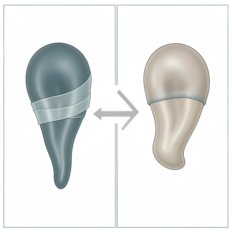
Anatomy of Persistent Penile Frenulum
AI-generated conceptual illustration (canine penis and prepuce). It explains the idea and does not show study data.
View source evidence ↗Figures & tables
From the original paper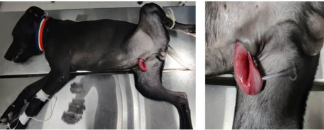 Figure 1
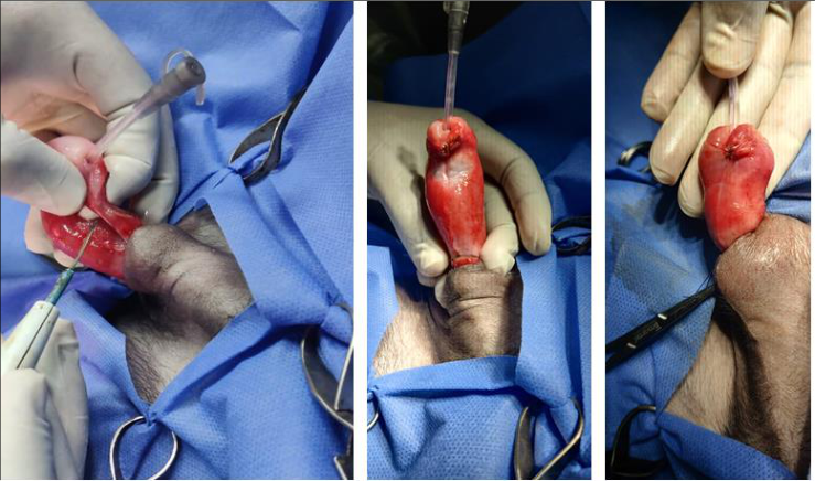 Figure 2
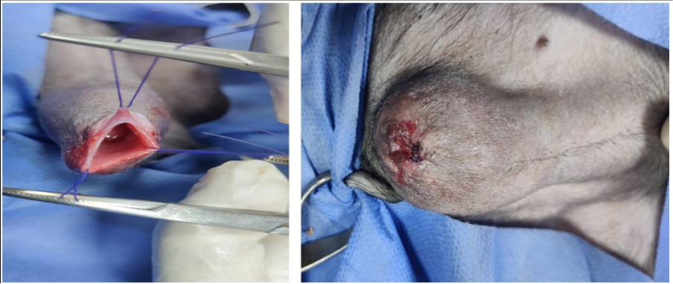 Figure 3
Introduction
Preputial and penile issues in young dogs arise either from developmental abnormalities or from accidents causing trauma. (Smith and Gourley, 1990) [6] . Anomalies of the external genitalia in small animals are infrequently documented. These abnormalities can stem from congenital or traumatic causes, leading to issues such as urinary dysfunction, breeding difficulties, or both. Examples of these abnormalities encompass acquired or congenital phimosis and paraphimosis, as well as malformations arising from irregular closure of the urogenital folds and ventral body wall. (Ader and Hobson, 1978; Smith and Gourley, 1990; Boothe, 1993) [1,5,2] . Reconstructive surgery may be necessary for congenital abnormalities to prevent recurring urinary issues. However, repairing these defects solely to facilitate successful breeding should be discouraged due to the insufficient data on the heritability of such conditions. (Olsen and Salwei, 2001) [4] . The penile frenulum is a slender band of connective tissue located on the underside of the penis, running from the prepuce to near the tip of the exposed part of the penis. (Papazoglou and Kazakos, 2002; Wolfe and Rodning, 2009) [5,8] . The spontaneous rupture of the penile frenulum typically occurs during the pubertal period in males, influenced by androgen levels. (Boothe, 2008; Papazoglou and Kazakos, 2002) [3,5] .
Clinical Examination and Diagnosis
A ten-month-old puppy, weighing 14.4 kg and of unidentified breed, was brought to Devashraya Animal Hospital by Sarvodaya Foundation in Faridabad, Haryana. The primary complaint was that its penis was protruding and unable to retract into the prepuce, accompanied by difficulty urinating over the past 8-12 hours. The clinical examination revealed normal physiological parameters. However, upon inspection of the penile region, it was observed that the puppy had a prominent thick connective tissue attaching from the tip of the penis just below the urethral opening to the preputial mucosa at the level of bulbus glandis area, causing the cranial portion of the penis to protrude. This condition was diagnosed as ventral deviation of the penis causing paraphimosis, attributed to a congenital penile frenulum that had not resolved (Fig. 1). The decision was made to proceed with surgical intervention to correct the cause of the penile deviation.
Surgical Intervention and Perioperative Procedure
The dog was preanesthetized with butorphanol and atropine sulfate at a dose rate of 0.2 mg/kg and 0.04 mg/kg body weight, respectively, administered intramuscularly. Anesthesia was induced with propofol at a rate of 2-4 mg/kg body weight slowly administered intravenously, followed by maintenance with isoflurane to achieve the desired effect via inhalation. The dog was positioned in dorsal recumbency and prepared for surgery. Perioperative intramuscular antibiotic and analgesic therapy included ceftriaxone @ 20 mg/kg body weight and meloxicam @ 0.2 mg/kg body weight, respectively. An infant feeding tube of size 10FG was used to catheterize the urethra to maintain its patency. The persistent frenulum was resected from two sites: near its attachment at the urethral orifice and at the mucosa of the prepuce near the bulbus glandis region. The tissue was thick and highly vascular, and bleeding was managed using direct pressure and judicious use of electrocautery. This freed the penis from its abnormal attachment, resolving the ventral deviation. The wounds resulting from transection of the persistent frenulum and abnormal tissue were meticulously repaired by suturing with synthetic absorbable suture (Polygactin-910, size 5-0) to minimize the risk of adhesion formation between the glans penis and the visceral preputial mucosa (Fig. 2). The prolapsed penis was then repositioned inside the preputial cavity, followed by the application of a temporary pursestring suture to narrow the preputial opening and secure the penis inside the prepuce until inflammation subsided (Fig. 3). The dog continued to receive intramuscular antimicrobial and analgesic therapy (ceftriaxone @ 20 mg/kg body weight and meloxicam @ 0.2 mg/kg body weight, respectively) and an antimicrobial steroid ointment infused into the preputial cavity for 5 days.
Outcome and Discussion
The dog recovered without complications, with the condition resolved and normal urination occurring after removal of the purse-string suture on the 3rd day post-operation. Diagnosis of persistent penile frenulum typically involves visual inspection, noting connective tissue along the ventral penile region extending from the prepuce towards the tip of the exposed part of the penis. When spontaneous rupture does not occur, surgical intervention is necessary for treatment.
In dogs and ruminants, persistent penile frenulum often presents asymptomatically, though it can lead to dysuria and difficulty with copulation (Volpato et al., 2010) [7] . Persistent penile frenulum is considered a heritable condition. Therefore male with a repaired persistent frenulum should be castrated.
Acknowledgement
AcknowledgmentsThanks to the Managing Director and the team of Devashraya Animal Hospital by Sarvodaya Foundation, Faridabad, Haryana for providing support and facilities require for the surgical intervention and post-operative care.
References
5 in the original paper- Ader PL, Hobson HP. Hypospadias: A review of the veterinary literature and a report of three cases in the dog. J Am Anim. Hosp. Assoc. 1978;14(6):721-727.
- Olsen D, Salwei R. Surgical correction of a congenital preputial and penile deformity in a dog. J Am Anim. Hosp. Assoc. 2001;37(2):187-192.
- Papazoglou LC, Kazakos GM. Surgical conditions of the canine penis and prepuce. Compend Contin. Educ. Pract. Vet. 2002;24(3):204-219.
- Smith MM, Gourley IM. Preputial reconstruction in a dog. J Am Vet Med Assoc. 1990;196(9):1493-1496.
- Volpato R, Ramos RS, Magalhaes LCO, Lopes MD, Souza DB. Review on disorders of the penis and prepuce in dogs. Vet Zootec. 2010;17(3):312-323.
Cited by 1
From OpenAlex, OpenCitations and reference lists read by Scinovex
- Male Dromedary Reproductive Emergencies: Clinical Presentation, Diagnosis, Management and Prognosis ↗
Ahmed Ali, Derar R. Derar · Animals · 2026
) threaten fertility, health, and welfare but remain poorly documented. This review consolidates knowledge on the diagnosis, management, and prognosis of acute reproductive pathologies, including traumatic injuries (testicular trauma, penile hematoma, and preputial laceration), obstructive conditions (urethral obstruction, phimosis, and paraphimosis), vascular emergencies (spermatic cord torsion, priapism), inflammatory diseases (orchitis, epididymitis, pizzle rot), congenital anomalies (persistent frenulum), iatrogenic complications, and…
❝ 0PDF