
Research article · 2024 · Full text
International Journal of Veterinary Sciences and Animal HusbandryManagement of dystocia and faecolith in cat
Volume 9(3) · Pages 609–610 · DOI: 10.22271/veterinary.2024.v9.i3i.1483 ↗
Research areasVeterinary Medicine and SurgeryIntestinal Malrotation and Obstruction DisordersPediatric Urology and Nephrology Studies
Abstract
Author-writtenA 3-year-old cat was presented to Department of veterinary surgery and radiology, Veterinary Clinical complex, Mhow with the history of bleeding from vagina and constipation since 4 days. X-ray examination revealed presence of fetus in uterus and fecoliths inside intestine. Surgical intervention includes C-section and enterotomy to remove fecoliths. Cat recovered after post-operative care of 7 days.
Summary
Source-checkedPlain-language reading of the full paper, prepared with AI and checked against the text. Each point links to the authors' own words.
Why it matters
- Dystocia in cats is characterized by difficulty delivering kittens through the birth canal during the 6 to 12 hours of labor. View evidence ↗
- The occurrence of dystocia is reported to be between 3.3% and 5.8% in certain cat breeds. View evidence ↗
- Maternal factors are linked to 67.1% of dystocia cases, while fetal factors account for 29.7%. View evidence ↗
The question
- The study aimed to manage a case of dystocia and concurrent faecoliths through combined surgical intervention (C-section and enterotomy). View evidence ↗
What they did
- The subject was a 3-year-old cat treated at the Veterinary Clinical Complex in Mhow. View evidence ↗
- Diagnostics included sonography, which identified a dead fetus, and X-rays, which showed the fetus in caudal presentation and multiple dehydrated faecoliths. View evidence ↗
- Preoperative care included intravenous Ceftriaxone (20 mg/kg BW) and intramuscular meloxicam (0.2 mg/kg BW). View evidence ↗
- The C-section involved a longitudinal incision along the uterine horn to remove the fetus and placenta. View evidence ↗
- An enterotomy was performed on the anti-mesenteric border of the colon to extract six large faecoliths. View evidence ↗
What they found
- The cat achieved complete recovery without complications, resuming feeding and defecation after 7 days of post-operative care. View evidence ↗
What it means
- The authors conclude that a C-section is the best treatment method for feline dystocia when the fetus is dead. View evidence ↗
Visual summary
1 visualsIllustrations explain the paper; they do not show study data.
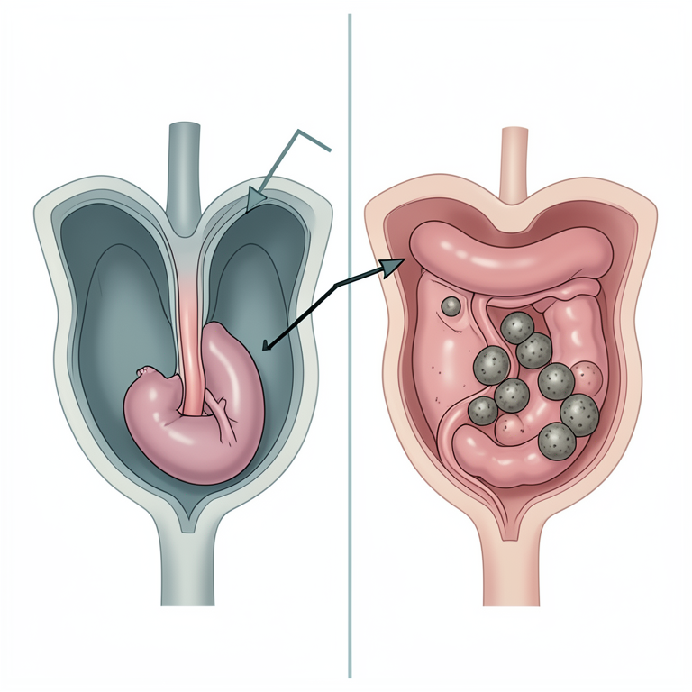
Combined Surgical Approach
AI-generated conceptual illustration (domestic cat abdominal cavity). It explains the idea and does not show study data.
View source evidence ↗Introduction
Dystocia in cat (queen) is defined as difficulty in the exploding of the fetus or delivery of the kitten through the birth canal at the time (6-12 hours) of labor (Dar et al., 2015) [1] . The causes of dystocia are classified as maternal, fetal and in some cases, both (Stedile et al., 2011) [5] . The incidence of dystocia in cats is around 3.3-5.8% in some breeds of cats (Pretzer, 2008) [3] . Overall, 67.1 percent of instances have been linked to maternal factors, while 29.7% have been linked to fetal factors (Jyothi and Rajesh, 2018) [2] .
Materials and Methods
A 3-year-old cat was presented to Veterinary Clinical Complex, College of Veterinary Science and Animal Husbandry, Mhow. Cat has delivered one fetus. Clinical examination revealed restlessness, bleeding from vagina, anorexia and straining to expel the fetus. Per vaginal examination showed no fetus in birth canal. Sonographic examination revealed the presence of dead fetus. X-ray examination was done to rule out the possibility of no. of fetus; which showed fetus in caudal presentation along with severely dehydrated multiple faecolith. On consultation with the owner regarding faecolith, owner insisted to remove the faecolith as well during the Caesarean section as the cat did not defecate since a long time. It was then decided to perform surgical intervention including C-section and enterotomy.
Surgical Treatment
The surgery was performed under general anesthesia using combination of atropine @0.02 mg/kg BW, xylazine @1 mg/kg BW intramuscularly as pre-anaesthetic medication. Patient was put into dorsal recumbency and mid ventral midline abdominal site was aseptically prepared for surgery. Patient was maintained during the surgery with zolazepam and tiletamine combination (Zoletil™) @15 mg/kg BW intramuscularly. After following all the aseptic measures and precautions, preoperative administration of antibiotic Ceftriaxone @20 mg/kg BW intravenously and meloxicam @0.2 mg/Kg BW IM was administered intravenously. Fluid therapy during the entire operative period was given. A 4 cm long incision was given on skin followed by incising the Linea alba and parietal peritoneum. Uterine incision was closed in two layers using inversion suturing technique. While exploring of uterus, multiple stone like fecoliths were felt in the large colon which would not be corrected by therapeutic management; hence it was decided to perform colotomy as discussion with the owner.
An incision was given on anti-mesenteric border of colon at the site of fecolith. Six large sized fecoliths were extracted after gentle manipulation up to the site of incision. Incision was closed using Lembert technique using 3-0 vicryl suture material. Abdominal cavity was lavaged using normal saline and laparotomy wound was closed as per standard method. Owner was advised for fluid therapy for 2-3 days postoperation followed by liquid diet after 24 hrs in multiple small boluses. After 48 hours, owner was advised to feed semiliquid diet in multiple small boluses. Semi-solid diet was started after 5 days.
Uterus was grasped and pulled out from cavity and longitudinal incision along the long axis of uterine horn was given. Fetus along with placenta was removed. Uterus was ligated in two layers with absorbable polyglactin 910 no. 2-0 (vicryl) using Lembert technique. Repositioning of uterus to its original position was done. After C-section, it was decided to perform enterotomy to get rid of fecoliths. Large intestine was pulled out and was incised proximal to fecolith and fecoliths were removed followed by flushing of intestine with metronidazole. Intestine was sutured in two layers with absorbable Polyglactin 910 (Vicryl 2-0) using Lembert technique. Intestine was repositioned and abdominal cavity was flushed with diluted betadine with Normal saline. Linea alba along with parietal peritoneum was sutured using absorbable polyglactin 910 no. 2-0 (vicryl), Subcutaneous tissue was sutured separately using absorbable Polyglactin 910 no 2-0 (Vicryl). Lastly, skin was sutured using Silk no 1 (mersilk). Antiseptic Dressing of suture line was done using liquid povidone-iodine followed by its ointment. Post-operative care included administration of ceftriaxone @20 mg/kg BW intravenously for 7 days, meloxicam @0.2 mg/kg BW intramuscularly for 3 days, intravenous fluid therapy for 5 days and syrup Duphalac 1 tsp bid for 3 days and anti-septic dressing of suture line on alternate days. Owner was advised to keep the cat off-fed for 2 days and was advised to put E-collar to prevent self-mutilation of surgical wound.
Results and Discussion
Cat recovered completely without any complications and started feeding and defecating after 7 days of post operative care. The queen cat is a sensitive and subtle animal, so all steps to treat dystocia need gentleness and complete all physical examination before deciding the dystocia treatment method.
Conclusion
The best method for treating dystocia in cats was C-section when fetus was found dead.
References
5 in the original paper- Dar KH, Ansari MM, Qadri SA, Baba MA, Kumar M. Dystocia and its surgical management in Siamese queen. The blue cross book. 2015;31:40-41.
- Jyothi S, Rajesh K. Caesarean section I canine: case report. The pharma innovation J. 2018;7(4):561-562.
- Pretzer SD. Medical management of canine and feline dystocia. Theriogenology. 2008;70:332-336.
- Saha C, Mukherjee P, Ghosh D, Nandi SK. Surgical Management of Fecolith in a cat. Indian Journal of Animal Health. 2016;55(1):89-90.
- Stedile R, Olivira ST, Muccillo MDS, Contesini EA, Beck CAD. Dystocia in cat due to an ectopic artery. Vet. Recorded. 2011;169(21):1-1.