
Research article · 2024 · Full text
International Journal of Veterinary Sciences and Animal HusbandryUltrasonographic evaluation of fetal echo biometry and serum progesterone changes in normal and aborted pregnancies in bitches
Volume 9(3) · Pages 629–632 · DOI: 10.22271/veterinary.2024.v9.i3i.1489 ↗
Research areasVeterinary Medicine and SurgeryAnimal Virus Infections StudiesCardiovascular Conditions and Treatments
Abstract
Author-writtenThirty bitches of different breeds, weighing 5 to 40 kg and aged between 2 to 6 years presented to the Small Animal Gynaecology unit of Madras Veterinary College, Chennai for pregnancy diagnosis (after 30-35 days of mating) and were divided into three groups of 10 bitches each as Group I (Abortion), II (Foetal Resorption) and III (Normal pregnancy). Foetal echo biometry and serum progesterone estimation were done. Fetal heart rate from the 5th week of pregnancy was significantly lower in abortion (235.2±9.75) and resorption (203±5.49) groups. As gestation age advances, a significant reduction in the fetal heart rate was observed in group I. Gestational sac diameter during the 7th week of gestation was significantly higher (p
Summary
Source-checkedPlain-language reading of the full paper, prepared with AI and checked against the text. Each point links to the authors' own words.
Why it matters
- Accurately predicting pregnancy outcomes in dogs is clinically important because it allows for timely interventions to reduce reproductive losses. View evidence ↗
- Pregnancy loss in bitches can occur through embryonic death, fetal resorption, or abortion, often driven by infectious causes. View evidence ↗
- Progesterone is essential for maintaining pregnancy; a sharp decline in this hormone often signals an impending abortion and threatens fetal viability. View evidence ↗
The question
- The study aimed to evaluate changes in fetal growth, heart rate, and hormone levels using ultrasonography to compare normal pregnancies with those resulting in abortion. View evidence ↗
What they did
- The study included 30 bitches (aged 2-6 years, weighing 5-40 kg) divided into three equal groups of 10: abortion, fetal resorption, and normal pregnancy. View evidence ↗
- Serum progesterone levels were measured using commercial Radioimmunoassay (RIA) kits. View evidence ↗
What they found
- At the 5th week, fetal heart rates were significantly lower in the abortion (235.2±9.75 bpm) and resorption (203±5.49 bpm) groups compared to normal pregnancies (277.1±4.07 bpm). View evidence ↗
- In the abortion group, fetal heart rate showed a pronounced decline from the 5th to 7th week (235.2±9.75 to 170.9±2.96 bpm), which the authors associate with fetal and maternal stress. View evidence ↗
- By the 7th week, the gestational sac diameter was significantly larger in normal pregnancies (4.02±0.41 cm) than in the abortion group (3.75±0.09 cm). View evidence ↗
- At the 5th week, placental diameter was significantly higher in normal pregnancies (0.49±0.03 cm) than in the abortion (0.37±0.01 cm) or resorption (0.33±0.02 cm) groups. View evidence ↗
- During the 6th week, serum progesterone was significantly lower in the abortion group (8.81 ng/ml) compared to the normal pregnancy group (10.32 ng/ml). View evidence ↗
- The resorption group showed a drastic reduction in progesterone (1.72±0.32 ng/ml) during the 6th week, indicating fetal death and the onset of resorption. View evidence ↗
What it means
- The authors conclude that reduced fetal heart rates and lower maternal serum progesterone are indicators of fetal or maternal stress that lead to abortion or resorption. View evidence ↗
- The study highlights that monitoring gestational sac diameter, fetal heart rate, placental thickness, and progesterone levels is useful for distinguishing normal pregnancies from high-risk ones. View evidence ↗
Visual summary
2 visualsIllustrations explain the paper; charts are replotted from the paper's own table values.
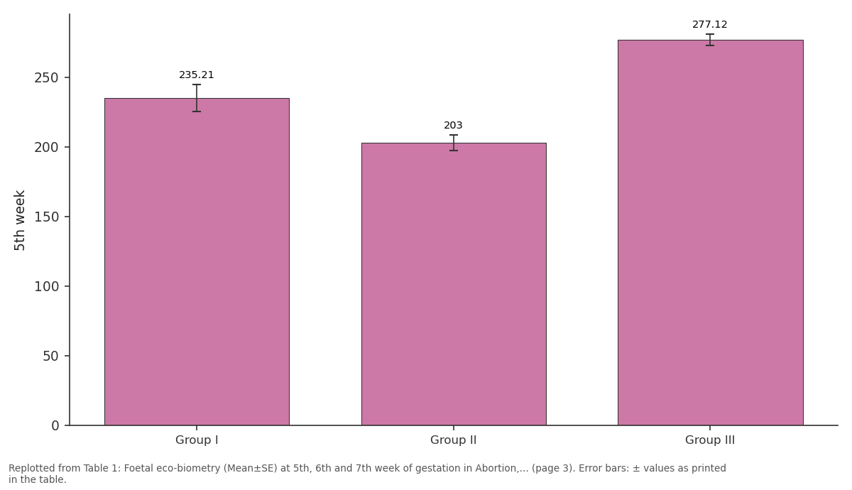
5th week: Group I vs Group II vs Group III
Replotted from Table 1 (page 3) of the original paper. Every bar is the paper's own table value.
View source evidence ↗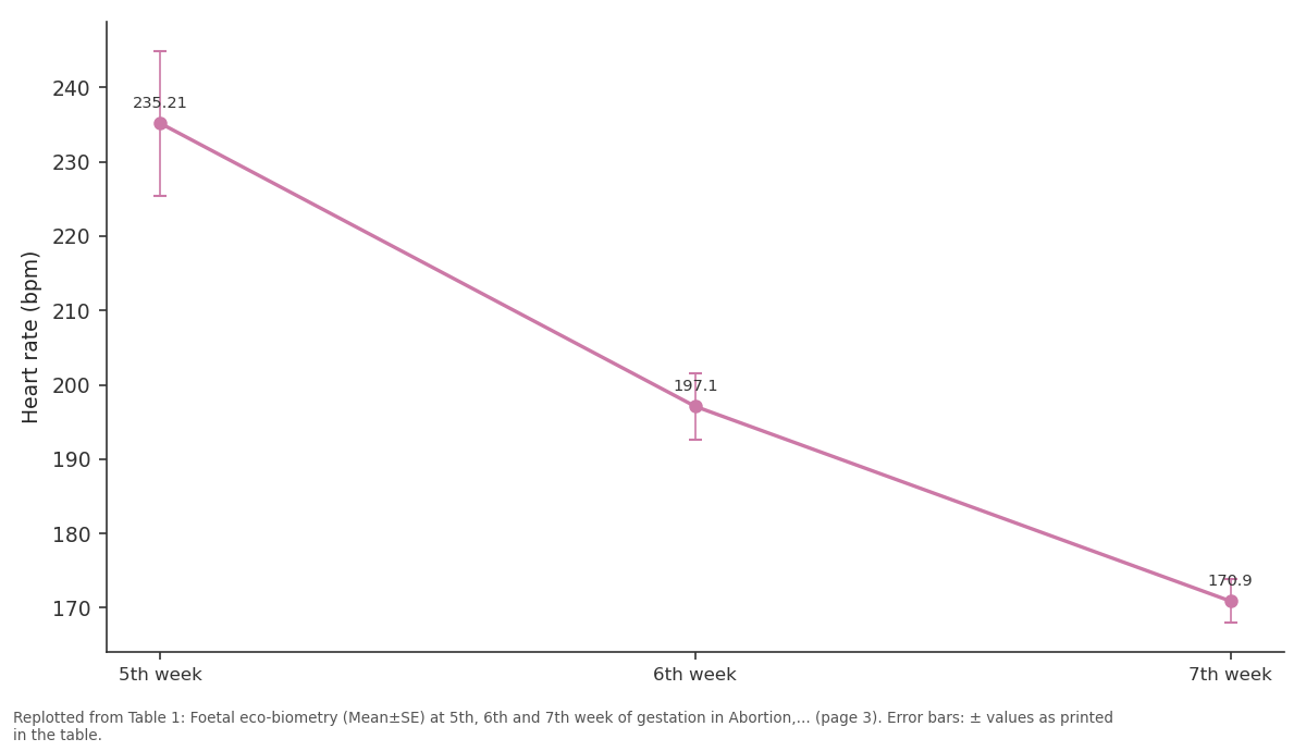
Group I: 5th week vs 6th week vs 7th week
Replotted from Table 1 (page 3) of the original paper. Every bar is the paper's own table value.
View source evidence ↗Figures & tables
From the original paper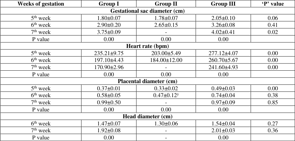 Table 1
 Table 2
Introduction
Accurate prediction of pregnancy is clinically useful to prevent or curtail reproductive losses by timely intervention. Traditionally, two-dimensional ultrasonography is considered to be a safe and simple method for tracking foetal growth and assessing viability (Yeager and Concannon, 1990). In Canine, the pregnancy losses are due to embryonic death, foetal resorption, abortion at any stage of pregnancy and infectious causes are the major reasons for it. Apart from infection, hormonal imbalances, hypoluteoidism, trauma, genetic abnormalities, adverse environmental conditions, uterine pathologies, maternal stress, nutritional deficiencies, drugs and toxic agents (Purswell, 1991; Verstegen et al., 2008; Gorlinger et al., 2005) [1,2,3] are responsible for the pregnancy loss in canines. Compared with other reproductive disorders, the incidence of abortion and resorption were reported to be 4.83 per cent and 11 to 25.9 per cent respectively, in canines (Dhurvey et al., 2022) [4] . Progesterone is the hormone of pregnancy and is essentially needed for the maintenance of pregnancy and a drastic reduction in progesterone indicates that the animal is going for abortion and the liveability of the fetus is compromised (Uchanska, et al., 2022) [5] . Considering the above facts, the present study was carried out to assess the ultrasonographic changes in foetal growth, heart rate, and hormonal changes in normal and abortion during pregnancies in bitches.
Materials and Methods
Female dogs of different breeds, weighing 5 to 40 kg and aged between 2 to 6 years presented to the Small Animal Gynaecology unit of Madras Veterinary College Teaching Hospital in Chennai for pregnancy diagnosis (after 30-35 days of mating) were utilized for this study.
Pregnancy diagnosis by ultrasound
Pregnancy was confirmed by ultrasound examination of bitches from the 5 th week of mating/breeding using a sector probe of 5-7.5 MHz. (EDAN). After confirmation of pregnancy, the selected pregnant bitches were monitored for foetal growth and well-being using two-dimensional ultrasonography at weekly intervals (5 th week onwards) throughout gestation until abortion/resorption/ normal parturition. Based on the outcome of pregnancy, the selected bitches were divided into three groups of 10 bitches each as Group I (Abortion), II (Foetal Resorption) and III (Normal pregnancy).
Gestational sac diameter
The gestational sac diameter (GSD) or inner chorionic cavity diameter (ICCD) was measured to estimate the age of the foetus during the 5 th week of pregnancy as per the formula of Yeager et al. (1992) [6] .
Head diameter
The head diameter (HD) or biparietal diameter (BPD) was measured to confirm the age of the foetus during 6 th , 7 th and 8 th week of pregnancy as per the formula of Yeager et al. (1992) [6] .
Heart rate
The viability of the foetus was evaluated based on visualization of foetal heartbeat. The frequency of heartbeat was calculated by capturing number of beats per minute or by measuring the distance between two systolic peaks using Mmode ultrasonography.
Placental thickness
The thickness of the zonary placenta within the gestational sac was measured by B-mode ultrasonography and the average placental thickness in centimetres was evaluated (Feliciano et al., 2015) [7] .
Serum progesterone estimation
Blood samples were collected in all selected pregnant bitches at weekly intervals, beginning from the 5 th week of gestation until abortion/resorption/normal delivery of the foetus for hormonal analysis. After separating serum, samples were stored at -20 °C until utilised for progesterone estimation. The estimation of progesterone concentration in group I, II and III bitches were performed using specific commercial Radioimmunoassay kits (RIA, Progesterone, Beckman Coulter, Czech Republic: Cat # IM188) according to the manufacturer's instructions.
Statistical analysis
The data collected in the study were analysed using one-way analysis of variance (ANOVA) and repeated measures of oneway (ANOVA) for ultrasonography and hormonal parameters between groups. A paired t-test was performed for the echobiometric parameters to correlate their association with gestational age with the aid of SPSS (20.0 version) statistical software.
Results and Discussions
In the present study, foetal eco-biometric parameters like gestational sac diameter (GSD), placental diameter (PD), heart rate (HR) and head diameter (HD) were recorded by 2D ultrasonographic examination at the 5 th , 6 th and 7 th week of gestation in abortion, resorption and normal pregnancy bitches (Table 1 and2).
| Weeks of gestation | Group I | Group II | Group III | ‘P’ value |
|---|---|---|---|---|
| Gestational sac diameter (cm) | ||||
| 5th week | 1.80±0.07 | 1.78±0.07 | 2.05±0.10 | 0.06 |
| 6th week | 2.90±0.20 | 2.65±0.15 | 3.26±0.08 | 0.41 |
| 7th week | 3.75±0.09 | - | 4.02±0.41 | 0.02 |
| P value | 0.00 | 0.00 | 0.00 | |
| Heart rate (bpm) | ||||
| 5th week | 235.21±9.75 | 203.00±5.49 | 277.12±4.07 | 0.00 |
| 6th week | 197.10±4.43 | 184.00±12.00 | 260.70±5.67 | 0.00 |
| 7th week | 170.90±2.96 | - | 241.60±4.93 | 0.00 |
| P value | 0.00 | 0.00 | 0.00 | |
| Placental diameter (cm) | ||||
| 5th week | 0.37±0.01 | 0.33±0.02 | 0.49±0.03 | 0.00 |
| 6th week | 0.58±0.05 | 0.47±0.12y | 0.74±0.04 | 0.38 |
| 7th week | 0.99±0.50 | - | 0.97±0.09 | 0.85 |
| P value | 0.00 | 0.00 | 0.00 | |
| Head diameter (cm) | ||||
| 6th week | 1.47±0.07 | 1.30±0.06 | 1.54±0.04 | 0.27 |
| 7th week | 1.92±0.08 | - | 2.01±0.03 | 0.36 |
| P value | 0.00 | - | 0.00 |
Table text extracted from the original PDF; superscript letters are shown inline. The original table image is below.
View original table image
Gestational sac diameter
In the present study, as pregnancy age advances there is a gradual, significant increase in the diameter of gestational sac in normal pregnancy as well as in the abortion group. Gestational sac diameter during the 7 th week of gestation was significantly higher (P<0.05) in normal pregnancy (G-III) bitches (4.02±0.41 cm) when compared to the bitches (3.75±0.09 cm) which aborted their foetus (G-I). Similarly, Alonge et al., (2016) [8] recorded the gestational sac diameter of 6.9 to 42.4 mm and 8.4 mm to 41.3 mm in large and giant size breeds of dogs from 42 to 26 days and 40 to 25 days respectively, before parturition. Whereas, Mattoon and Nyland (1995) [9] reported that at 20 days of post-breeding detection of a gestational sac was considered as the first sign of confirming pregnancy using ultrasonography.
Fetal heart rate
The mean heart rate from the 5 th week of pregnancy was significantly lower in abortion (235.2±9.75) and resorption (203±5.49) groups when compared to normal pregnancy bitches (277.1±4.07). England (1998) [10] recommended foetal heart rate as a useful parameter to estimate the survival possibilities of foetuses. The cardiac activity was deducted as early as 25 days after LH surge (Yeager et al. 1992) [6] . As pregnancy age advances a gradual significant reduction in the fetal heart rate was observed in normal pregnancy. However, the reduction was more pronounced in abortion groups (235.2±9.75, 197.1±4.43 and 170.9±2.96 at 5 th , 6 th and 7 th week of gestation), indicating fetal and maternal stress. Normal fetal heart rate is 220 to 240 bpm while rates of < 180 bpm are indicative of fetal distress (Zone and Wanke, 2001) [11] . Hypoxia-induced fetal stress, which presents as a drop-in heart rate, can happen during an abnormal pregnancy (Kutzler et al., 2003) [12] .
Placental diameter
During 5 th week of gestation, bitches with normal pregnancy (G-III) had significantly higher (P<0.01) placental diameter (0.49±0.03 cm) than the bitches (0.37±0.01 cm) which aborted their foetus (G-I) and bitches (G-II) which shown resorption (0.33±0.02 cm) of the foetus (Table 1). A significant increase in the placental diameter was observed between 5 th to 7 th week of normal pregnancy and the abortion groups. At any stage of pregnancy, the normal canine placenta should not exceed 1.2 cm at its centremost point regardless of the breed in dogs. The thickness of the placenta may assist in the diagnosis of gestational age in bitches. Maldonado et al., (2012) [13] and Babiker and Eisa (2014) [14] reported that the placental thickness of 0.34 cm, 0.44 cm, 0.67 cm and 0.89 cm would relate to 30.5 days, 35.2 days, 46 days and 56.3 days of pregnancy. The results of the present study were in agreement with them.
Head diameter
Head diameter observed during the 6 th and 7 th week of pregnancy in the present study ranged from 1.30±0.06 cm to 2.01±0.03 cm. Alonge et al. (2016) [8] recorded the HD of 5.2 to 28.1mm and 2 mm to 29.4 mm in large and giant size breeds of dogs from 30 to 2 days and 35 to 1 day respectively, before parturition. The head of the fetus showed significant growth during the 6 th to 7 weeks of pregnancy in both groups I and III bitches. While in the resorption group, the head diameter was slightly lower than the groups I and III during the 6 th week. Zone and Wanke (2001) [11] found that head diameters of < 2 cm from days 48 to term are indicative of growth retardation, resulting in significantly reduced average birth weight for the breed and are also at risk for abortion or early neonatal loss.
Serum Progesterone
The progesterone concentration was found to be within normal ranges (25.77±1.90, 27.40±2.53 and 28.10±2.30 ng/ml) in all the groups during the 5 th week of pregnancy in the present study (Table 2). However, the progesterone concentration during the 6 th week in the abortion group was 8.81 ng/ml which was found to be significantly lower than that of the normal pregnancy group (10.32 ng/ml). This decrease in progesterone concentration in the 6 th week coincident with clinical signs or signals of threatened or imminent abortion during the 7 th week of pregnancy in bitches, wherein the progesterone concentration has further reduced during the 7 th week (1.72±0.32 ng/ml).
| Stages of Pregnancy | Serum level of Progesterone (ng/dl) | — | — | — |
|---|---|---|---|---|
| Group I (Abortion) | Group II (Resorption) | Group III (Normal pregnancy) | P value | |
| 5th week | 25.77±1.90 | 27.40±2.53 | 28.10±2.31 | 0.75 |
| 6th week | 8.81±0.55 | 1.72±0.31 | 10.32±0.74 | 0.00 |
| 7th week | 1.15±0.17 | - | 5.73±0.60 | 0.00 |
Table text extracted from the original PDF; superscript letters are shown inline. The original table image is below.
View original table image
In pregnant bitches, with the induction of prepartum luteolysis and a steep reduction in P 4 concentration around day 60 of the CL lifespan. This active termination of CL function is associated with increased production of uteroplacental PGF 2 α (Kowalewski et al., 2020) [15] . The progesterone concentration in the resorption group is drastically (1.72±0.32 ng/ml) reduced than other groups during the 6 th week indicating death of the fetus and resorption has started within the 6 th week. Similar to the current study, Thuroczy et al., (2016) [16] have reported that a decrease in serum progesterone below 10 ng/ml at 4 th week of pregnancy may signal an impending abortion in bitches. According to experimental research, keeping a pregnancy in the bitch requires a plasma progesterone concentration of at least 6-9 nmol/L (2-3 ng/mL), and concentrations below this threshold for longer than 24-48 hours result in pregnancy loss. (Onclin et. al., 2002) [19] . The progesterone concentrations observed in the present study indicate that the animals suspected of hypoluteoidism can have their abortions predicted by serum progesterone estimations.
References
16 in the original paper- Purswell BJ. Management of apparent luteal insufficiency in a bitch. J Am Vet Med Assoc. 1991;199:902-903.
- Verstegen OK, Verstegen J. Endocrinology of pregnancy in the dog: a review. Theriogenology. 2008;70:291-299.
- Görlinger S, Galac S, Kooistra HS, Okkens AC. Hypoluteoidism in a bitch. Theriogenology. 2005;64:2139.
- Dhurvey M, Nema SP, Shivhare M, Gupta VK. Incidence of various reproductive disorders in different breeds of bitches. The Pharma Innovation Journal. 2022;11(3):886888.
- Uchanska O, Ochota M, Eberhardt M, Nizanski W. Dead or Alive? A Review of Perinatal Factors That Determine Canine Neonatal Viability. Animals. 2022;12:1402.
- Yeager AE, Mohammed HO, Meyers-Wallen V. Ultrasonographic appearance of the uterus, placenta, fetus and fetal membranes throughout accurately timed pregnancy in Beagles. American Journal of Veterinary Research. 1992;3:342-351.
- Feliciano MAR, Maciel GS, Coulinho LN, Aleida VT, Vicente WRR. Gestational echo biometry in brachycephalic pregnant bitches. Cienc. anim. bras. 2015;16(3):419-427.
- Alonge SM, Beccaglia M, Melandri G, Luvoni C. Prediction of whelping date in large and giant canine breeds by ultrasonography foetal biometry. Journal of Small Animal Practice. 2016, DOI: 10.1111/jsap.12534
- Mattoon JS, Nyland TG. Ultrasonography of the genital system: In Veterinary Diagnostic Ultrasound. Eds. Nyland, T.G. and Mattoon, J.S., W.B. Saunders, USA. 1995, 146-148.
- England GC. Ultrasonographic assessment of abnormal pregnancy. The Veterinary clinics of North America. Small animal practice. 1998;28:849-868.
- Zone MA, Wanke MM. Diagnosis of canine fetal health by ultrasonography. J Reprod Fertil. 2001;57:215-9.
- Kutzler AM, Yeager AE, Mohammed HO, MeyersWallen VN. Accuracy of canine parturition date prediction using fetal measurements obtained by ultrasonography. Theriogenology. 2003;60:1309-17.
- Maldonado AL, Junior EA, Mendonla DS, Nardozza LM, Ajzen SS. Ultrasound Determination of Gestational Age Using Placental Thickness in Female Dogs: An Experimental Study Veterinary Medicine International Article. 2012, ID 850867, doi:10.1155/2012/850867
- Babiker MS, Eis RA. Placenta thickness measurements during gestational age progress. Journal of Applied Medical Sciences. 2014;3(6):31-37.
- Kowalewski MP, Pereira MT, Kazemian A. Canine conceptus-maternal communication during maintenance and termination of pregnancy, including the role of species-specific decidualization. Theriogenology. 2020;150:329-338.
- Thuróczy J, Müller L, Kollár E, Balogh L. Thyroxin and progesterone concentrations in pregnant, nonpregnant bitches, and bitches during abortion. Theriogenology. 2016;85:1186-1191.