
Research article · 2024 · Full text
International Journal of Veterinary Sciences and Animal HusbandryTibiotarsal fracture repair in a duckling using a spinal needle
Volume 9(3) · Pages 438–439 · DOI: 10.22271/veterinary.2024.v9.i3f.1456 ↗
Research areasDiabetic Foot Ulcer Assessment and ManagementFoot and Ankle SurgeryBone fractures and treatments
Abstract
Author-writtenA 5-month old female duckling weighing 300 g was brought with a history of non-weight bearing lameness in the right foot. History suggested a possible stone pelting. Clinical examination revealed deformity, muscle damage and crepitation in the distal tibiotarsal region. X-ray revealed, distal third tibiotarsal transverse fracture. Under ketamine induction and isoflurane maintenance, normograde intramedullary pinning was done with a 20 G sterile spinal needle. There was no post-operative complication and normal weight bearing was regained in 2 weeks.
Summary
Source-checkedPlain-language reading of the full paper, prepared with AI and checked against the text. Each point links to the authors' own words.
Why it matters
- Tibiotarsal fractures occur frequently in birds because the bone is long and more exposed. View evidence ↗
- The thin cortices of avian bones make them susceptible to shattering if drilling is used during repair. View evidence ↗
- External skeletal fixation is risky in birds because it often requires drilling at multiple sites, increasing the chance of bone shattering. View evidence ↗
The question
- The study aimed to treat a distal third tibiotarsal transverse fracture in a 300 g, 5-month-old female duckling presenting with non-weight bearing lameness. View evidence ↗
What they did
- The procedure used ketamine (20 mg/kg i/m) for induction and 0.8% isoflurane in 100% oxygen (1 L/minute) for maintenance, employing a 20 G sterile spinal needle for normograde intramedullary pinning. View evidence ↗
- The spinal needle was inserted through the cranial aspect of the proximal right tibiotarsus and advanced into the distal fragment following fracture reduction. View evidence ↗
- A soft cotton bandage was applied after the surgery to protect the intramedullary pin. View evidence ↗
What they found
- The duckling regained normal weight bearing in the right foot within 2 weeks, with no post-operative complications reported. View evidence ↗
- The intramedullary spinal needle pin was removed 20 days after the procedure. View evidence ↗
- Following the removal of the pin, the owner reported that the duck was actively moving with proper weight bearing in the affected limb. View evidence ↗
What it means
- The authors conclude that using a 20G spinal needle for intramedullary pinning is an effective method for treating transverse tibiotarsal fractures in ducklings, providing necessary stability for recovery. View evidence ↗
- The case demonstrates the overall effectiveness of normograde intramedullary pinning for avian tibiotarsal fractures. View evidence ↗
Visual summary
1 visualsIllustrations explain the paper; they do not show study data.
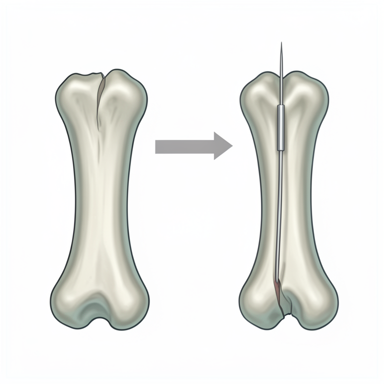
Normograde Pinning Concept
AI-generated conceptual illustration (duckling tibiotarsus bone). It explains the idea and does not show study data.
View source evidence ↗Figures & tables
From the original paper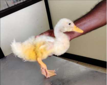 Figure 1
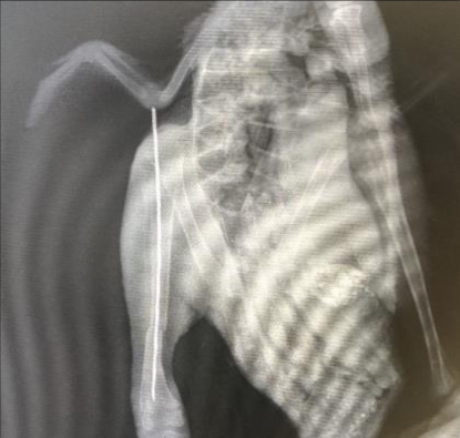 Figure 2
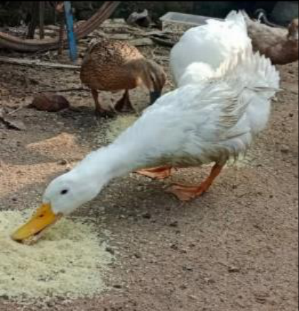 Figure 3
Introduction
Tibiotarsal fractures are common in birds since it is long and more exposed bone (Wright et al., 2018) [7]. Transverse fractures are most common. Thin cortices of bird bones may predispose them to easily shatter during drilling. Only normograde pinning is advisable in tibiotarsal fractures.
External skeletal fixation may need drilling the bone at multiple sites which may predispose to shattering. The muscle damage at the site also was another reason for choosing an intramedullary fixation technique. Tape splints, external skeletal fixation and interlocking nail were used for repair of tibiotarsal fractures in birds. History and Clinical findings A 5-month old female duckling weighing 300 g was brought with a history of non-weight bearing lameness in the right foot.
History suggested a possible stone attack. Clinical examination revealed deformity, muscle damage and crepitation in the distal tibiotarsal region. X-ray revealed, distal third tibiotarsal transverse fracture.
Materials And Methods
Under ketamine (@ 20 mg/kg b. wt. i/m) induction and isoflurane (0.8%) in 100% oxygen maintenance @ 1 L/minute, normograde intramedullary pinning was done with a 20 G sterile spinal needle. The spinal needle was introduced into the cranial aspect of proximal right tibiotarsus and advanced into the distal fragment after reduction of the fracture fragments. This could be easily done by the grasping the hub of the spinal needle rather than using a chuck under C-arm. Following the procedure, a soft cotton bandage was applied to protect the intramedullary pin.
Follow up treatment included oral Enrofloxacin @ 10 mg/kg b. wt. once daily for 7 days and Meloxicam @ 1 mg/kg b. wt. once daily for three days along with daily wound dressings.
Results And Discussion
The adopted approaches to repair tibiotarsal fracture in avian species are splints, casts, bone plates, external fixator, intramedullary pinning, circlage and hybrid combination of these
Methods
, depending upon the type of fracture and condition of bone and animal health status. Splints are difficult to apply and manage bone because of soft tissue and musculature. (Kavanagh, 1997; Helmer and Redig, 2006; Duer, 2010; Carrasco, 2019; Fernandes et al., 2021) [5, 4, 2, 1, 3]. VET 2024; 9(3): 438-439 AR Ninu Department of Veterinary Surgery and Radiology, Veterinary College and Research Institute, Tirunelveli, Tamil Nadu Veterinary and Animal Sciences University, Tamil Nadu, India N Krishnaveni Department of Veterinary Surgery and Radiology, Veterinary College and Research Institute, Tirunelveli, Tamil Nadu Veterinary and Animal Sciences University, Tamil Nadu, India AR Ninu Department of Veterinary Surgery and Radiology, Veterinary College and Research Institute, Tirunelveli, Tamil Nadu Veterinary and Animal Sciences University, Tamil Nadu, India https://www.veterinarypaper.com Use of intramedullary pin alone may lead to rotation of the pin, but combined with external coaptation it will provide stability as in our case. The presence of the hub necessitated proper grip and accurate pressure while inserting the spinal needle.
The duckling was able to bear weight in the right foot after 2 weeks. The pin was removed after 20 days. Owner reported that the duck is now actively moving around with proper weight bearing in the affected limb.
Acknowledgement
The authors are thankful for the help and support extended by the Dean, VCRI, Tirunelveli, Director of Clinics, Chennai and Professor and Head, VCC, Tirunelveli.
Conclusions
Tibiotarsal fractures are common in birds, especially transverse fractures due to their long and exposed bones. Thin cortices can lead to shattering during drilling, making normograde pinning the preferred method for these fractures. In a 5-month-old duckling, a transverse tibiotarsal fracture was successfully treated with intramedullary pinning using a 20G spinal needle. This method provided stability and allowed for weight-bearing within two weeks.
The pin was removed after 20 days, and the duckling showed full recovery with proper weight-bearing. This case highlights the effectiveness of normograde intramedullary pinning in avian tibiotarsal fractures.
Figures and tables from the paper
References
7 in the original paper- Carrasco DC. Fracture management in Avian species. Vet. Clin. North Am. Exot. Anim. Pract. 2019;22(2):223- 238.
- Duerr R. Splinting avian fractures. 2nd Edition, Fig 1: The duckling on the day of presentation International Bird Rescue Research Center, Cordelia, CA, 2010.
- Fernandes MC, Mota da Costa AL, Da Silva Machado Camargo TF, Cotes LC, Santos LS, Silva RC, et al. Use of reconstruction plate and intramedullary pin for repair of a tibiotarsus fracture in a red-legged seriema (Cariama cristata Linnaeus, 1766): Case report. Int J Vet Sci Res. 2021;7(2):057-059.
- Helmer P, Redig PT. Surgical Resolution of Orthopedic Disorders. In Clinical Avian Medicine, Volume II; c2006. p. 761-774.
- Kavanagh M. Tibiotarsal fracture repair in a scarlet macaw using external skeletal fixation. Journal of Small Animal Practice. 1997;38:296-298.
- Siqueira DB, Costa Neto EO, Silva MA. Tibiotarsal Fig 2: Fracture immobilization with spinal needle fracture repair by using intramedullary pin and cerclage in bluefronted-amazon (Amazona aestiva) kept in domiciliary captivity: case report. Clínica Veterinária. 2009;14(82):94-96.
- Wright L, Mans C, Olsen G, Doss G, Amene EW, Britsch G, et al. Retrospective evaluation of tibiotarsal fractures treated with tape splints in birds: 86 cases (2006-2015). J Avian Med. Surg. 2018;32(3):205-209. How to Cite This Article Ninu AR, Krishnaveni N. Marine bacteria: A pandora box against cancer. International Journal of Veterinary Sciences and Animal Husbandry. 2024;9(3):438-439. Creative Commons (CC) License This is an open-access journal, and articles are distributed under the terms of the Creative Commons Attribution-Non Commercial-Share Alike 4.0 International (CC BY-NC-SA 4.0) License, which allows others to remix, tweak, and build upon the work non-commercially, as long as appropriate credit is given and the new creations are licensed under the identical terms. Fig 3: Post -operatively after 4 months-normal weight bearing in the right foot