
Research article · 2024 · Full text
International Journal of Veterinary Sciences and Animal HusbandryHistomorphological developmental study of tongue in local awassi sheep fetuses (Ovis aries): Prenatal period
Volume 9(3) · Pages 345–350 · DOI: 10.22271/veterinary.2024.v9.i3e.1437 ↗
Research areasRangeland Management and Livestock EcologyCongenital Anomalies and Fetal Surgery
Abstract
Author-writtenThis study was conducted at Al-Qassim Green University, Collage of Veterinary Medicine, Anatomy and Histology Branch. The experiment was designed to study the tongue of awassi sheep fetuses: perinatal period samples (20 male and female) were collected from healthy females uterine and divided according to age groups into two groups by crown rump length formula (CRL) for the prenatal stage. Group I (50-55) days of gestation, Group II (70-75) days of gestation containing 10 foetii each to record the developmental changes in the tongue and its papillae. In the first group (50-55), the tongue was in the process of development, as it was not fully development and The tongue appeared elongated tape cord-like shape structure with a flat surface and almost uniform in thickness and width throughout its length. While histologically showed three layers: tunica mucosa with primitive of lingual papillae, undifferential mesenchymal tissue (pluripotiantial tissue) and tunica muscularis. In the second group (70-75), the tongue appear more development of the pervious stage occupies the greater part of proper oral cavity and extends into the oropharynx, dorsal surface of the tongue appeared rough showing beginning of formation of lingual papillae scattered mostly at the tip and on the dorsum-linguae and very shallow elevation of tours linguae; while histologically, The stratification of epithelium on dorsum of tongue in both stage were thick and it thickness continues in the different height level reaching to tip and lateral borders and surfaces of the tongue at different places, while at ventral surface of the tongue, the degree of stratification was highly increase.
Summary
Source-checkedPlain-language reading of the full paper, prepared with AI and checked against the text. Each point links to the authors' own words.
Why it matters
- The tongue is a muscular organ essential for moving food within the mouth. View evidence ↗
- Lingual papillae on the tongue are important for the processes of food intake, transport, and swallowing. View evidence ↗
- In certain animals, the torus linguae likely compensates for a defective chewing process caused by the absence of upper incisors. View evidence ↗
The question
- The study aimed to describe the histomorphological development of the tongue in prenatal local Awassi sheep fetuses. View evidence ↗
What they did
- The research used 20 sheep fetuses collected from pregnant ewes slaughtered in Najaf provinces. View evidence ↗
- Fetuses were divided into two groups of 10 based on crown rump length: Group I (50-55 days gestation) and Group II (70-75 days gestation). View evidence ↗
- Tongue samples were fixed in 10% buffered formalin, sectioned at 5-6 μm, and stained using H&E, periodic acid Schiff, and trichrome masons stains. View evidence ↗
What they found
- At the first stage of gestation, the tunica muscularis contained immature, not fully developed muscle fibers appearing as strands within the mesenchymal tissue. View evidence ↗
- At 70-75 days, the tunica muscularis showed well-developed myoblasts and skeletal muscle fibers organized into longitudinal, vertical, and transverse bundles. View evidence ↗
What it means
- The authors conclude that the study successfully detailed the morphological changes and the onset of lingual papillae formation in sheep embryos during the first and second trimesters. View evidence ↗
- The authors state that this understanding of oral anatomy is essential for veterinary and medical research and provides a basis for further embryonic development studies. View evidence ↗
Visual summary
2 visualsIllustrations explain the paper; they do not show study data.
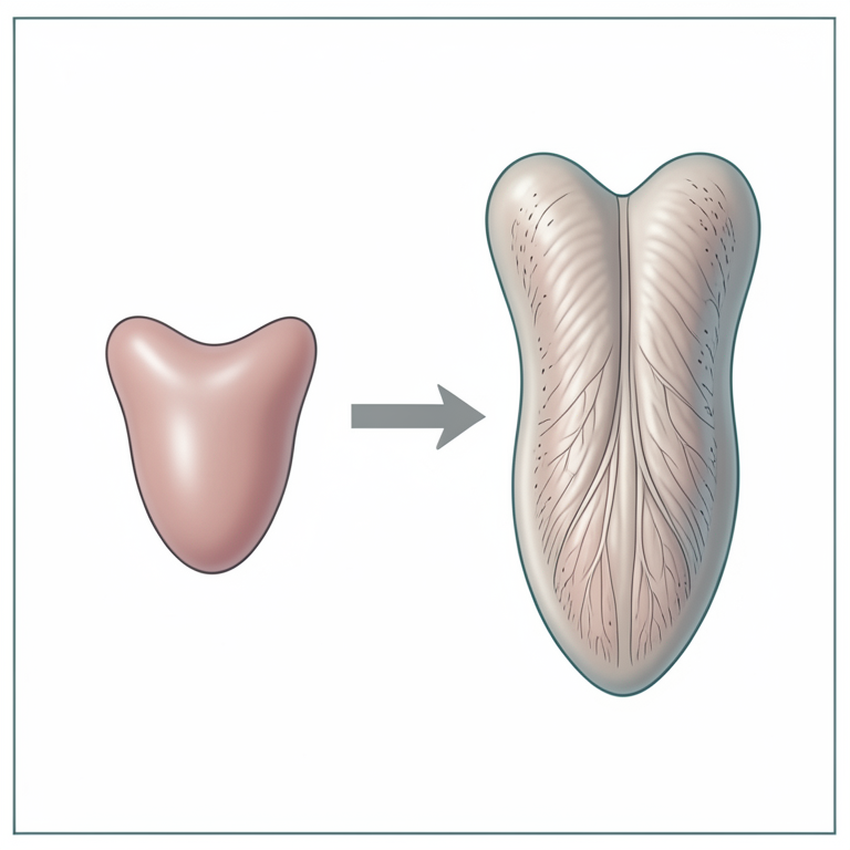
Tongue Growth Timeline
AI-generated conceptual illustration (Awassi sheep fetuses (Ovis aries) tongue tissue). It explains the idea and does not show study data.
View source evidence ↗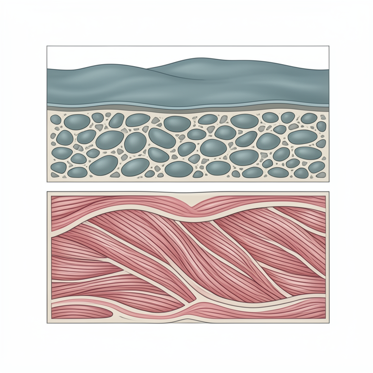
Histological Layering
AI-generated conceptual illustration (Awassi sheep fetuses (Ovis aries) tongue tissue). It explains the idea and does not show study data.
View source evidence ↗Figures & tables
From the original paper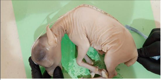 Figure 1
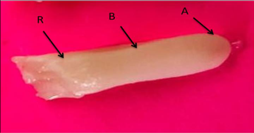 Figure 2
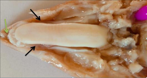 Figure 3
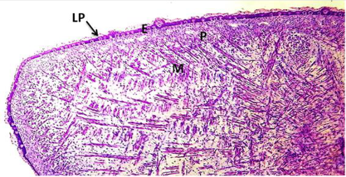 Figure 4
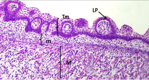 Figure 5
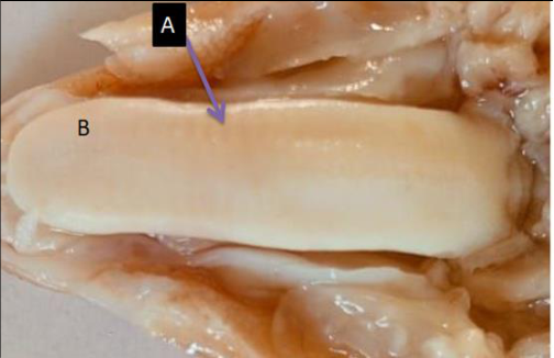 Figure 6
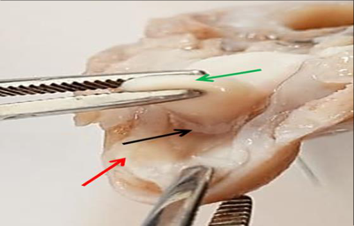 Figure 7
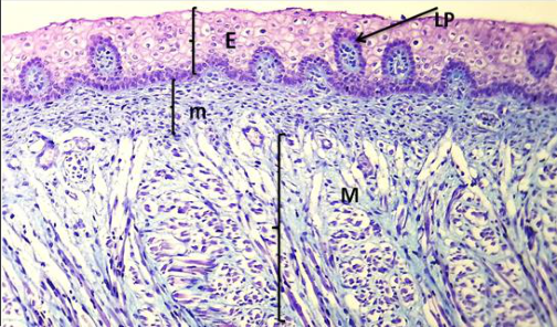 Figure 8
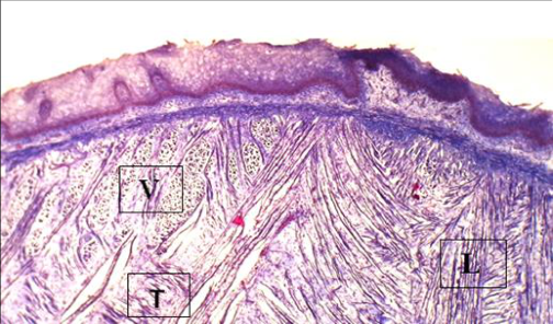 Figure 9
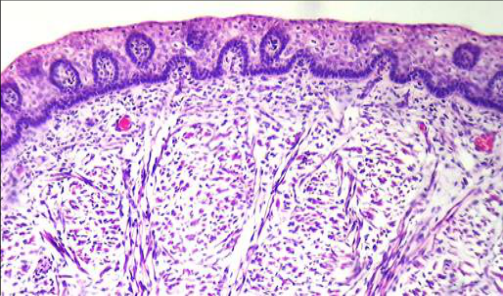 Figure 10
Introduction
The digestive system's organs are able to receive nutrition, break it down chemically and mechanically into its component particles and then absorb them. Lastly, it must remove excreted and unabsorbed remnants. The digestive system is responsible for interrupting nutrition into small portions so that it can be used for energy, growth, and cellular regeneration. Digestion-related cells are essential to this process and may have hormonal functions. Nervous system, lymphatics, and blood arteries are all crucial to digestion. The digestive system consists of the alimentary duct, which runs from the mouth to the anus. Salivary glands, the liver, the pancreas, and auxiliary glands discharge digestive products into the alimentary channel. The pharynx and mouth, stomach and esophagus, large intestine, small intestine, and anal canal are the five sections that make up the alimentary canal. Embryology, The tongue development initiates from fourth week of embryo. The tongue is formed as five edges originating from the pharyngeal arches (TW Sadler, 2004) [26] . The tongue is a muscular organ used to movement the food mass within the mouth. The tongue can be differentiated microscopically from other muscle tissues because it has fibers oriented in three directions. The tongue not only moves food to the table surfaces of the cheek teeth but also serves as a plunger to move food into the esophagus. It assists some animals in seizing food and bringing it to the mouth. There are three parts to the tongue: the corpus linguae, the radix linguae, and the apex (apex linguae). A mucosal fold called the frenulum (frenulum linguae) connects the tongue's body to the oral floor. A median groove, or sulcus medianus, that extends a septum into the tongue, longitudinally identifies the dorsal portion of the canine tongue. In carnivores, A rod-shaped fibrous structure called the lyssa is located in the median plane beneath the ventral mucosa of the tongue in carnivores. It does not reach the hyoid bone, but it does stretch from nearly the tip to the root of the tongue. It is surrounded by a thick layer of connective tissue that is made up of striated muscle, adipose tissue, and occasionally islands of cartilage. On the dorsal surface of the tongue, the lingual mucosa was thrown into projections called the lingual papillae (Dyce, Sack, & Wensing, 2010; Sisson & Grossman, 2010) [8,23] . The lingual papillae perform a variety functions during food intake, transport and swallowing (Abbate et al., 2009; Skieresz-Szewczyk & Jackowiak, 2017) [1,24] . The lingual papillae in camels, like other mammalian species, were classified into mechanical and gustatory papillae. Mechanical papillae, as previously described, include filiform, lentiform, conical and wart-like papillae (Qayyum, Fatani, & Mohajir, 1988) [21] .
Gustatory papillae with taste buds are fungiform and circumvallate (Erdunchaolu et al., 2001; Qayyum et al., 1988; Salehi et al., 2010) [21] . Skeletal muscle, which makes up the majority of the tongue, is organized into three layers that are perpendicular to one another (Sisson and Grossman, 1975; Reece, 2017) [23,29] . Allowing it some flexibility for food prehension and mastication (Dyce et al., 2010; Getty, 1975) [8] . According to (Yamane, 2005) [32] , the majority of tongue muscles have a somatic origin. It has been documented that the tongues of ox and sheep (Habel, 1975) [11] , goats (Qayyum and Beg, 1975) [21] , and buffalo (Dhingra and Barnwal, 1979; Prakash and Rao, 1980) have conspicuous torus linguae with a crescentic depression, or fossa linguae. The lack of incisors in the upper jaw results in a defective masticatory process, which is likely compensated for by the torus linguae (Labh and Mitra, 1969) [17] .
Material and Methods
This study was designed to described the Histomorphological and development study of tongue in local awassi sheep fetuses (Ovis aries) at prenatal. A sample are collected in (September to November) of year (2023). The study is performed on (20) sheep fetuses, that are collected from pregnant ewes slaughtered in the abattoirs of Najaf provinces for prenatal study. Which is determined depending on the crown rump length (CRL) using of following formula (Y=2.74X+30.15) where 'Y' is developmental age of fetus in days and 'X' is the crownrump length in cm (Veeresh et al., 2022) [27] . The tongue of sheep's fetuses were fixed in (10%) buffered formalin, dehydrated in a graged series of alcohol, cleared in xylene the embedded in paraffin wax. The blocks were sectioned at (5-6) um thickness of slice using a rotary microtome. Histological sections were stained with Haematoxylin and Eosin (H&E), periodic acid Schiff and trichrome masons stain (Suvarna et al., 2018) [25] . The sections were studied using the Olympus light microscope with the digital camera, which was connected to the computer
Result and Discussion First trimester at (50-55) days of gestation
The body weight of sheep embryo in this study about (39.4±1.02) grams and crown rump length in about (10.6±0.509) cm (Figure 1). The body weight of camel embryo at the stage of first trimester (below 130 days) about (0.18±0.05) and the crown rump length in about (15.75±4.42) (Bello et al., 2015) [4] . [20] . At this age of embryo, The tongue appeared elongated tape cord-like shape structure with a flat surface and almost uniform in thickness and width throughout its length. the tongue comprised of three parts the cranial part called apex (tip); The apex of the tongue was nearly rounded in shape which formed by meeting of the dorsal and the ventral surfaces, middle part called body (corpus); The body was the largest and the longer part of the tongue located between the apex and root and finally the caudal part was root (radix); The root was the last and the shorter part of the tongue lies caudally to body of the tongue and slopes ventrally and caudally toward the base of the epiglottis. And the tongue at this stage where appeared whitish or colorless and opaque in appearance with small in size relatively compare with the advanced stages with irregular and an uneven dorsal surface (Figure ,2). The pervious result agree with (Bello et al., 2015) [4] in camel who mentioned that the tongue at first trimester, were seen as smooth muscle mass, with almost uniform width and thickness throughout the length. They were uniformly pinkish, no pigmentation and no visual evidence of lingual papillae.
The dorsal surface of the tongue is smooth and slop relatively and the shape of the tongue is a vertical bar stable in the floor of the mouth cavity between ram of mandible set in proper mouth cavity and tours linguae is not well development with very low elevation without lingual fossa.
The current results crosseponding with (Arti, A., 2021) [3] in (goat) who mentioned that The tongue of foetal goat was located on the floor of mouth between the two rami of mandible, Three distinct parts viz cranial apex, middle body and caudal root were identified from 43 days onwards. The cranial part, apex was free anteriorly and attached with the body posteriorly, the apex was thin, narrow and rounded with sharp borders in group I, free part of tongue was formed by meeting of the dorsal and the ventral surfaces. The shape of apex was almost spatula shaped during entire study. Primordium lingual papillae were appear very few localization along the dorsal surface of tongue fetuses and appeared like dome-shape protrusion scattered on dorsum of body and apex but absent on dorsal surface of root and there is no papillae along lateral margins of tongue (Fig. 3). This result agreement with (Verma et al., 2011) [28] in buffalo who mentioned that the first indication of formation of circumvallate papillae was observed at 10.7 cm CVR length (77 days). While histological observations showed that the tongue at this age has three layers: tunica mucosa with primitive of lingual papillae, un-differential mesenchymal tissue (pluripotiantial tissue) and tunica muscularis (Figure 4). Tunica mucosa covered by epithelial cells which was stratified cuboidal or stratified squamous cells (nonkeratinized) about 7-8 cellular layer (Figure 5) and this current study agreement with (Hejazi, S., & Baroughi, R., 2013) [12] in sheep who mention that the epithelial lining of the tongue is cubic in this age of gestation without indication of differentiation of the tongue mucosa epithelium. The layer of mesenchymal tissue of tongue sheep fetuses at 50-55 days of gestation showed be consists of undifferentiated mesenchymal cells that will eventually differentiate into various types of connective tissue like fibroblast, collagen fiber and myoblast rich with blood vessels. This result is agreement with (Uppal et al., 2006) [28] in buffalo who mention that the Connective tissue underneath the epithelium was composed of mesenchymal tissue differentiating into different types of cells and fibers. The tunica muscularis at first stage of gestation composed of primitive muscle fiber not fully developed or prominent in the tongue tissue where noticed firstly the long and short strands of immature muscle fibers appear among the undifferentiation of mesenchymal tissue (Figure ,5). The current study agree with (Dar et al., 2015) [31] in goat mentioned that differentiation of skeletal muscle was first noticed among the mesenchymal cells at 40 days of foetal age (CRL=3.40cm) while The lingual intrinsic muscle differentiation has been reported in mouse at E15 of gestation (Yamane, 2005) [32] and at 44th day (Verma, 2008) [28] and 79 th day (Uppal et al. 2006) [28] in buffalo foetii. Long strands of skeletal muscles with large, elongated nuclei were observed lying at the periphery of the muscle fibers. [4] .
Second trimester at (70-75) days of gestation
Second trimester at (70-75) days of gestation
Morphologically
The morphometric measurement of tongue appear as the following, the total length of the tongue in sheep (30.40±0.509) mm. The length and width of the apex was (8.36±0.246) mm, and (6.20±0.375) mm, the length and width part of the body was (14.67±0.509) mm and (7.26±0.359) mm, whereas in the root, the width was (6.4700±0.240) mm and length was (2.44±0.171) mm in the part of root. Comparatively with mean values of length, width and thickness of tongue in goat foetuses about 3.04, 0.94, 0.66 cm in group 2 respectively (Parmar et al., 1999) [20] .
Comparatively with mean values in goat the average weight of the tongue in this group was 0.566±0.10 gm and length was 18.73±1.03cm. The tongue appeared more elongated flat organ with a flat surface and almost uniform in thickness and width throughout its length whitish or creamy opaque in color and consisting from three parts: apex (apex linguae), body (corpus linguae) and root (radix linguae) more development of the pervious stage occupies the greater part of proper oral cavity and extends into the oropharynx, dorsal surface of the tongue appeared rough showing beginning of formation of lingual papillae scattered mostly at the tip and on the dorsum-linguae and this disagree with result (Abou-elhamd et al., 2015) [2] in camel who mentioned that the first sign of lingual papillae was demonstrated on the surface of the lingual root at 4.7cm CVRL (78 days) and then after became visualized on the body and apex of the tongue at 8.2 cm CVRL (88 days). The transverse edges of hard palate became appear impression on the dorsal surface of the body and tip for tongue sheep fetuses (Figure 6). This result agree with (Arti, A., 2021) [3] in goat fetuses who mentione at second group (59 days of gestation) and (Kumar et al., 2015) [16] in goat who that mentioned the transverse ridges of hard palate became evident on the dorsum. Impressions of palatine ridges were very faint in first group and gradually became evident from 49 days onward. The thickness of the tongue at this age is thicker in root and body and less thickness in apex. Also the tongue appear very shallow elevation of the tours linguae on the dorsal surface of the tongue. The body of the tongue is joined to the oral floor by a mucosal fold, called the frenulum (frenulum linguae) (Fig. 7) this current study is agree with (Bello et al., 2015) [4] camel who mentioned that the tongue of camel was elongated and dorso-ventrally flattened along its cranial two-thirds on the floor of the buccal cavity between the rami of the mandible from first trimester to third trimester with a rounded apex and a well-developed torus.
Histological results
Show that the development of the tongue sheep fetuses at the second trimester (70-75) days of gestation consist of epithelium tissue, un-differential mesenchymal tissue layer, and muscularis (Figure 8). The tongue was covered by epithelial cells which was stratified sequamous epithelial tissue. The epithelium on both surface serves (dorsal and ventral) to protect the underlying tissues of the tongue and may show some differences in thickness between two surface. In contrast to (Chudasama, M. M. (2016) [5] . In goat who mention that the tongue goat fetuses at the same age relatively (67) days gestation, the lamina epithelialis was distinctly stratified and divided into dark basal (cuboidal or columnar type of cells), light middle and superficial flattened cells layer without keratinization, while at this age (119 days) onwards epithelium of dorsal surface revealed a slight degree of keratinization. Mesenchymal layer, Beneath the superficial epithelium tissue is layer of un-differential mesenchymal cell layer called connective tissue with highly vascularize rich with blood capillaries. This result of pervious study agree with (Jaji et al.,
2016) [15] in camel who reported that the few lingual glands were found at the propria submucosa, which had also increased in vascularization.
The tunica muscularis of the tongue sheep fetuses at (70-75 day) was well-developed myoblasts and mesenchymal tissue differentiating into skeletal muscles fibers and become more development and clear than the previous stage to form bundle of muscles cell with different direction of these fibers may longitudinal, vertical and transverse direction among collagenous fibers (Figure ,9). and this is in accordance with the findings of (Dar et al., 2015) [31] in goat who reported that the Bundles of muscle cells were first noticed at 93 days of foetal age,which were separated by delicate collagenous fibers, The thickness of lingual muscle mass increased with advancing foetal age. (Eurell & Frappier, 2006) [9] had described the tunica muscularis appear vertical, longitudinal and transverse) of skeletal muscle directions. [14] in goat who mention that the foetuses of about 80-day there was further maturation of the papillae; primordial of filiform, vallate, fungiform were very apparent.
Figures and tables from the paper
References
6 in the original paper- Arti A. Macroscopic Anatomy of Prenatal Goat Tongue. Indian J Vet Anat. 2021;33(2).
- Cho CH, Kim CS, Koh PO, Kang BI, Lee JH, Won CK. Morphological study on the development of the tongue in fetuses and neonates of Korean native goat (Capra hircus). Korean J Vet Sci. 2005;45(4):477-84.
- Sisson S, Grossman JD. The Anatomy of Domestic Mammals. 4th ed. Philadelphia: W.B. Saunders Co.; 1975.
- Suvarna KS, Layton C, Bancroft JD. Bancroft's Theory and Practice of Histological Techniques. Elsevier Health Sciences; 2018.
- Sadler TW. Langman's Medical Embryology. 9th ed. Lippincott; 2004.
- Dar TA, Uddin M, Khan MM, Hakeem KR, Jaleel H. Jasmonates counter plant stress: a review. Environmental and experimental Botany. 2015 Jul 1;115:49-57.
Cited by 1
From OpenAlex, OpenCitations and reference lists read by Scinovex
- Lingual Papillae in Hamdani Sheep During Foetal Periods: Gross, Scanning Electron Microscopy, Histochemical and Immunohistochemical Analysis ↗
Barış Can Güzel, İhsan Işbilir, Senem Esin Yavaş, Fatma İşbilir · Veterinary Medicine and Science · 2025
This study aimed to investigate the developmental, macroscopic, microscopic and immunohistochemical characteristics of tongue tissues in sheep foetuses collected at three gestational periods. The study was carried out with foetuses collected from pregnant ewes slaughtered in private slaughterhouses. For this purpose, a total of 15 foetuses, 5 each from the first trimester (38-48 days), second trimester (56-73 days) and third…
❝ 2PDF