
Research article · 2024 · Full text
International Journal of Veterinary Sciences and Animal HusbandryInterferons and interferon stimulated genes - role in RNA viral infections
Volume 9(3S) · Pages 214–219 · DOI: 10.22271/veterinary.2024.v9.i3sd.1444 ↗
Research areasViral Infections and Immunology Research
Abstract
Author-writtenInterferons and interferon-stimulated genes (ISGs) play a crucial role in the immune response against viral infections. Interferons are signaling proteins that are released by infected cells to alert neighboring cells of the presence of a viral invader. Upon binding to their receptors on neighboring cells, interferons induce the expression of ISGs, which have various antiviral functions. These ISGs can inhibit viral replication, enhance antigen presentation, and activate immune cells to eliminate the virus. Understanding the intricate interplay between interferons and ISGs is essential for developing effective antiviral therapies.
Summary
Source-checkedPlain-language reading of the full paper, prepared with AI and checked against the text. Each point links to the authors' own words.
Why it matters
- Interferons and interferon-stimulated genes (ISGs) are critical components of the host's immune response to viral infections. View evidence ↗
- Interferons serve as a first line of defense in the innate immune system, acting as signaling proteins produced when a host is invaded by a virus. View evidence ↗
- ISGs are genes triggered by interferon signaling that produce proteins designed to limit the spread and replication of viruses. View evidence ↗
The question
- The paper aims to discuss the various components of the innate immune system that contribute to active immunity against viral infections. View evidence ↗
What they did
- The innate immune response begins when pattern recognition receptors (PRRs) detect viral nucleic acids and pathogen-associated molecular patterns (PAMPs). View evidence ↗
- Toll-like receptors (TLRs) and RIG-I-like receptors (RLRs) are the primary PRRs responsible for sensing RNA viral genomes. View evidence ↗
- Regardless of their specific receptor types, all interferons utilize the JAK-STAT signaling pathway to function. View evidence ↗
What they found
- Different TLRs target different RNA structures: TLR3 recognizes double-stranded RNA, while TLR7 and TLR8 detect single-stranded RNA. View evidence ↗
- In BHK-21 cells, the expression of bovine Mx1 protein significantly inhibited FMDV O serotype, reducing viral yields by 90% and VP1 mRNA by 60%. View evidence ↗
- The soluble oxysterol 25-hydroxycholesterol (25HC) prevents viral infection by blocking the fusion of the viral membrane with the host cell membrane. View evidence ↗
- Interferon-induced transmembrane proteins (IFITM) restrict viral entry by preventing viruses from crossing the cell's lipid bilayer into the cytoplasm. View evidence ↗
- ISG-15 enhances the production of IFNβ by protecting the transcription factor IRF3 from being degraded by the virus. View evidence ↗
- Tetherin inhibits the egress of enveloped viruses, such as HIV, by physically trapping new viral particles at the cell surface. View evidence ↗
What it means
- The authors conclude that the combined action of interferons and ISGs creates a powerful defense mechanism against viral infections. View evidence ↗
- The authors state that understanding these pathways is essential for creating effective new antiviral therapies. View evidence ↗
Visual summary
1 visualsIllustrations explain the paper; they do not show study data.
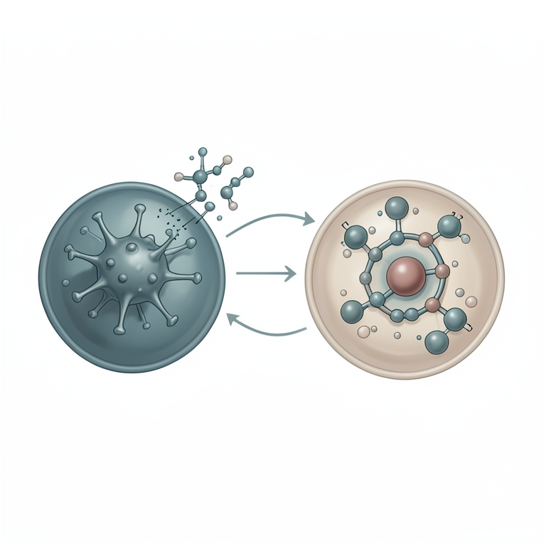
The Interferon Signaling Cascade
AI-generated conceptual illustration (mammalian cell and RNA virus). It explains the idea and does not show study data.
View source evidence ↗Introduction
Interferons and interferon-stimulated genes (ISGs) play a crucial role in the immune response against viral infections. Interferons are a group of signaling proteins that are produced by host cells in response to viral invasion. They are part of the innate immune system and act as a first line of defense against viruses. Upon detection of viral components, infected cells release interferons to alert neighboring cells of the impending threat. These interferons bind to specific receptors on surrounding cells, triggering a cascade of events that result in the expression of various ISGs. ISGs are genes that are upregulated in response to interferon signaling. They encode proteins that have antiviral properties and play a key role in limiting viral replication and spread. ISGs can be broadly categorized into two groups: those that directly inhibit viral replication and those that modulate the immune response. Some ISGs encode enzymes that target different steps of the viral life cycle, such as viral entry, replication, and assembly. These enzymes can interfere with essential viral processes, preventing the virus from replicating efficiently. Other ISGs are involved in the activation and regulation of immune cells, enhancing the immune response against the virus. The induction of interferons and ISGs is tightly regulated to ensure an appropriate immune response. The production of interferons is triggered by the recognition of viral components by pattern recognition receptors (PRRs) within infected cells. PRRs can detect various viral molecules, such as viral RNA or DNA, and initiate a signaling pathway that leads to interferon production. Once released, interferons bind to their receptors on neighboring cells, activating intracellular signaling pathways that result in the upregulation of ISGs. The role of interferons and ISGs in viral infections extends beyond their direct antiviral effects. They also play a crucial role in shaping the adaptive immune response. Interferons can enhance antigen presentation by infected cells, leading to a more efficient recognition of viral antigens by T cells. Additionally, they can regulate the differentiation and function of various immune cell types, such as dendritic cells and natural killer cells, further contributing to the elimination of viruses. Different components of innate immune system involved in active immunity against viral infections are discussed here.
Pattern recognition receptors
The presence of viral nucleic acids and PAMPs (pathogen associated molecular patterns) in the host cells are recognized by PRRs (Pattern recognition receptors). The PRRs are distributed in the cytoplasm, endosomes and on the cellular surface. The recognition of PAMPs by PRRs leads to activation of a cascade of reactions leading to the increased expression of type I Interferons as well as inflammatory cytokines and chemokines. Of all the PRRs involved in sensing RNA viral genomes, Toll-like receptors (TLRs) and RLRs have remarkable role. TLRs such as TLR2, TLR4 and TLR5 are expressed on the surface, while TLR3, TLR7, TLR8 and TLR9 are present on the endosomal compartments of some cell types. Retinoic acid-inducible gene-I (RIG-I)-like receptors (RLRs) are ubiquitous and recognize cytosolic RNA helicases. TLR3 recognises double stranded RNA (dsRNA) where as TLR7 and 8 detect single-stranded RNA (Said et al., 2018) [31] . TLRs, which belong to type I transmembrane proteins, have extracellular N-terminal with three major domains: Nterminal extracellular domain (ECD), which is rich in leucinerich repeats (LRRs) undergoes dimerization upon binding with the dsRNA of the pathogen and initiates the signal transduction cascade that is mediated through the transmembrane alpha helix domain. The third domain, Cterminal cytoplasmic toll-interleukin 1 receptor (TIR) domain-contains adaptor protein-inducing IFN-β (TRIF) is then recruited, which, upon undergoing slight conformational changes form a signalling complex with TNF receptorassociated factor 6 (TRAF6), TRAF3 and TBK1. This signalling complex activates NF-κB pathway leading to the production of type 1 IFNs and cytokines, respectively (Said et al., 2018) [31] . RLRs, which form the second group of PRRs involved in sensing viral RNA, belong to the family of cytosolic RNA helicases that specifically detects either double stranded or single stranded RNA species derived from viruses in the cytoplasm. Till date, three RLRs such as RIG-I (Retinoic acid inducible gene I), MDA-5 (Melanoma differentiation associated factor 5) and LGP2 (Laboratory of Genetics and Physiology 2) have been identified. Basal or low levels of RLR expression is maintained in normal uninfected cells; however, the expression is upregulated following exposure to IFN or viral infection (Yoneyama et al., 2004) [45] . They are involved in activating the innate immune response in myeloid cells, epithelial cells, and cells of central nervous system. Further, MDA-5 is reported to be upregulated in cells lacking IFN receptor upon virus infection indicating that the virus can directly upregulate RLR expression (Yount et al., 2007) [46] . Once viral RNA PAMPs is recognized by the C terminal domain, RIG-I or MDA-5 undergoes a conformational change and the latent CARD domains are exposed, leading to an association with CARD adaptor protein mitochondrial antiviral signalling (MAVS) through homotypic CARD-CARD interactions leading to dimerization of MAVS. The dimerization of MAVS recruits TNF receptor-associated factors (TRAFs), resulting in the activation of transcription factors such as nuclear factor-κB (NF-κB) and IFN regulatory factor (IRF)-3/7. Homodimerized IRFs and activated NF-κB translocate to the nucleus and activate the production of proinflammatory cytokines and type I IFN transcripts (Chiba et al., 2015) [5] . Members of RLR family are involved in recognizing a variety of viruses. For instance, RIG-I senses hepaciviruses and the members of Paramyxoviridae, Rhabdoviridae and Orthomyxoviridae. Both MDA-5 and RIG-I are involved in recognition of certain viruses like West nile virus, Dengue virus and Reo viruses (Kato et al., 2006) [17] . Studies have suggested that innate immune responses in Picornavirus infection is triggered by MDA-5. MDA-5 recognises large dsRNA fragments, representing either part of a viral genome or a viral replication product (Feng et al., 2012) [10] . During the course of viral replication, negative strand RNA and double stranded RNA (dsRNA) are formed, known as the replicative form (RF), triggers a significant IFN α/β response (Kato et al., 2006) [17] .
Interferons
Interferons (IFNs), the first line of host innate immune defenses against viral infections, were discovered by Issacs and Lindenmann in 1957 [16] during the infection of chick chorio-allantoic cells with influenza virus. The authors demonstrated that infected cells produced a secreted factor that induced a virus resistant state. IFNs belong to a large family of multifunctional, secreted proteins involved in antiviral defense, cell growth, differentiation, apoptosis and modulation of immune function through autocrine and paracrine mechanisms (Samuel, 2001) [32] . The IFNs are classified into three distinct types depending on the receptor complex they signal through-type I, II and III. Type I IFNs, also called viral IFNs include many subtypes like IFN-α, IFN-β, IFN-ε, IFN-κ and IFN-ω, that signals through the type I IFN heterodimeric receptor complex comprising IFNAR1 and IFNAR2. Type I IFNs are primarily induced by virus infections and can be synthesized by almost all cell types particularly upon viral infection. They have antiproliferative and immunomodulatory activities along with the antiviral activity. Type II IFNs are also known as immune IFN with IFN γ as the only member. They are stimulated by mitogen or antigen stimuli and are synthesized by cells of immune system. Type II IFNs mainly have pro-inflammatory and immunomodulatory functions that are distinct from type I and type III IFN functions (Lee and Ashkar, 2018). Type III IFNs include IFN λ1, IFN λ2and IFN λ3 and are stimulated by similar pathogen sensing pathway as of type I IFN and elicits antiviral, anti-proliferative and immunomodulatory activities. These class of IFNs confer antiviral activity at epithelial surfaces and provides a front-line defence that confers less collateral damage than type I IFNs (Lazear et al., 2019) [22] . Type III IFNs signal through heterodimeric receptor, IFNLR, and forms a signalling-component ternary complex upon binding. Despite the differences in their receptors, all the IFNs signal through JAK-STAT pathway. Upon binding, IFN induces phosphorylation of JAK1/TYK2 kinases that in-turn activate the latent cytoplamsic proteins STAT1 and STAT2. The phosphorylated STATs heterodimerize and bind to IRF9 forming the ISGF3 complex, which translocate to the nucleus to activate IFN stimulated genes through ISRE dependent promoters (Dussurget et al., 2014) [8] . Though type III IFNs binding to their cognate receptor, IFNLR1, induces similar signalling cascade, it activates GAS-dependent promoters in the nucleus. Expression of genes during IFN signalling include ISGs, the Interferon stimulated genes, which are involved in wide range of activities (Schneider et al., 2014) [33] .
Interferon Stimulated Genes
Interferon stimulated genes (ISGs), the effectors of IFN signalling, are engaged in a wide array of activities in the cell especially during viral infection. Over 300 ISGs are identified till date and often detects viral molecules and modulate signalling pathways. ISGs activate transcription factors of JAK-STAT pathway to augment IFN production and protection from virus (Sadler and Williams, 2008) [30] . Some ISGs act in synergistic manner to augment specific PRR signalling pathways. Some important ISG candidates pertaining to antiviral activity are OAS/RNaseL, viperin, PKR (Protein kinase R), ISG-15, Mx-proteins, Cholesterol 25 hydroxylase (Ch25H), TRIM, IFIT, IFITM. Some ISGs like OAS and PKR can recognize double stranded RNA in the cells. OAS is expressed at constitutive levels in the cytosol and are also induced by IFNs. Cytosolic PAMP (viral RNA) detection by OAS leads to the synthesis of 2'-5'oligoadenylates, which then act as intracellular second messengers to activate latent RNaseL (Hovanessian and Justesen, 2007) [14][15] resulting in widespread cleavage of both host and viral RNA. And, the production of additional PAMPs reinforce the innate immune response. In addition to the OAS/RNaseL system, activation of protein kinase R (PKR) by cytosolic PAMPs leads to a dramatic reduction in host cell translation as well as to the degradation of the inhibitor of κB (ΚB) that results in activation of the NF-κB signalling pathway.
Conclusion
In conclusion, interferons and interferon stimulated genes play a crucial role in the immune response against viral infections. Interferons are cytokines that are produced by infected cells as a defense mechanism to limit viral replication and spread. They act by inducing an antiviral state in surrounding cells, which helps to inhibit viral replication and prevent the spread of infection. Additionally, interferon stimulated genes are activated by interferons and play a key role in the antiviral response. These genes produce proteins that have various antiviral activities, such as inhibiting viral replication, degrading viral RNA, and activating immune cells. Together, interferons and interferon stimulated genes form a potent defense system against viral infections. In summary, understanding the role of interferons and interferon stimulated genes in viral infections is critical for the development of effective antiviral therapies. Further research is needed to explore the specific mechanisms by which these molecules exert their antiviral effects and to identify potential interventions that can enhance their activity. By harnessing the power of interferons and their downstream targets, we can hope to improve our ability to combat viral infections and protect public health.
References
44 in the original paper- Anafu AA, Bowen CH, Chin CR, Brass AL, Holm GH. Interferon-inducible transmembrane protein 3 (IFITM3) restricts reovirus cell entry. J Biol Chem. 2013;288(24):17261-17271. https://doi.org/10.1074/jbc.M112.438515
- Bailey CC, Zhong G, Huang IC, Farzan M. IFITMfamily proteins: The cell's first line of antiviral defense. Annu Rev Virol. 2014;1:261-283. https://doi.org/10.1146/annurev-virology-031413-085537
- Brennan-Laun SE, Ezelle HJ, Li XL, Hassel BA. RNaseL control of cellular mRNAs: roles in biologic functions and mechanisms of substrate targeting. J Interferon Cytokine Res. 2014;34(4):275-288. https://doi.org/10.1089/jir.2013.0147
- Cai KJ, Meng QL, Qiao J, Huang J, Zhang ZC, Wang GC, et al. Expression of bovine Mx1 protein inhibits the replication of foot-and-mouth disease virus in BHK-21 cells. Acta Virol. 2013;57(4):429-434. https://doi.org/10.4149/av_2013_04_429
- Chiba Y, Matsumiya T, Satoh T, Hayakari R, Furudate K, Xing F, et al. Retinoic acid-inducible gene-I-like receptor (RLR)-mediated antiviral innate immune responses in the lower respiratory tract: roles of TRAF3 and TRAF5. Biochem Biophys Res Commun. 2015;467(2):191-196. https://doi.org/10.1016/j.bbrc.2015.10.010
- Doms A, Sanabria T, Hansen JN, Altan-Bonnet N, Holm GH. 25-Hydroxycholesterol production by the cholesterol-25-hydroxylase interferon-stimulated gene restricts mammalian reovirus infection. J Virol. 2018;92(18)
- Dussurget O, Bierne H, Cossart P. The bacterial pathogen Listeria monocytogenes and the interferon family: type I, type II and type III interferons. Front Cell Infect Microbiol. 2014;4:50. https://doi.org/10.3389/fcimb.2014.00050
- Einav S, Glenn JS. Prenylation inhibitors: a novel class of antiviral agents. J Antimicrob Chemother. 2003;52(6):883-886. https://doi.org/10.1093/jac/dkg490
- Feng Q, Hato SV, Langereis MA, Zoll J, Virgen-Slane R, Peisley A, et al. MDA5 detects the double-stranded RNA replicative form in picornavirus-infected cells. Cell Rep. 2012;2(5):1187-1196. https://doi.org/10.1016/j.celrep.2012.10.005
- He F. Bradford protein assay. Bio-101. 2011;e45. https://doi.org/10.21769/BioProtoc.45
- Hinson ER, Cresswell P. The antiviral protein, viperin, localizes to lipid droplets via its N-terminal amphipathic alpha-helix. Proc Natl Acad Sci U S A. 2009;106(48):20452-20457. https://doi.org/10.1073/pnas.0911679106
- Hinson ER, Cresswell P. The N-terminal amphipathic alpha-helix of viperin mediates localization to the cytosolic face of the endoplasmic reticulum and inhibits protein secretion. J Biol Chem. 2009;284(7):4705-4712. https://doi.org/10.1074/jbc.M807261200
- Hovanessian AG. On the discovery of interferoninducible, double-stranded RNA activated enzymes: the 2'-5'oligoadenylate synthetases and the protein kinase PKR. Cytokine Growth Factor Rev. 2007;18(5-6):351361. https://doi.org/10.1016/j.cytogfr.2007.06.003
- Hovanessian AG, Justesen J. The human 2'- 5'oligoadenylate synthetase family: unique interferoninducible enzymes catalyzing 2'-5' instead of 3'-5' phosphodiester bond formation. Biochimie. 2007;89(67):779-788. https://doi.org/10.1016/j.biochi.2007.02.003
- Isaacs A, Lindenmann J. Virus interference. I. The interferon. Proc R Soc Lond B Biol Sci. 1957;147(927):258-267. https://doi.org/10.1098/rspb.1957.0048
- Kato H, Takeuchi O, Sato S, Yoneyama M, Yamamoto M, Matsui K, et al. Differential roles of MDA5 and RIGI helicases in the recognition of RNA viruses. Nature. 2006;441(7089):101-105. https://doi.org/10.1038/nature04734
- Ke W, Fang L, Jing H, Tao R, Wang T, Li Y, et al. Cholesterol 25-hydroxylase inhibits porcine reproductive and respiratory syndrome virus replication through enzyme activity-dependent and -independent mechanisms. J Virol. 2017;91(19)
- Kuhl BD, Cheng V, Wainberg MA, Liang C. Tetherin and its viral antagonists. J Neuroimmune Pharmacol. 2011;6(2):188-201. https://doi.org/10.1007/s11481-0109256-1
- Kumar A, Haque J, Lacoste J, Hiscott J, Williams BR. Double-stranded RNA-dependent protein kinase activates transcription factor NF-kappa B by phosphorylating I kappa B. Proc Natl Acad Sci U S A. 1994;91(14):62886292. https://doi.org/10.1073/pnas.91.14.6288
- Lazear HM, Schoggins JW, Diamond MS. Shared and distinct functions of type I and type III interferons. Immunity. 2019;50(4):907-923. https://doi.org/10.1016/j.immuni.2019.03.025
- Lee AJ, Ashkar AA. The dual nature of type I and type II interferons. Front Immunol. 2018;9:2061. https://doi.org/10.3389/fimmu.2018.02061
- Li W, Mao L, Cao Y, Zhou B, Yang L, Han L, et al. Porcine viperin protein inhibits the replication of classical swine fever virus (CSFV) in vitro. Virol J. 2017;14(1):202. https://doi.org/10.1186/s12985-0170868-4
- Li XL, Blackford JA Jr, Judge CS, Liu M, Xiao W, Kalvakolanu DV, et al. RNase-L-dependent destabilization of interferon-induced mRNAs. A role for the 2-5A system in attenuation of the interferon response. J Biol Chem. 2000;275(12):8880-8888. https://doi.org/10.1074/jbc.275.12.8880
- Liu SY, Aliyari R, Chikere K, Li G, Marsden MD, Smith JK, et al. Interferon-inducible cholesterol-25-hydroxylase broadly inhibits viral entry by production of 25hydroxycholesterol. Immunity. 2013;38(1):92-105. https://doi.org/10.1016/j.immuni.2012.11.005
- McMahon HT, Gallop JL. Membrane curvature and mechanisms of dynamic cell membrane remodelling. Nature. 2005;438(7068):590-596. https://doi.org/10.1038/nature04396
- Okumura F, Zou W, Zhang DE. ISG15 modification of the eIF4E cognate 4EHP enhances cap structure-binding activity of 4EHP. Genes Dev. 2007;21(3):255-260. https://doi.org/10.1101/gad.1521607
- Perez-Caballero D, Zang T, Ebrahimi A, McNatt MW, Gregory DA, Johnson MC, et al. Tetherin inhibits HIV-1 release by directly tethering virions to cells. Cell. 2009;139(3):499-511. https://doi.org/10.1016/j.cell.2009.08.039
- Sadler AJ, Williams BR. Interferon-inducible antiviral effectors. Nat Rev Immunol. 2008;8(7):559-568. https://doi.org/10.1038/nri2314
- Said EA, Tremblay N, Al-Balushi MS, Al-Jabri AA, Lamarre D. Viruses seen by our cells: the role of viral RNA sensors. J Immunol Res. 2018;2018:9480497. https://doi.org/10.1155/2018/9480497
- Samuel CE. Antiviral actions of interferons. Clin Microbiol Rev. 2001;14(4):778-809. https://doi.org/10.1128/CMR.14.4.778-809.2001
- Schneider WM, Chevillotte MD, Rice CM. Interferonstimulated genes: a complex web of host defenses. Annu Rev Immunol. 2014;32:513-545. https://doi.org/10.1146/annurev-immunol-032713120231
- Shi HX, Yang K, Liu X, Liu XY, Wei B, Shan YF, et al. Positive regulation of interferon regulatory factor 3 activation by Herc5 via ISG15 modification. Mol Cell Biol. 2010;30(10):2424-2436. https://doi.org/10.1128/MCB.01466-09
- Silverman RH, Skehel JJ, James TC, Wreschner DH, Kerr IM. rRNA cleavage as an index of ppp(A2'p)nA activity in interferon-treated encephalomyocarditis virusinfected cells. J Virol. 1983;46(3):1051-1055. https://doi.org/10.1128/JVI.46.3.1051-1055.1983
- Sun L, Lv F, Guo X, Gao G. Glycogen synthase kinase 3β (GSK3β) modulates antiviral activity of zinc-finger antiviral protein (ZAP). J Biol Chem. 2012;287(27):22882-22888. https://doi.org/10.1074/jbc.M111.306373
- Tan KS, Olfat F, Phoon MC, Hsu JP, Howe J, Seet JE, et al. In vivo and in vitro studies on the antiviral activities of viperin against influenza H1N1 virus infection. J Gen Virol. 2012;93(Pt 6):1269-1277. https://doi.org/10.1099/vir.0.040824-0
- Tang YD, Na L, Zhu CH, Shen N, Yang F, Fu XQ, et al. Equine viperin restricts equine infectious anaemia virus replication by inhibiting the production and/or release of viral Gag, Env, and receptor via distortion of the endoplasmic reticulum. J Virol. 2014;88(21):1229612310. https://doi.org/10.1128/JVI.01379-14
- Teng TS, Foo SS, Simamarta D, Lum FM, Teo TH, Lulla A, et al. Viperin restricts chikungunya virus replication and pathology. J Clin Invest. 2012;122(12):4447-4460. https://doi.org/10.1172/JCI63120
- Van der Hoek KH, Eyre NS, Shue B, Khantisitthiporn O, Glab-Ampi K, Carr JM, et al. Viperin is an important host restriction factor in control of Zika virus infection. Sci Rep. 2017;7(1):4475. https://doi.org/10.1038/s41598017-04138-1
- Verhelst J, Hulpiau P, Saelens X. Mx proteins: antiviral gatekeepers that restrain the uninvited. Microbiol Mol Biol Rev. 2013;77(4):551-566. https://doi.org/10.1128/MMBR.00024-13
- Vonderstein K, Nilsson E, Hubel P, Nygård Skalman L, Upadhyay A, Pasto J, et al. Viperin targets flavivirus virulence by inducing assembly of noninfectious capsid particles. J Virol. 2018;92(1)
- Yoneyama M, Kikuchi M, Natsukawa T, Shinobu N, Imaizumi T, Miyagishi M, et al. The RNA helicase RIG-I has an essential function in double-stranded RNAinduced innate antiviral responses. Nat Immunol. 2004;5(7):730-737. https://doi.org/10.1038/ni1087
- Yount JS, Moran TM, López CB. Cytokine-independent upregulation of MDA5 in viral infection. J Virol. 2007;81(13):7316-7319. https://doi.org/10.1128/JVI.00545-07
- Yuan B, Fang H, Shen C, Zheng C. Expression of porcine Mx1 with FMDV IRES enhances the antiviral activity against foot-and-mouth disease virus in PK-15 cells. Arch Virol. 2015;160(8):1989-1999. https://doi.org/10.1007/s00705-015-2473-4
- Zheng X, Wang X, Tu F, Wang Q, Fan Z, Gao G. TRIM25 is required for the antiviral activity of zinc finger antiviral protein. J Virol. 2017;91(9)