
Research article · 2024 · Full text
International Journal of Veterinary Sciences and Animal HusbandryGastrotomy in a Checkered Keelback snake for the removal of a fishhook: A case report
Volume 9(3S) · Pages 174–176 · DOI: 10.22271/veterinary.2024.v9.i3sc.1431 ↗
Research areasTurtle Biology and ConservationRabies epidemiology and controlLeech Biology and Applications
Abstract
Author-writtenA Checkered Keelback snake was presented with a history of being caught on a fishing line and had swallowed the hook along with the bait. The fishhook was palpable in the middle third of the body. Based on the definitive history and confirmation on palpation, the decision for surgical retrieval of the hook was taken. The snake was anaesthetised using inj. ketamine @ 20 mg/kg and inj. diazepam @ 0.4 mg/kg intramuscularly. A ventral midline celiotomy followed by Gastrotomy were performed and the hook was removed. An antibiotic and an analgesic were administered. The snake was kept off feed for four days with supportive parenteral fluid therapy following which it was fed orally every two days. The snake recovered uneventfully and was released back into the wild.
Summary
Source-checkedPlain-language reading of the full paper, prepared with AI and checked against the text. Each point links to the authors' own words.
Why it matters
- Snakes may accidentally swallow inedible foreign objects if those objects are contaminated with smells that attract them. View evidence ↗
- Failure to pass ingested foreign bodies can lead to complete gastrointestinal obstruction, resulting in dehydration, muscle atrophy, and death. View evidence ↗
The question
- The study aims to report the successful surgical management of a Checkered Keelback snake that accidentally swallowed a fishhook. View evidence ↗
What they did
- The subject of the case report was a Checkered Keelback snake weighing 144 g. View evidence ↗
- Anaesthesia was induced using intramuscular injections of ketamine at 20 mg/kg and diazepam at 0.4 mg/kg. View evidence ↗
- The surgical procedure involved a ventral midline incision, a gastrotomy to remove the hook, stomach suturing with 3-0 chromic catgut, and skin closure with nylon. View evidence ↗
- Post-operative care included intramuscular injections of enrofloxacin (5 mg/kg) and meloxicam (0.25 mg/kg) every 24 hours for three days. View evidence ↗
- The snake received parenteral fluid therapy with Ringer’s lactate at 20 ml/kg/day and was kept off feed for four days. View evidence ↗
What they found
- The chosen anaesthetic protocol provided adequate muscle relaxation and a lack of response to noxious stimuli 30 minutes after administration. View evidence ↗
- The snake recovered from anaesthesia 2.5 hours after induction and returned to normal activity approximately 10 hours later. View evidence ↗
- Following the removal of sutures 20 days post-surgery, the snake showed normal eating and defecation, weight gain, and alertness, allowing for its release into the wild. View evidence ↗
What it means
- The authors conclude that prompt surgical removal of ingested fishhooks is critical for improving the prognosis and ensuring an uneventful recovery in snakes. View evidence ↗
- The authors note that while they achieved a favorable outcome, the anaesthetic protocols and suture materials used were chosen based on availability rather than being the most recommended options. View evidence ↗
Visual summary
1 visualsIllustrations explain the paper; they do not show study data.
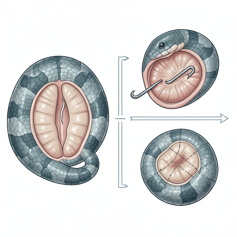
Surgical Process Flow
AI-generated conceptual illustration (Checkered Keelback snake). It explains the idea and does not show study data.
View source evidence ↗Figures & tables
From the original paper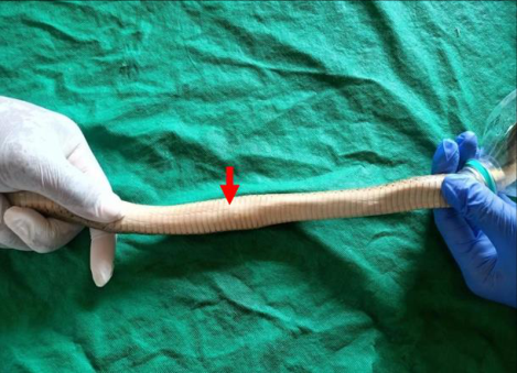 Figure 1
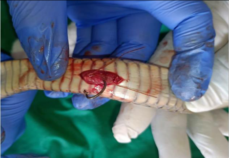 Figure 2
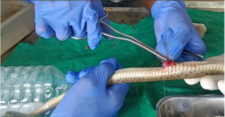 Figure 3
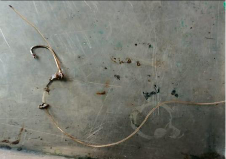 Figure 4
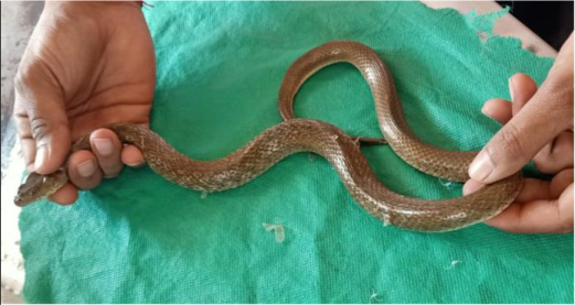 Figure 5
Introduction
Snakes exhibit great adaptability and resilience to various environmental conditions, allowing their extensive distribution across various habitats. They are carnivorous reptiles that rely on taste and olfaction to select their prey [1] . Hence, contamination of inedible foreign objects with some kind of smell may cause a snake to accidentally swallow them [1,2] . Such incidents wherein snakes have swallowed various foreign bodies like an artificial chicken egg, plastic bottle, piece of cloth, male contraceptive, socks, plastic bag and golf balls have been reported [3, 1, 2, 4, 5] . If a snake is unable to pass such foreign bodies by regurgitation or defecation, complete obstruction of the gastrointestinal tract may occur that may lead to chronic issues like dehydration and muscle atrophy eventually leading to death [1,5] . This case reports the accidental swallowing of a fishhook by a Checkered Keelback snake and its successful surgical management, a rare but noteworthy event that highlights the complex and sometimes hazardous intersection between natural habitats and recreational fishing.
Case report
A Checkered Keelback snake was presented with a history of being caught on a fishing line and had swallowed the hook along with the bait. The fishing line had been cut before the presentation of the snake. On clinical examination, the snake appeared active and alert. The fishhook was palpable in the middle third of the body (Fig 1). The snake with the ingested hook weighed 144 g. As the facility for taking radiographs was not available, the decision for surgical retrieval of the fishhook was taken based on the definitive history and confirmation on palpation. The snake was anaesthetised using inj. ketamine @ 20 mg/kg and inj. diazepam @ 0.4 mg/kg intramuscular (IM) [6] . The snake exhibited, adequate muscle relaxation and lack of response to noxious stimuli 30 minutes post-anaesthetic administration. Surgical site was prepared aseptically. A ventral midline incision was taken just above the palpable hook. The stomach was identified and incised. The hook was carefully removed and the stomach was sutured with 3-0 chromic catgut using Cushing followed by Lembert suture pattern (Fig 2 & 3). The body wall was apposed and the skin was closed with nylon using horizontal suture pattern [1] . The snake was administered inj. enrofloxacin @ 5 mg/kg and inj. meloxicam @ 0.25 mg/kg IM every 24 hours for three days [6] . The snake was kept off feed for four days with supportive parenteral fluid therapy with inj. Ringer's lactate @ 20 ml/kg/day [5] .
Following which it was fed half an egg orally every two days. The sutures were removed 20 days post-surgery. The snake ate normally, defecated normally, gained weight and was alert and active prompting its release back into the wild (Fig 5).
Discussion
The Checkered Keelback (Fowlea piscator), also known as the 'Asiatic water snake' is a non-venomous species commonly found in India [7] . They are a semi-aquatic, cathemeral species found in and around water bodies [8] . In the present case, the snake accidentally ingested the fishhook along with the bait which was retrieved surgically. Various anaesthetic combinations have been described for gastrointestinal surgery and endoscopic procedures in snakes. Souza et al. (2004) induced anaesthesia with 5% isoflurane after premedication with 1 mg/kg of inj. butorphanol tartrate IM followed by maintenance with 1.5-2% isoflurane whereas, Vasaruchapong and Chanhome (2013) utilized inj. ketamine @ 40 mg/kg and inj. xylazine @ 2 mg/kg IM. In a more recent study, Scott et al. (2021) evaluated four different induction protocols in rat snakes: (i) isoflurane after premedication with inj. morphine @ 0.66 mg/kg IM, (ii) inj. propofol @ 7.5 mg/kg intracardiac and inj. ketamine @ 15 mg/kg IM, (iii) inj. alfaxalone @ 5 mg/kg intravenous (IV), and (iv) a combination of inj. alfaxalone and dexmedetomidine (7.5 mg/kg + 100 µg/kg IV) followed by isoflurane in oxygen for maintenance. As snakes lack a diaphragm, their breathing is controlled by skeletal intercostal muscles, which prove insufficient at surgical plane of anaesthesia, necessitating the need for positive pressure ventilation [9] . Propofol and alfaxalone provide controlled and rapid induction but their use is limited by their intravenous route and requirement of positive pressure ventilation in case of alfaxolone whereas, isoflurane or sevoflurane are preferred for the maintenance of anaesthesia as they are safer, more controllable and facilitate faster recoveries [9] . In the present case, inj. ketamine @ 20 mg/kg and inj. diazepam @ 0.4 mg/kg were administered intramuscularly which provided adequate sedation and muscle relaxation enough for the completion of the surgical procedure. The snake recovered 2.5 h post induction and it resumed normal activity approximately 10 h post induction. In reptiles, ketamine when used alone provides marginal analgesia and muscle relaxation hence it is recommended to be used as a preanesthetic prior to inhalation anaesthesia with isoflurane, or in combination with butorphanol, diazepam or midazolam for better muscle relaxation [6] . A ventral midline incision was preferred in this case to minimize trauma during the removal of the broad fishhook (Fig 2 &4). A scalloped incision on the lateral aspect of the body where the scutes and scales meet is the preferred site for celiotomy as it enhances wound healing and the incision wound is spared from contact with the ground [10] . However, there are no differences observed between the lateral and the midline ventral incision for providing better surgical exposure [10] . Chromic catgut in the reptile patient is not readily absorbed as it is dependent on proteolysis for its dissolution and should be avoided. It is recommended to remove skin sutures 4-6 weeks post-surgery or post subsequent ecdysis [10] . Although better anaesthetic protocols and suture materials are recommended, in this case, ketamine and diazepam were used as anaesthetics and chromic catgut as the suture material due to their ready availability which yielded favourable outcomes.
Conclusion
The ingestion of fishhooks by snakes is rare but poses a significant threat to their lives. In the present case, the prompt surgical removal of the fishhook led to an early and uneventful recovery, emphasizing the importance of immediate veterinary intervention to improve the prognosis.
Acknowledgement
We acknowledge the Maharashtra Forest Department, Animal Sahara Foundation, Virbhadra Desai and Aditya Bhosale for their invaluable support and contributions thus making this study possible.
Figures and tables from the paper
References
9 in the original paper- Vasaruchapong T, Chanhome L. Surgical removal of foreign bodies in the gastrointestinal tract of Monocellate Cobra, Naja kaouthia. The Thai Journal of Veterinary Medicicne. 2013;43(2):297-300.
- Sharma V, Sayyed A, Bhandari R. Herbivory and inanimate objects in the diet of the Oriental Ratsnake, Ptyas mucosa (Linnaeus 1758). IRCF Reptiles and Amphibians Journal. 2016;23(2):102-103.
- Souza MJ, Hall KE, Wilson JD, Lewbart GA. Surgical removal of an artificial chicken egg from the gastrointestinal tract of a Black Rat Snake, Elaphe obsoleta. Journal of Herpetological Medicine and Surgery. 2004;14(4):4-5.
- Deshmukh RV, Deshmukh SA, Badhekar SA, Katgube SD. A plastic bag consumed by a Common Indian Krait, Bungarus caeruleus (Schneider 1801). IRCF Reptiles and Amphibians Journal. 2017;24(3):172-174.
- Scott G, Waffa BJ, DeVoe RS, Harms CA, Lewbart GA. Successful and unsuccessful non-surgical removal of ingested golf balls and an artificial egg in four rat snakes (Pantherophis alleghianensis). Journal of the American Veterinary Medical Association. 2021;258(10):11351141.
- Gibbons PM, Klaphake E, Carpenter JW. Reptiles. In: Exotic animal formulary. Edn 4. Elsevier Saunders, St. Louis, Missouri, 2013, p. 84-113.
- Chequered Keelback (Snakes of peninsular India). iNaturalist. https://www.inaturalist.org/guide_taxa/1331777. 25 May, 2024.
- Parmar DS. Notes on the Checkered Keelback, Xenochrophis piscator (Schneider 1799), in Gujarat, India. IRCF Reptiles and Amphibians Journal. 2018;25(2):115-119.
- Divers SJ. Reptiles. In: The Merck Veterinary Manual. Edn 11, Merck and co., Inc., Kenilworth, NJ, USA; c2016. p. 1978-1980.