
Research article · 2024 · Full text
International Journal of Veterinary Sciences and Animal HusbandryCongenital eventration and epitheliogenesis imperfecta in a male goat kid: A case report
Volume 9(3) · Pages 256–258 · DOI: 10.22271/veterinary.2024.v9.i3d.1408 ↗
Research areasMilk Quality and Mastitis in Dairy CowsCancer Cells and MetastasisTissue Engineering and Regenerative Medicine
Abstract
Author-writtenA four hours old goat kid was presented with protrusion of a transparent peritoneal sac containing congested stomach and intestines from a ventral abdominal defect at the umbilical region. Based on clinical examination the case was diagnosed as eventration with epitheliogenesis imperfecta. A small linear incision was made at the cranial aspect of the abdominal defect following which the kid started gasping. Despite immediate efforts to manage the condition the kid succumbed. This communication reports the occurrence of congenital eventration with epitheliogenesis imperfecta in a male goat kid.
Summary
Source-checkedPlain-language reading of the full paper, prepared with AI and checked against the text. Each point links to the authors' own words.
Why it matters
- Congenital anomalies are structural and functional deformities that are present when an animal is born. View evidence ↗
- The combination of congenital eventration and epitheliogenesis imperfecta is considered a rare anomaly in goats. View evidence ↗
- Congenital eventration occurs when the abdominal wall does not close completely during fetal development, causing visceral organs to protrude within a serous sac. View evidence ↗
- Epitheliogenesis imperfecta is characterized by a lack of skin in certain body areas at birth due to a congenital discontinuity of the squamous epithelium. View evidence ↗
The question
- The authors aim to report a case of congenital eventration with epitheliogenesis imperfecta in a male goat kid and analyze why the animal died. View evidence ↗
What they did
- The subject of the case report was a male goat kid, four hours old, presenting with protruding viscera at the umbilical region. View evidence ↗
- Diagnosis was based on clinical examination, which identified a transparent peritoneal sac attached at the umbilical region and protruding from a ventral abdominal defect. View evidence ↗
- Surgical correction was attempted using 2% lignocaine hydrochloride for local infiltration and a 1 cm linear incision at the cranial margin of the defect. View evidence ↗
What they found
- The protruding peritoneal sac contained a congested abomasum and intestines that exhibited no motility. View evidence ↗
- The kid initially suckled but later stopped, passed only a small amount of urine, and did not defecate. View evidence ↗
- Immediately after a 1 cm incision was made, the kid began gasping and subsequently died despite management efforts. View evidence ↗
- The authors suggest the kid's death was likely caused by a communication between the thoracic and abdominal cavities, which led to a loss of negative pressure in the thorax upon incision. View evidence ↗
- Unlike some reported trends where defects are common in multiparous does, this kid was born to a 1.2-year-old doe during her first pregnancy. View evidence ↗
What it means
- The authors conclude that congenital eventration with epitheliogenesis imperfecta is rare in goats and may occur alongside other conditions. View evidence ↗
- The authors recommend using X-ray and ultrasonography to differentiate coexisting conditions and improve diagnostic accuracy and clinical outcomes. View evidence ↗
- Because the owner did not allow a post-mortem examination, the authors could not rule out other developmental anomalies of the diaphragm or other organs as causes of death. View evidence ↗
Figures & tables
From the original paper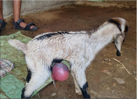 Figure 1
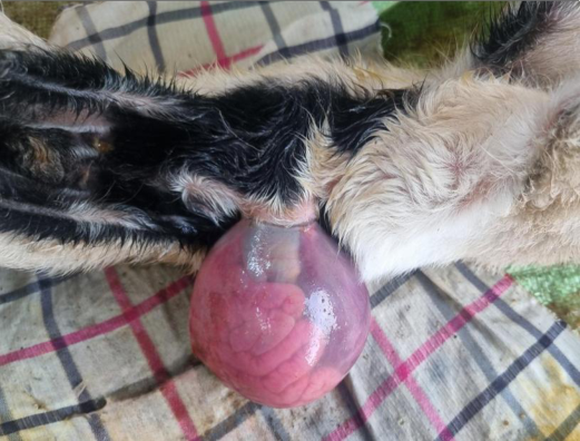 Figure 2
Introduction
Congenital anomalies are defined as the structural and functional deformities present at the time of birth [1] . While the precise reason behind congenital defects in small ruminants remains unclear, several factors have been suggested as probable causes during gestation, including inheritance, genetics, and the environment (Toxic, infectious, and nutritional) [2] . Congenital anomalies like atresia ani, umbilical hernia, dermoid cyst, urethral diverticulum, monster kid, epitheliogenesis imperfecta, eventration, urethral dilatation, ectopia cordis, double monster, etc. can be observed in small ruminants [2,3,4] . In goats, congenital eventration with epitheliogenesis imperfecta is a rare congenital anomaly [1] . Congenital eventration refers to the protrusion of visceral organs contained in a serous sac as a result of incomplete closure of the abdominal wall during fetal development [5] . Epitheliogenesis imperfecta is a congenital discontinuity of squamous epithelium which is seen as lack of skin in specific areas of the body at the time of birth [6,7] . It is commonly seen in pigs while it is uncommon in sheep, dogs and cats. It is well described in cattle (autosomal recessive trait) and horses [6] . Diagnosis is usually made on the basis of history and clinical findings. This communication reports the occurrence of congenital eventration with epitheliogenesis imperfecta in a male goat kid and discusses the probable reasons for its demise.
Case Report
A four hours old male goat kid was presented with protrusion of viscera from the umbilical region. The kid suckled initially but eventually it stopped. It passed scanty urine but showed absence of defecation. Clinical examination revealed the presence of a transparent peritoneal sac protruding from the defect in the ventral abdomen to which it was attached at the umbilical region. The sac had remnants of the umbilical cord ventrally and contained a congested abomasum and intestines which showed no motility (Fig 1 & 2). Based on clinical examination, the case was diagnosed as eventration with epitheliogenesis imperfecta. As these cases have a better prognosis when corrected promptly to prevent contamination and injury to the organs [5] , the decision for surgical correction was made. The surgical site was prepared aseptically and 2% lignocaine hydrochloride was infiltrated at the site of the abdominal defect. A 1 cm linear incision was made at the cranial margin of the abdominal defect following which the kid started gasping.
Despite immediate efforts to manage the condition the kid succumbed. Gasping immediately after making a small abdominal incision might be suggestive of communication between thoracic and abdominal cavities.
Discussion
Umbilical hernia refers to the displacement of organs or part thereof through a defect in the abdominal wall at the umbilical region where the skin overlying the protruded organs is intact [8] whereas in congenital eventration there is protrusion of visceral organs with their serous sac from a ventral abdominal defect because of incomplete closure of the abdominal wall at the stage of fetal development [5] . Congenital defects were observed to be more common in calves and lambs born of dams from their first pregnancy while they were common in goat kids born from second pregnancy. Most small ruminants with congenital defects were produced by multiparous does or ewes [9] . Conversely, in the present case, the kid was born to a 1.2 years old doe in her first gestation. In the present case, despite aggressive efforts to manage the condition, the goat kid did not survive as it started gasping immediately upon making a small abdominal incision which might be suggestive of communication between thoracic and abdominal cavities, resulting in fall of negative pressure in the thoracic cavity. As post-mortem could not be performed due to noncompliance of the goat owner, the possibility of other developmental anomalies involving the diaphragm or other organ systems which might have caused the kid's demise could not be ruled out. This is the first reported case of congenital eventration with epitheliogenesis imperfecta in goat kid in Gondia district of Maharashtra, India.
Conclusion
Congenital eventration with epitheliogenesis imperfecta is a rare congenital anomaly in goats that might coexist with other conditions. Diagnostic techniques such as X-ray and ultrasonography will help differentiate these conditions and provide accurate diagnosis, ultimately improving clinical outcomes.
Figures and tables from the paper
References
11 in the original paper- Giggin T, Deny J, Anoop S, Sudheesh SN, Soumya R, John Martin KD. Congenital eventration with epitheliogenesis imperfecta in a day-old kid (Capra hircus) and its surgical management. Journal of Indian Veterinary Association, Kerala. 2022;20(2):81-84.
- Senna NA, Abu-seida AM, Gadallah SM, El-husseiny IN, Rakha GM. Congenital anomalies in native breeds of sheep and goats: a report on 120 cases of 24 varieties. Veterinary Medical Journal, Giza. 2003;51(3):363-380.
- Samad MA. A systematic review of congenital anomalies in calves and kids reported during the period from 1975 to 2021 in Bangladesh. Journal of Veterinary Medical and One Health Research. 2021;3(2):129-153.
- Upadhye SV, Dhoot VM. A case of ectopia cordis in a goat. Indian Veterinary Journal. 2001;78:1028-1029.
- Veena P, Sankar P, Kokila S, Kumar RVS, Lakshmi DN. Congenital umbilical defect with visceral eventration in a buffalo calf-a case report. Buffalo Bulletin. 2011;30(3):165-167.
- Benoit-Biancamano M-O, Drolet R, D'Allaire S. Aplasia cutis congenita (Epitheliogenesis imperfecta) in swine: Observations from a large breeding herd. Journal of Veterinary Diagnostic Investigation. 2006;18(6):573-579.
- Doijode V. Umbilical hernia in ruminant calves: A review. The Pharma Innovation Journal. 2019;8(4):164167.
- Polat E. Descriptive study of congenital anomalies encountered in ruminants in Elazig region of Turkey. Revista FAVE. Sección Ciencias Veterinarias, 2022;21:1-1.
- Saradamma T, Anoop S, Sooryadas S, Jose S, Nayar KNM. Congenital umbilical defect with visceral eventration in a kid. Indian Journal of Veterinary Surgery. 2000;21:112.
- Gangwar AK, Devi KS, Singh AK, Katiyar N, Patel G, Srivastava S. Congenital anomalies and their surgical correction in ruminants. Advances in Animal and Veterinary Sciences. 2014;2:369-376.
- Praharee TP. Surgical management of epitheliogenesis imperfecta with congenital eventration in a neonatal calf. https://www.pashudhanpraharee.com/surgicalmanagement-of-epitheliogenesis-imperfecta-withcongenital-eventration-in-a-neonatal-calf/. 11 February, 2024
Cited by 1
From OpenAlex, OpenCitations and reference lists read by Scinovex
- Emergency management of necrotic rectal prolapse in a domestic cat ↗
Fadeli Bermani, Septiyani · ARSHI Veterinary Letters · 2025
A case of rectal prolapse in a domestic cat was treated at Veterinary Teaching Hospital, Universitas Padjadjaran, Indonesia. The cat presented with a six-day history of a reddish mass protruding from the anus. Physical examination revealed a prolapsed rectum with areas of necrosis, pale mucous membranes, and decreased skin turgor, which was consistent with dehydration. The fecal examination was negative…
❝ 0PDF