
Research article · 2024 · Full text
International Journal of Advanced Biochemistry ResearchEffect of 5-fluorouracil drug and Annona Muricta seed extract on induced tumors in laboratory rats
Volume 8(5) · Pages 10–17 · DOI: 10.33545/26174693.2024.v8.i5a.1049 ↗
Research areasTraditional and Medicinal Uses of AnnonaceaeCancer Treatment and PharmacologyNatural product bioactivities and synthesis
Abstract
Author-writtenCancer is one of the diseases that rapidly appear worldwide, affecting 82% of the world's population. It is a complex disorder involving modifications in the body's physiological conditions. This disease is considered a global issue, primarily affecting developing countries. According to research surveys, 63% of cancer-related deaths are reported in developing countries. Some studies describe it as an abnormal condition where a group of physiological cells ignore cell divisions and grow uncontrollably. These cells do not respond to signals that activate the natural cell cycle, exhibiting a degree of self-sufficiency leading to unregulated growth and spread in transformed cells. 90% of cancer cell spread is fatal due to their migration to adjacent tissues.Objectives: Study the effect of 5-Fluorouracil drug and Annona muricata seed extract on chemically induced tumors by Thioacetamide on the livers of laboratory rats and determine the changes occurring in the tissues. Method of work: Taking (48) male rats and dividing them into eight groups, each group consisting of six animals. (The first group) the control group, the natural control group, for a period of 28 days. (The second group) treated with 5-fluorouracil once a week for 28 days. (The third group) treated with the plant extract of seeds for 28 days. (The fourth group) treated with the plant extract of seeds and 5-fluorouracil for 28 days. (The fifth group) received daily doses of thioacetamide for 5 days. (The sixth group) received daily doses of thioacetamide for 5 days, then treated with 5-fluorouracil for 23 days as mentioned above. (The seventh group) received daily doses of thioacetamide and then treated with the extract. (The eighth group) received daily doses of thioacetamide for 5 days and then treated with 5-fluorouracil and the plant extract of seeds for 23 days. Results and Conclusion: Histological sections of the liver and chemical tests showed clear positive effects of the plant extract of seeds in rats treated with thioacetamide. Positive effects were also observed in rats treated with thioacetamide and injected with 5-Fluorouracil, and in rats treated with thioacetamide and the plant extract of seeds along with 5-Fluorouracil.
Summary
Source-checkedPlain-language reading of the full paper, prepared with AI and checked against the text. Each point links to the authors' own words.
Why it matters
- Cancer is a rapidly spreading global health issue that affects approximately 82% of the world's population. View evidence ↗
- Developing countries are disproportionately affected, accounting for 63% of cancer-related deaths. View evidence ↗
The question
- The study aimed to evaluate how 5-Fluorouracil and Annona muricata seed extract affect liver tumors induced by Thioacetamide in laboratory rats. View evidence ↗
What they did
- The experiment used 48 male rats divided into eight groups of six animals each. View evidence ↗
- Tumors were induced using Thioacetamide administered at 100 mg/kg per day for five days. View evidence ↗
- Treatments included 5-Fluorouracil injections at 75 mg/kg and oral Annona muricata seed extract at 500 mg/kg daily. View evidence ↗
What they found
- Thioacetamide significantly reduced glutathione levels to 1.77±0.61 µmole/l compared to 5.20±0.39 µmole/l in the control group, but combined treatment (Seed+TAA+Flu) restored it to 4.15±0.29 µmole/l. View evidence ↗
- Malondialdehyde (MDA) levels increased to 11.35±1.17 µmole/l in the Thioacetamide group compared to 4.30±0.53 µmole/l in controls, but decreased to 5.51±0.55 µmole/l with combined treatment. View evidence ↗
- Alpha Feto Protein (AFP) levels surged to 90.16±1.47 ng/ml in the Thioacetamide group versus 4.76±0.72 ng/ml in controls, with the seed extract alone reducing this to 47.65±1.04 ng/ml. View evidence ↗
- Alkaline phosphatase activity peaked at 150.026±4.36 IU/L in the Thioacetamide group, while the seed extract treatment reduced it to 81.29±2.48 IU/L. View evidence ↗
- ALT levels were highest in the Thioacetamide group (146.78±7.047 IU/L) compared to the control (49.74±3.24 IU/L), with combined treatment lowering it to 70.45±3.68 IU/L. View evidence ↗
- Histological analysis of Thioacetamide-treated livers revealed hepatocellular cancer cells, nuclear division, inflammatory cell infiltration, and sinusoid congestion. View evidence ↗
- Treatment with Fluorouracil after Thioacetamide reduced the presence of tumor cells, though some nuclear pleomorphism and inflammation remained. View evidence ↗
What it means
- The authors conclude that both 5-Fluorouracil and Annona muricata seed extract are effective in reducing liver tumor damage and improving biochemical markers. View evidence ↗
- The authors recommend integrated therapies combining these treatments to increase efficacy and minimize side effects in liver tumor management. View evidence ↗
Visual summary
3 visualsIllustrations explain the paper; charts are replotted from the paper's own table values.
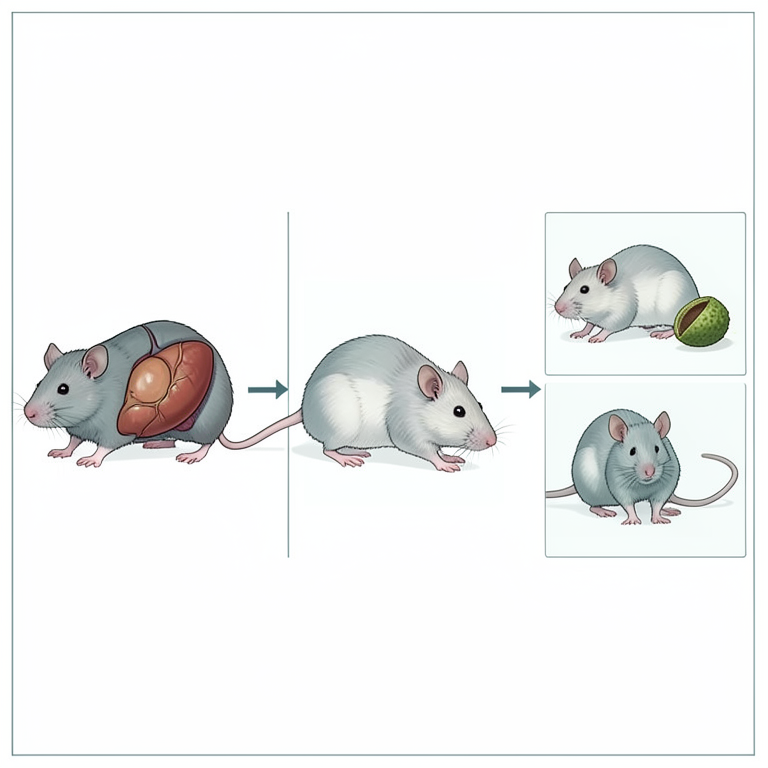
Experimental Treatment Groups
AI-generated conceptual illustration (laboratory rats with liver tumors). It explains the idea and does not show study data.
View source evidence ↗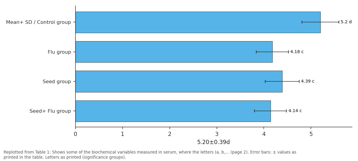
5.20±0.39d: Mean+ SD / Control group vs Flu group vs Seed group vs Seed+ Flu group
Replotted from Table 1 (page 2) of the original paper. Every bar is the paper's own table value.
View source evidence ↗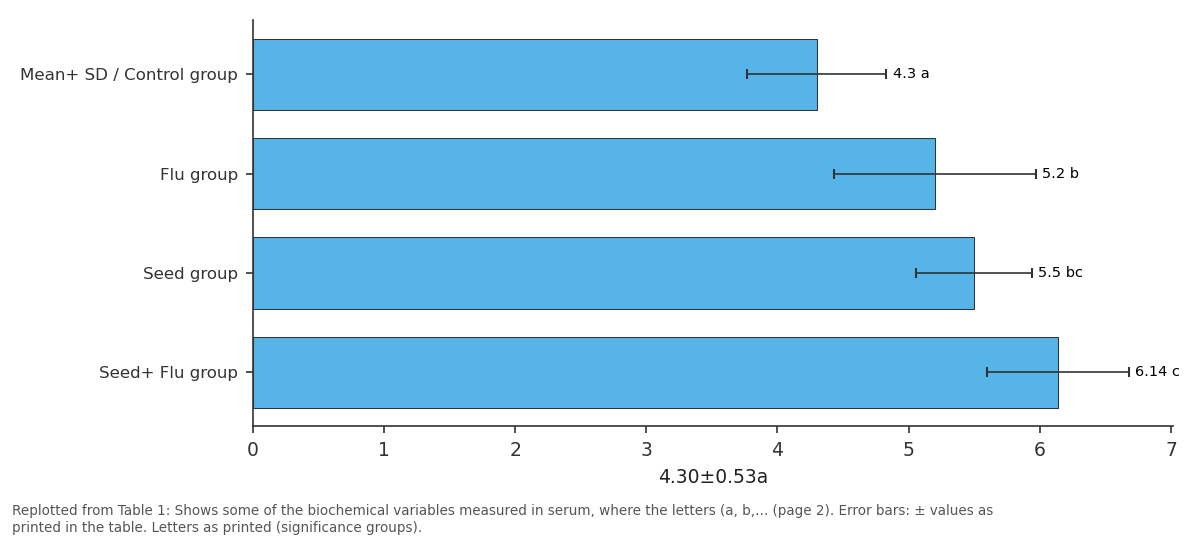
4.30±0.53a: Mean+ SD / Control group vs Flu group vs Seed group vs Seed+ Flu group
Replotted from Table 1 (page 2) of the original paper. Every bar is the paper's own table value.
View source evidence ↗Figures & tables
From the original paper Table 1
 Table 2
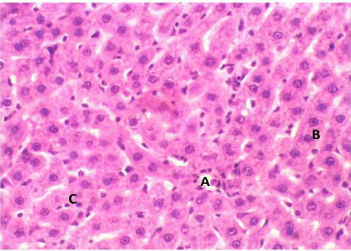 Figure 1
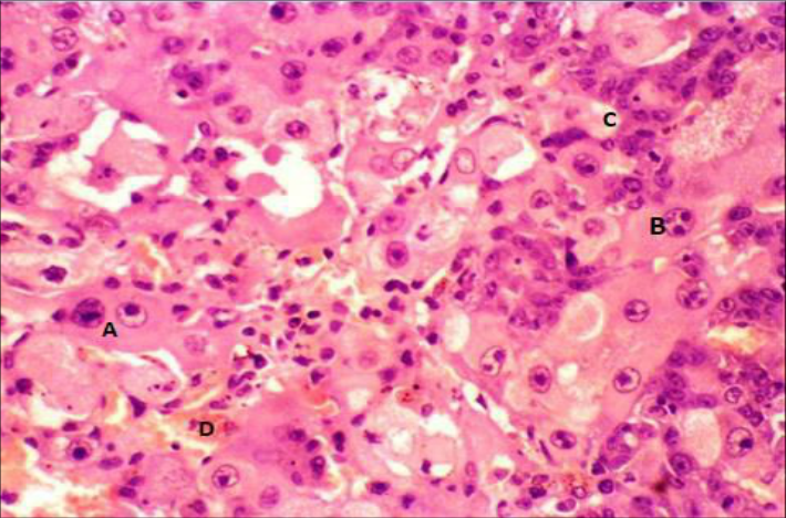 Figure 2
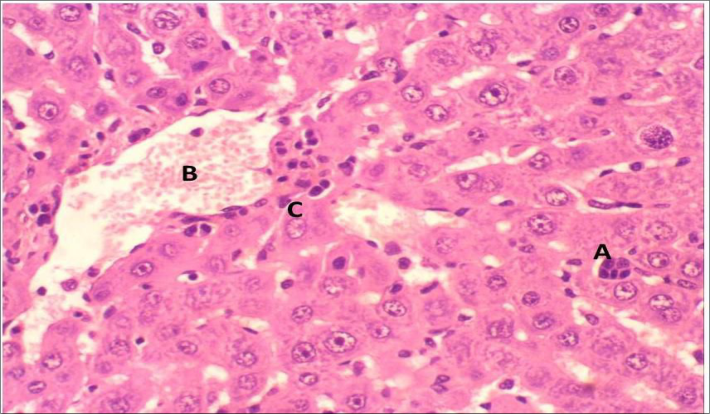 Figure 3
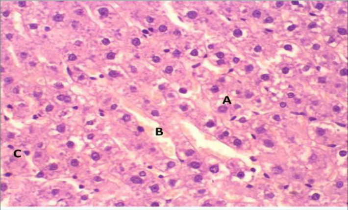 Figure 4
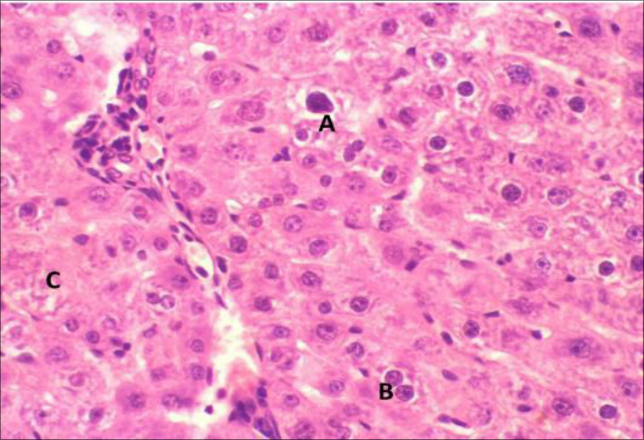 Figure 5
Introduction
Fluorouracil was first introduced as an anticancer agent 30 years ago and is still widely used in the treatment of common malignant tumors including liver, colon, breast, skin, rectum, ovary, pancreatic, and stomach cancers. It is metabolized through metabolic pathways in the body, and despite identifying several potential sites of antitumor activity, the precise mechanism of action and the contribution of each of these sites to tumor suppression or host cell toxicity remain unclear. Various methods are available for measuring fluorouracil in serum, plasma, and other biological fluids, and this drug is poorly absorbed after oral administration, with incorrect bioavailability, hence it is primarily used as an injectable dose, intravenously in humans and intraperitoneally in mice. Additionally, this drug is used to treat malignant skin cancers [2] . This medication is considered a primary chemotherapy treatment for many glandular tumors, urinary tract, and digestive system [3] . Despite its proven high therapeutic efficacy, this medication has many side effects that affect the body. The most common symptoms associated with fluorouracil include diarrhea, mucosal membrane inflammation, body infections, ventricular arrhythmias, arterial spasms, heart muscle atrophy, heart failure, heart shock, and vascular angina [3] . Due to the importance of plants in medical treatments, Annona muricata plant was used to study its effects on liver tumors. It is a straight tree with low branching, reaching a height of 8-10 meters.
The tree has shiny green leaves that are evergreen, and flowers appear anywhere on the trunk or any branch and is usually grown from seeds that can be stored for several months before planting. Seed germination usually takes 3 weeks, but under less than ideal conditions, it can be delayed for 2-3 months. It produces tropical fruits due to the strict environmental requirements for tree cultivation and the short life after harvest of its fruits [4] . The Annonaceae family consists of about 120 genera and is distributed in tropical and subtropical regions around the world. The Annona genus is the most important genus in this family with about 50 other species in Brazil [5] . It is a strange fruit with inedible skins, and the plant is covered with many soft thorns, and the pulp is white. The unripe fruit is green, and when ripe, the skin becomes soft and turns greenish-yellow, and the pulp becomes soft, with a number of black seeds and fibrous membranes around the pulp. Its taste is described as a mix of strawberry, pineapple, and citrus, with a distinctive scent similar to guava fruit [6] . This plant is of great importance due to its various biological effects such as antioxidants, anti-mutagenic, antimicrobial properties, and treatment for diabetes [7] .
The materials and methods
A plant (Annona muricata) was obtained from the local market, 1 kilogram of it was taken, cleaned from impurities and dust, placed in dry and clean bags, and stored in the refrigerator until use. The chemical materials used in this study were standard ready-made Kit packages.
The animals used
Male rats weighing (200-250) grams were taken from the animal house at the College of Veterinary Medicine / University of Mosul for experimental investigation in this study. They were placed in prepared cages equipped with water and their specific animal feed. They were divided into eight groups, left for one week to acclimatize to the laboratory conditions of light and temperature.
Method of extracting seeds from the Annona Muricuta plant
The seeds are removed from the Annona muricata plant, then washed, ground, and dried in an electric oven at 45 degrees Celsius for two weeks, where the seeds become ground into coarse particles. Then, 100 grams of the coarse form are mixed in 500 ml of distilled water and placed in a magnetic stirrer for 48 hours to create the mixture. The mixture is then filtered through a Whatman filter paper and concentrated using a rotary evaporator, followed by drying in an electric oven at 40 degrees Celsius and cooling the extract to 4 degrees Celsius before use [8] .
Doses 1. Thioacetamide
Dissolve thioacetamide in distilled water and administer this substance to rats at a concentration of 100 mg/kg per day for five days [9] .
Fluorouracil
Rats were given 5-Fluorouracil by injection into the peritoneal membrane at a dose of (75 mg/kg), as chemotherapy is given in standard doses to show the minimum weight loss, as large doses have caused weight loss or death in animals [10] .
Plant extracts
After preparing the plant extracts from the seeds of the Annona muricata plant, the rats were given the plant extracts orally at a dose of (500) mg/kg daily for 28 days [8] .
Experimental design
Taking (48) male rats and dividing them into eight groups, each group consisting of six animals. Group 1: Control group (natural control group) for 28 days. Group 2: Treated with (5-Fluorouracil) once a week for 28 days. Group 3: Treated with plant extract (Seeds) for 28 days. Group 4: Treated with plant extract (Seeds) and 5-Fluorouracil for 28 days. Group 5: Receiving daily doses of Thioacetamide for five days. Group 6: Receiving daily doses of Thioacetamide for 5 days then treated with 5-Fluorouracil for 23 days as mentioned above. Group 7: Receiving daily doses of Thioacetamide and then treated with plant extract (Seeds) for 23 days. Group 8: Receiving daily doses of Thioacetamide for five days and then treated with Fluorouracil and plant extract (Seeds).
Blood samples collection
After the specified period, blood was drawn from the rats from the eye socket using special capillary tubes by blood dripping [11] and collected in dry and clean tubes. The tubes were then left in a water bath for 10 minutes at a temperature of 37 degrees Celsius, followed by serum separation using a centrifuge for 15 minutes at a speed of 5000 revolutions per minute and stored in special tubes at (-20) degrees Celsius for biochemical tests. This may be attributed to the side effects resulting from the drug use, as a study indicated that some medications have a negative effect on antioxidant levels. A decrease in glutathione levels was observed during patient treatment, as glutathione removes the free radicals produced by chemotherapy, thereby reducing the toxic effects of the treatment [12] . While there were no significant differences in the concentrations of glutathione in rats injected with 5-Fluorouracil and treated with plant seed extract compared to the group treated with plant seed extract alone, the results also show a significant decrease in glutathione concentrations in the group of rats treated with thioacetamide compared to the group treated with thioacetamide and injected with 5-Fluorouracil. The reason for the decrease in glutathione concentrations in rats treated with thioacetamide may be attributed to a deficiency in the essential precursors necessary for its synthesis during oxidative stress, including NADPH produced by the pentose phosphate pathway, which serves as the cofactor for the enzyme Glutathione reductase, responsible for converting glutathione from its inactive disulfide form to its active form [13] . The decrease in glutathione concentrations is accompanied by a decrease in the levels of other antioxidants in general and an increase in cell sensitivity to oxidative damage, leading to lipid peroxidation. The results also indicated a significant increase in GSH concentrations in rats treated with thioacetamide and injected with 5-Fluorouracil and plant seed extract compared to rats treated with thioacetamide alone. This may be attributed to the fact that glutathione is formed by two enzymes involved in its synthesis, the first enzyme being glutamate-cysteine ligase and the second enzyme being γ-glutamyl transpeptidase, which depend on stimulation to provide the initial substrates for glutathione synthesis, which may be provided by the seeds [14] .
Results and Discussion
Results and Discussion
Malondialdehyde
The results shown in the previous table indicate a significant increase in MDA concentrations at a probability level of p≤0.05 in rats injected with 5-Fluorouracil compared to the control group. This may be attributed to the fact that chemical treatments affect both tumor and normal cells, resulting in toxic secretions and increased free radicals, leading to increased oxidative stress. MDA is one of the products of lipid oxidative stress. Additionally, chemotherapy stimulates enzymes such as Nitric oxide-Synthase and cyclooxygenase enzymes, while increasing ROS [15] . However, no significant differences were observed in MDA levels in rats injected with 5-Fluorouracil and treated with plant seed extract compared to the group treated with plant seed extract. The results indicate a significant increase in MDA concentrations in the group of rats treated with thioacetamide compared to rats treated with thioacetamide and injected with Fluorouracil. This may be attributed to the fact that many oxidative stress-related conditions, including cancerous tumors, exhibit increased activity of free radicals beyond the capacity of antioxidants to neutralize or remove them. This leads to lipid peroxidation, elevation of MDA levels, and an imbalance between the effectiveness of free radicals and antioxidant activity causing oxidative stress and an increase in MDA formation, which leads to loss of cellular membrane elasticity and permeability, thereby increasing the proportion of free radicals [15,16] . The results also revealed a significant decrease in MDA concentrations in rats treated with thioacetamide and injected with 5-Fluorouracil and plant extract from seeds compared to the group of rats treated with thioacetamide and plant extract from seeds, as well as the group treated with thioacetamide alone. This decrease may be attributed to the seeds containing various vital components, including flavonoids and polyphenolics, which are compounds that may inhibit free radicals in the body, resulting in decreased MDA concentrations [17] . The reason may also be attributed to the increase in levels of antioxidant enzymes in the plant extract and the decrease in MDA concentration by inhibiting the growth of cancer cells through the arrest of the G1 cell cycle phase, leading to programmed cell death of cancer cells, in addition to its regulation of mitochondrial function [18] , The drug Fluorouracil, used as a chemotherapeutic treatment, is a pyrimidine analogue with a stable fluorine atom instead of a hydrogen atom at position 5 of the uracil ring. The fluorine interferes with the conversion of Deoxyuridylic acid to Thymidylic acid, depriving the cell of thymidine base. This drug is one of the essential types for DNA synthesis and is primarily used in the treatment of slow-growing tumors [19] . Such properties found in seeds and Fluorouracil may work to reduce MDA concentrations in the body.
Total Protein
The results shown in the table indicate a significant decrease in total protein levels at a probability level of p≤0.05 in rats injected with 5-Fluorouracil compared to the control group. There was also a significant decrease in total protein concentration in rats injected with 5-Fluorouracil and treated with plant seed extract compared to the group treated with plant seed extract alone. This may be attributed to the side effects of the chemical treatment Fluorouracil which has been shown to have adverse effects on the body and may lead to a reduction in blood serum protein concentration [20] . The results also indicate a significant increase in the total protein concentration in rats treated with thioacetamide and injected with Fluorouracil compared to the group treated with thioacetamide alone, as the concentration reached. The results also pointed to a significant increase in the total protein concentration in rats treated with thioacetamide and plant seed extract and injected with Fluorouracil compared to the group treated with thioacetamide and plant seed extract. The reason for the increase in total protein concentration may be attributed to the presence of Fluorouracil, which acts through the chemical treatment response by suppressing the activity of Thymidylate Synthase and its involvement in the metabolic process of DNA and RNA synthesis resulting from cell death [21] , thereby potentially increasing protein concentration in the blood. The plant seed extract contains several amino acids that may help increase total protein concentration in the body [22] .
Alpha Feto Protein
An increase in the level of significance at p≤0.05 was observed in rats injected with Fluorouracil compared to the control group. Additionally, a significant increase was noted in rats treated with the plant seed extract and injected with Fluorouracil, where the AFP concentration was found to be comparable to the group treated with the plant seed extract. This may be attributed to the negative effects of Fluorouracil, which can lead to inflammation in the body [20] . Previous studies have indicated a significant increase in AFP concentration due to the occurrence of liver inflammation, especially acute and chronic inflammation. AFP concentration is also used as a diagnostic marker for liver inflammation [23] . The results also demonstrate a significant increase in AFP concentration in rats treated with thioacetamide compared to the group treated with thioacetamide and injected with Fluorouracil. This may be attributed to the increase in gene expression (AFP-mRNA), leading to its elevation in the bloodstream. This increase is typically observed in liver cancers [24] Studies also indicate a correlation between elevated AFP levels in patients with non-tumorous testicular cancer, making it important for diagnosis, classification, monitoring of progression, and response to chemotherapy [25] . The results also indicated a significant decrease in AFP levels in rats treated with thioacetamide and seed plant extract compared to the group treated with thioacetamide, Fluorouracil, and seeds, and the group treated with thioacetamide alone. The decrease in AFP levels may be attributed to the extent of the impact of phenolic compounds present in the plant and their support and assistance to liver cells in regeneration. The plant components, rich in phenolic compounds, led to a reduction in AFP formation [26] . Therefore, genetic expression is inhibited by the formation of hepatic tumors [27] . As known, AFP is formed early during the S1 phase of the cell and is secreted before the M phase [28] . It is directly proportional to the amount of mRNA produced during the gene synthesis phase, with a half-life of 5-6 days in adult cancer. After that, gene transcription, and as the tumor progresses, it is cleared from the bloodstream by Asialo glycoprotein present on the surface of Kupffer cells, which remove the sugar protein from the bloodstream [29] .
Alkaline Phosphates
The results in Table (2) indicated a significant increase at a probability level of p≤0.05 in the activity of the enzyme ALP in rats injected with the anticancer drug Fluorouracil compared to the control group. This may be attributed to the side effects of Fluorouracil, which cause tissue changes affecting the liver of rats, especially the inner parenchymal cells [30] , and the drug also causes necrosis in liver cells with changes in hepatocytes leading to an increase in enzyme activity in the blood serum [31] . No significant differences in the enzymatic activity of ALP were observed in rats injected with Fluorouracil and treated with plant seed extract compared to the group treated with plant seed extract alone. The results indicate a significant increase in ALP enzyme activity in the group of rats treated with thioacetamide compared to the group injected with Fluorouracil and treated with thioacetamide. This was attributed to a study suggesting that enzyme activity increases in cases such as disturbances in enzyme secretion activity, changes in enzyme receptors, or genetic abnormalities in enzyme formation that may affect its effectiveness [32] . The reason for the increase may also be attributed to obstruction in the bile ducts leading to a failure in enzyme secretion through them, and the relatively narrow bile ducts lead to its accumulation and an increase in enzyme activity in the blood serum. Such an increase occurs in cases of liver cancers, as indicated by previous studies [33,34] . It is also noted from the results a significant decrease in enzyme activity in rats treated with thioacetamide and plant seed extract compared to rats treated with thioacetamide injected with Fluorouracil and plant seed extract. This may be attributed to the plant seed extract containing phenols and flavonoids that may improve liver damage, as well as for the anticancer drug Fluorouracil [35] .
Paraoxonase
The results shown in Table (2) indicate a significant decrease at the probability level p≤0.05 in rats injected with Fluorouracil compared to the control group. This may be attributed to the enzyme resisting the action of chemotherapy through enzyme reactions present in the mitochondria membranes, as it performs the same function to resist chemotherapy in a number of cancer cells. The enzyme plays a vital and defensive role for the cell in an attempt to keep it alive [37,36] . It is inversely proportional to the weight of the animal's body, and this is consistent with previous studies [38] , and no significant differences were found in the enzyme activity in rats injected with Fluorouracil and treated with plant seed extract and the group of rats treated with plant seed extract. The results show a significant decrease in the activity of the paroxonase enzyme in the group of rats treated with thioacetamide compared to the group of rats treated with thioacetamide injected with Fluorouracil, and the reason for the decrease in the activity of the paroxonase enzyme may be that cancer patients suffer from oxidative stress and that cancer has multiple strategies for tumor spread, affecting respiratory chain pathways, increasing free radicals in the body, and affecting enzyme levels [39] . It is also noted that there is a significant increase in enzyme activity in rats treated with thioacetamide and treated with plant seed extract injected with Fluorouracil compared to the group of rats treated with thioacetamide and plant seed extract, and the group of rats treated with thioacetamide, and the reason for this may be that fenugreek seeds possess enzymatic and non-enzymatic antioxidants, which may work to maintain or increase enzyme levels in the body [40] . Flavonoids present in the seeds, such as active polyphenols, act as if they were drug compounds with biological roles and have been used as therapeutic agents, and drugs and chemical compounds may bind to the chemical structure of the enzyme, leading to a reduction in enzyme activity in the blood serum [41] .
Aspartate Transaminase
The results showed a significant increase in rats treated with Fluorouracil drug at a probability level of p≤0.05 compared to the control group. This may be attributed to liver damage caused by Fluorouracil drug, as it is eliminated by approximately 90% through metabolic processes in the liver [42] . The enzyme AST correlates directly with the extent of cell damage, especially in the liver and kidneys [43] . There were no significant differences in rats injected with Fluorouracil and treated with plant seed extract compared to the control group. The results indicate a significant increase in AST enzyme activity in rats treated with thioacetamide compared to the group treated with thioacetamide and injected with Fluorouracil. This could be due to the chemical substances, including thioacetamide, causing damage to the inner area of the liver by increasing levels of Cytochrome P450 Oxidase enzymes, leading to the formation of highly toxic compounds. This is followed by an increase in the production of reactive oxygen species (ROS) and the release of cytokines that assist in inflammation [44] . There were no significant differences in the enzymatic activity in rats treated with thioacetamide and plant seed extract injected with Fluorouracil and the group treated with thioacetamide and plant seed extract. The results also indicate a significant decrease in enzyme activity in rats treated with thioacetamide and plant seed extract injected with Fluorouracil compared to the group treated with thioacetamide. This may be attributed to the chemotherapy that may improve liver function, in addition to the plant seed extract, which contains bioactive compounds that may play an important role in improving liver function [45] .
Alanine Transaminase
The results shown in Table (2) indicated a significant increase at a probability level of p≤0.05 in rats injected with the drug Fluorouracil compared to the control group. Recent studies have confirmed that chemotherapy causes liver toxicity, manifesting in various patterns including hepatic inflammation, cholestasis, and the occurrence of what is known as fatty degeneration, which is a disturbance in the fat metabolism process within the cell. These disturbances may lead to liver damage, resulting in disturbances in liver enzymes and elevated levels [46] . No significant differences were observed in the enzyme activity in rats treated with plant seed extract injected with Fluorouracil compared to the control group treated with plant seed extract. The results indicate a significant increase in enzyme activity in the group of rats treated with Thioacetamide compared to the group treated with Thioacetamide injected with Fluorouracil. This may be attributed to the role of Thioacetamide in increasing coagulation pathways, exacerbating liver cell damage and enlargement, which may increase enzyme activity in the blood serum [47] . No significant differences were observed in enzyme activity in rats treated with Thioacetamide and plant seed extract injected with Fluorouracil compared to the group treated with Thioacetamide and plant seed extract. However, a significant decrease in enzyme activity was observed in rats treated with Thioacetamide and plant seed extract injected with Fluorouracil compared to the group treated with Thioacetamide. This may be attributed to the important role of seeds in prolonging the assumed lifespan of the drug Fluorouracil and supporting it through its components that have the ability to scavenge free radicals, reduce their effects on cells, and this depends on the decrease in the activity of the measured ALT enzyme [26] .
Histopathology
Histopathology
Conclusion
This study demonstrates the effectiveness of 5-Fluorouracil and Annona Muricata seed extract in mitigating liver tumor damage in rats, with improved histology and biochemical markers. The combination of both treatments enhances protective effects, suggesting potential for integrated therapies to enhance efficacy and reduce side effects in liver tumor management.
Figures and tables from the paper
| Clinical Variables | Mean+ SD | — | — | — | — | — | — | — |
|---|---|---|---|---|---|---|---|---|
| Control group | Flu group | Seed group | Seed+ Flu group | TAA group | Flu +TAA | Seed +TAA | Seed+ TAA +Flu | |
| Glutathione (µmole/l) | 5.20±0.39d | 4.18±0.34c | 4.39±0.36c | 4.14±0.34c | 1.77±0.61a | 3.33±0.59b | 3.44±0.35b | 4.15±0.29c |
| Malondialdehyde (µmole/l) | 4.30±0.53a | 5.20±0.77b | 5.50±0.44bc | 6.14±0.54c | 11.35±1.17e | 7.19±0.20d | 7.38±0.65d | 5.51±0.55bc |
| Total Protein (g/dl) | 16.01±3.43e | 12.98±1.14c | 15.35±1.71e | 13.55±0.86cd | 6.85±1.10a | 12.65±0.24c | 9.63±0.073b | 14.89±0.66de |
| AFP (ng/ml) | 4.76±0.72a | 8.26±0.83b | 8.90±0.43b | 13.90±0.43c | 90.16±1.47g | 67.53±1.34f | 47.65±1.04d | 52.10±3.94e |
Table text extracted from the original PDF; superscript letters are shown inline. The original table image is below.
View original table image
| Clinical Variables | Mean + SD | — | — | — | — | — | — | — |
|---|---|---|---|---|---|---|---|---|
| Control group | Flu group | Seed group | Seed +Flu | TAA group | Flu + TAA | Seed +TAA | Seed+ TAA + Flu | |
| Alkaline phosphatase (IU/L) | 47.19±4.91a | 99.64±7.24d | 75.55±3.12bc | 72.26±16.14b | 150.026±4.36f | 111.39±0.23e | 81.29±2.48c | 94.18±3.61d |
| Paroxonas (IU/L) | 126.64±14.59f | 62.092±4.32b | 66.41±10.97bc | 74.14±3.58cd | 35.64±1.57a | 81.60±4.10d | 72.99±2.97cd | 98.06±8.48e |
| AST (IU/L) | 43.19±2.12a | 99.95±3.73e | 62.23±6.10bc | 65.17±3.52c | 107.80±4.11f | 92.41±3.73d | 62.08±1.52bc | 60.17±4.36b |
| ALT (IU/L) | 49.74±3.24a | 80.02±7.05d | 62.37±6.84b | 63.56±3.50b | 146.78±7.047f | 100.16±4.93e | 73.62±1.95c | 70.45±3.68c |
Table text extracted from the original PDF; superscript letters are shown inline. The original table image is below.
View original table image
References
36 in the original paper- Diasio RB, Harris BE. Clinical pharmacology of 5 fluorouracil. Clin Pharmacokinet. 1989;16(4):215-237.
- Sorrentino MF, Kim J, Foderaro AE, Truesdell AG. 5fluorouracil induced cardiotoxicity: review of the literature. Cardiol J. 2012;19(5):453-457.
- Bento EB, Matias EF, Brito Jr FE, Oliveira DR, Coutinho HD, Sawant TP, et al. Sawant TP, Dongre RS. Bio-chemical compositional analysis of Annona muricata: a miracle fruit's review. Int. J Univ. Pharm Bio Sci. 2014;3(2):82-104.
- Okoro-Shekwaga C, Osunde ZD. Physical properties of soursop (Annona muricata) seeds. Int. J Eng. Res. Technol. 2013;2(3):1-5.
- Agbai E, Njoku C, Nwanegwo C, Nwafor A. Effect of aqueous extract of Annona muricata seed on atherogenicity in streptozotocin-induced diabetic rats. Afr. J Pharm Pharmacol. 2015;9(30):745-755.
- Abdel-Hamid N, El-Moselhy M, Fawzy M. Novel Panel of Early Diagnostic Markers for Experimental Hepatocellular Carcinoma. J Health Sci. 2012;2(2):1418.
- Ahles TA, Saykin AJ, Furstenberg CT, Cole B, Mott LA, Skalla K, et al. Neuropsychologic impact of standard-dose systemic chemotherapy in long-term survivors of breast cancer and lymphoma. J Clin. Oncol. 2002;20(2):485-493.
- Stillinger D, Helland K, Van Atta C. Experiments on the transition of homogeneous turbulence to internal waves in a stratified fluid. J Fluid Mech. 1983;131:91122.
- Weijl N, Elsendoorn T, Lentjes E, Hopman G, Wipkink-Bakker A, Zwinderman A, et al. Supplementation with antioxidant micronutrients and chemotherapy-induced toxicity in cancer patients treated with cisplatin-based chemotherapy: a randomized, double-blind, placebo-controlled study. Eur. J Cancer. 2004;40(11):1713-1723.
- Latif YA, El-Bana M, Hussein J, El-Khayat Z, Farrag AR. Effects of resveratrol in combination with 5fluorouracil on N-methylnitrosourea-induced colon cancer in rats. Comp Clin. Pathol. 2019;28(5):13511362.
- Ikediobi CO, Badisa VL, Ayuk-Takem LT, Latinwo LM, West J. Response of antioxidant enzymes and redox metabolites to cadmium-induced oxidative stress in CRL-1439 normal rat liver cells. Int. J Mol. Med. 2004;14(1):87-92.
- Pourghassem-Gargari B, Ebrahimzadeh-Attary V, Rafraf M, Gorbani A. Effect of dietary supplementation with Nigella sativa L. on serum lipid profile, lipid peroxidation and antioxidant defense system in hyperlipidemic rabbits. J Med Plants Res. 2009;3(10):815-821.
- Moghadamtousi SZ, Fadaeinasab M, Nikzad S, Mohan G, Ali HM, Kadir HA. Annona muricata (Annonaceae): A review of its traditional uses, isolated acetogenins and biological activities. Int. J Mol. Sci. 2015;16(7):15625-15658.
- Jensen SA, Sørensen JB. Risk factors and prevention of cardiotoxicity induced by 5-fluorouracil or capecitabine. Cancer Chemother Pharmacol. 2006;58(4):487-493.
- Longley DB, Harkin DP, Johnston PG. 5-fluorouracil: mechanisms of action and clinical strategies. Nat Rev Cancer. 2003;3(5):330-338.
- Kuhajda FP. Fatty acid synthase and cancer: new application of an old pathway. Cancer Res. 2006;66(12):5977-5980.
- Liu Y-r, Lin B-b, Zeng DW, Zhu YY, Chen J, Zheng Q, et al. Alpha-fetoprotein level as a biomarker of liver fibrosis status: A cross-sectional study of 619 consecutive patients with chronic hepatitis B. BMC Gastroenterol. 2014;14(1):145.
- Chen W, Peng J, Ye J, Dai W, Li G, He Y. Aberrant AFP expression characterizes a subset of hepatocellular carcinoma with distinct gene expression patterns and inferior prognosis. J Cancer. 2020;11(2):403.
- Wu M, Liu H, Liu Z, Liu C, Zhang A, Li N. Analysis of serum alpha-fetoprotein (AFP) and AFP-L3 levels by protein microarray. J Int. Med Res. 2018;46(10):42974305.
- Rezaei-Moghadam A, Mohajeri D, Rafiei B, Dizaji R, Azhdari A, Yeganehzad M, et al. Effect of turmeric and carrot seed extracts on serum liver biomarkers and hepatic lipid peroxidation, antioxidant enzymes and total antioxidant status in rats. BioImpacts: BI. 2012;2(3):151.
- Sell S, Leffert H, Muller-Eberhard U, Kida S, Skelly H. Relationship of the biosynthesis of α-fetoprotein, albumin, hemopexin, and haptoglobin to the growth state of fetal rat hepatocyte cultures. Annals of the New York Academy of Sciences. 1975;259(1):45-58.
- Bajin-Katić K, Stankov K, Đolai M, Kovačević Z. Intestinal alkaline phosphatase activity as a molecular marker of enterotoxicity induced by single dose of 5fluorouracil and protective role of orally administered glutamine. Archive of Oncology. 2006;14(3-4):101105.
- Al-Hamdany M, Al-Hubaity A. The histological and histochemical changes of the rat's liver induced by 5fluorouracil. Iraqi Journal of Veterinary Sciences. 2014, 28(2).
- Bailey T, Berg P, Sandy C. The effect of highperformance work practices on employee earnings in the steel, apparel, and medical electronics and imaging industries. ILR Review. 2001;54(2A):525-543.
- Gopal DV, Rosen HR. Abnormal findings on liver function tests: Interpreting results to narrow the diagnosis and establish a prognosis. Postgraduate medicine. 2000;107(2):100-114.
- Yeo W, Mo F, Koh J, Chan A, Leung T, Hui P, et al. Quality of life is predictive of survival in patients with unresectable hepatocellular carcinoma. Annals of Oncology. 2006;17(7):1083-1089.
- Ukwubile CA. Phytochemical screening and antiovarian cancer properties of Annona muricata Linn (Annonaceae) Seed Ethanol Extract. Int. J Pharm. Front. Res. 2012;2:09-17.
- Okuturlar Y, Gunaldi M, Kocoglu H, Hursitoglu M, Gedikbasi A, Acarer D, et al. Serum paraoxonase and arylesterase can be useful markers to predict neoadjuvant chemotherapy requirement in patients with breast cancer. Journal of cancer research and therapeutics. 2018;14(9):362.
- Vijayameena C, Subhashini G, Loganayagi M, Ramesh B. Original Research Article Phytochemical screening and assessment of antibacterial activity for the bioactive compounds in Annona muricata. Int. J Curr. Microbiol. Appl. Sci. 2013;2:01-08.
- Kıyıcı A, Okudan N, Gökbel H, Belviranlı M. The effect of grape seed extracts on serum paraoxonase activities in streptozotocin-induced diabetic rats. Journal of medicinal food. 2010;13(3):725-728.
- Tural D, Akar E, Öztürk MA, Yıldız Ö, Turna H, Serdengeçti S. Severe liver dysfunction and safe use of 5-fluorouracil leucovorin and oxaliplatin in one patient with metastatic colorectal carcinoma. Journal of Cancer Research and Therapeutics. 2014;10(3):745.
- Huang X-J, Choi Y-K, Im H-S, Yarimaga O, Yoon E, Kim H-S. Aspartate aminotransferase (AST/GOT) and alanine aminotransferase (ALT/GPT) detection techniques. Sensors. 2006;6(7):756-782.
- de David C, Rodrigues G, Bona S, Meurer L, GonzálezGallego J, Tuñón MJ, et al. Role of quercetin in preventing thioacetamide-induced liver injury in rats. Toxicologic pathology. 2011;39(6):949-957.
- Orabi SH, Shawky SM. Effect of date palm (Phoenix dactylifera) seeds extracts on hematological, biochemical parameters and some fertility indices in male rats. Int J Sci Basic Appl Res. 2014;17:137-147.
- Grigorian A, O'Brien CB. Hepatotoxicity secondary to chemotherapy. Journal of clinical and translational hepatology. 2014;2(2):95.
- Lin Y-Y, Hu C-T, Sun DS, Lien T-S, Chang H-H. Thioacetamide-induced liver damage and thrombocytopenia is associated with induction of antiplatelet autoantibody in mice. Scientific reports. 2019;9(1):01-11.